

The DOI matches its Crossref record; 215 of the 215 references it cites were found in a registry or an index.
TREM2 dependent and independent functions of microglia in Alzheimer’s disease
Open accessAbstract
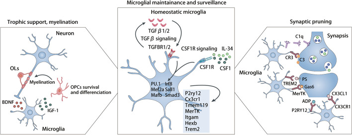
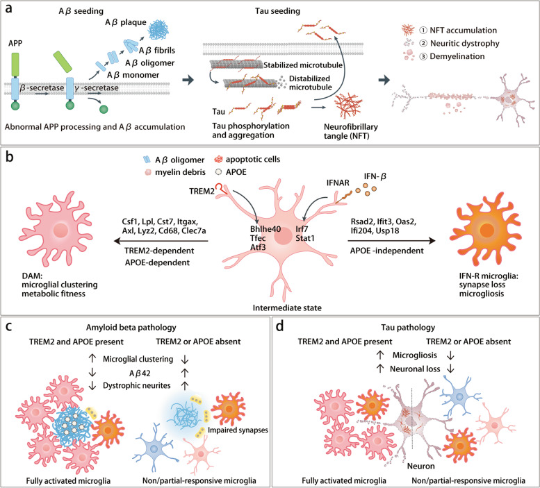
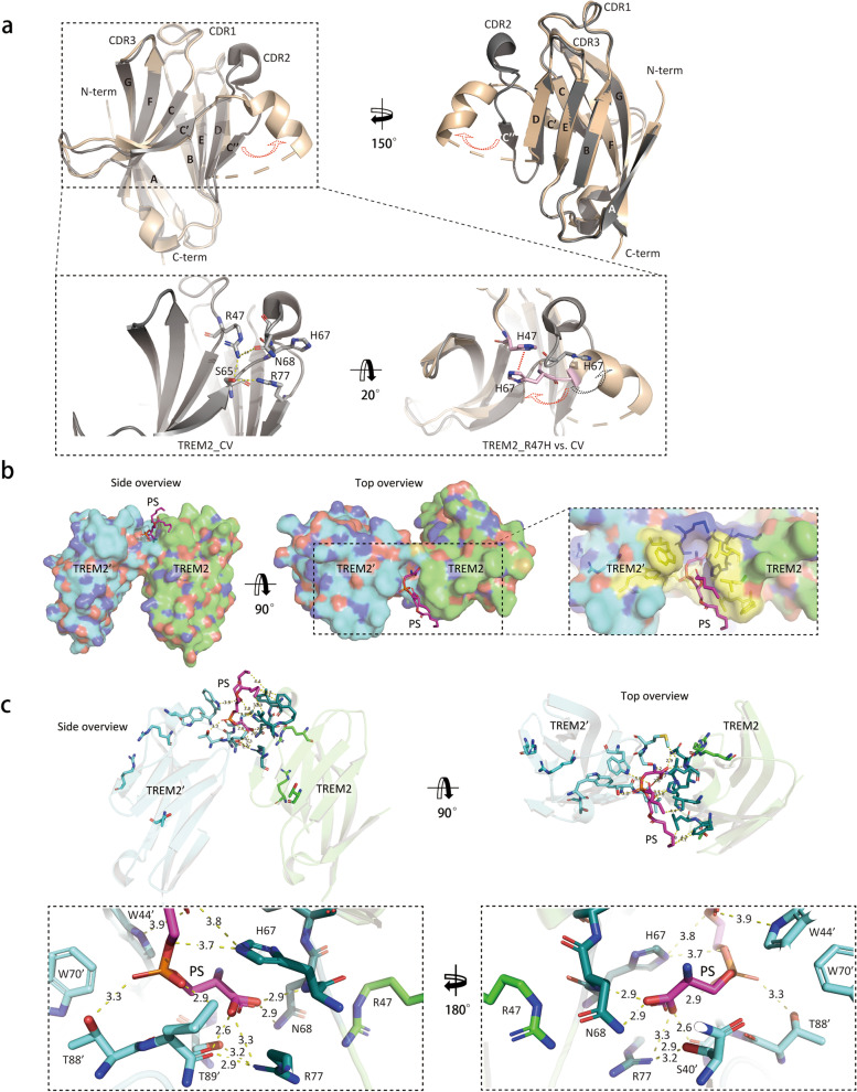
Microglia are central players in brain innate immunity and have been the subject of extensive research in Alzheimer's disease (AD). In this review, we aim to summarize the genetic and functional discoveries that have advanced our understanding of microglia reactivity to AD pathology. Given the heightened AD risk posed by rare variants of the microglial triggering receptor expressed on myeloid cells 2 (TREM2), we will focus on the studies addressing the impact of this receptor on microglia responses to amyloid plaques, tauopathy and demyelination pathologies in mouse and human. Finally, we will discuss the implications of recent discoveries on microglia and TREM2 biology on potential therapeutic strategies for AD.
Overview
The claims and statistics of this paper haven’t been checked yet.
Run the check — free, no account needed. It reads the open-access text and checks the sources it cites.
Integrity signals
Registry
Cites
Statistics
Figures
Verification
Claims the paper makes
The claims and statistics of this paper haven’t been checked yet.
Run the check — free, no account needed. It reads the open-access text and checks the sources it cites.
Claims other papers make about this one
Each is checked against this paper’s own text: does it say what the citing paper says it does?
The source backs part of the claim, not all of it — or backs it for a different population, condition or comparison.
In the citing paper
“Processing of TREM2 by γ -secretase generates soluble TREM2, which modulates microglial sur vival and activation in the AD context [14].”
From Is there a role for B cells in Alzheimer's disease? (2026) · cites this paper as [14]
Experimental — accuracy not yet measuredThe cited source says what the claim attributes to it.
In the citing paper
“TREM2 ligands include zwitterionic and anionic phospholipids, glycolipids, apoptotic cells, lipoproteins such as HDL, LDL, APOE and APOJ/CLU, as well as Aβ [30, 31].”
From Microglial function, INPP5D/SHIP1 signaling, and NLRP3 inflammasome activation: implications for Alzheimer’s disease (2023) · cites this paper as [30]
Self-check
Percentages, sums and reported differences re-computed from the paper’s own numbers.
The self-check needs the paper’s full text.
Citations
Cites
Amyloid oligomers exacerbate tau pathology in a mouse model of tauopathy
Selenica, M.-L. B., Brownlow, M., Jimenez, J. P., Lee, D. C., Pena, G., Dickey, C. A., Gordon, M. N., & Morgan, D. (2013) · Neurodegenerative Diseases · doi:10.1159/000337230 · Cited by 10,359
Alzheimer's disease: the amyloid cascade hypothesis
Hardy, J. A., & Higgins, G. A. (1992) · Science · doi:10.1126/science.1566067 · Cited by 7,484
Macrophage activation and polarization: nomenclature and experimental guidelines
Murray, P. J., Allen, J. E., Biswas, S. K., Fisher, E. A., Gilroy, D. W., Goerdt, S., Gordon, S., Hamilton, J. A., Ivashkiv, L. B., Lawrence, T., et al. (2014) · Immunity · doi:10.1016/j.immuni.2014.06.008 · Cited by 6,337
The amyloid hypothesis of Alzheimer's disease at 25 years
Selkoe, D. J., & Hardy, J. (2016) · EMBO Molecular Medicine · doi:10.15252/emmm.201606210 · Cited by 6,164
Lecanemab in early Alzheimer's disease
van Dyck, C. H., Swanson, C. J., Aisen, P., Bateman, R. J., Chen, C., Gee, M., Kanekiyo, M., Li, D., Reyderman, L., Cohen, S., et al. (2023) · New England Journal of Medicine · doi:10.1056/nejmoa2212948 · Cited by 5,678
A Unique Microglia Type Associated with Restricting Development of Alzheimer’s Disease
Keren-Shaul, H., Spinrad, A., Weiner, A., Matcovitch-Natan, O., Dvir-Szternfeld, R., Ulland, T. K., David, E., Baruch, K., Lara-Astaiso, D., Toth, B., et al. (2017) · Cell · doi:10.1016/j.cell.2017.05.018 · Cited by 5,493
Self-citationFate Mapping Analysis Reveals That Adult Microglia Derive from Primitive Macrophages
Ginhoux, F., Greter, M., Leboeuf, M., Nandi, S., See, P., Gokhan, S., Mehler, M. F., Conway, S. J., Ng, L. G., Stanley, E. R., et al. (2010) · Science · doi:10.1126/science.1194637 · Cited by 5,175
Synaptic Pruning by Microglia Is Necessary for Normal Brain Development
Paolicelli, R. C., Bolasco, G., Pagani, F., Maggi, L., Scianni, M., Panzanelli, P., Giustetto, M., Ferreira, T. A., Guiducci, E., Dumas, L., et al. (2011) · Science · doi:10.1126/science.1202529 · Cited by 4,244
Oakley, H., Cole, S. L., Logan, S., Maus, E., Shao, P., Craft, J., Guillozet-Bongaarts, A., Ohno, M., Disterhoft, J., Van Eldik, L., et al. (2006) · The Journal of Neuroscience · doi:10.1523/jneurosci.1202-06.2006 · Cited by 3,530
Apolipoprotein E and Alzheimer disease: risk, mechanisms and therapy
Liu, C.-C., Kanekiyo, T., Xu, H., & Bu, G. (2013) · Nature Reviews Neurology · doi:10.1038/nrneurol.2012.263 · Cited by 3,459
Research works printed but not linked: 1
- Depp C, Sun T, Sasmita AO, Spieth L, Berghoff SA, Steixner-Kumar AA, Subramanian S, Möbius W, Göbbels S, Saher G et al: Ageing-associated myelin dysfunction drives amyloid deposition in mouse models of Alzheimer’s disease. 2021. BioRixv. Available from https://www.biorxiv.org/content/biorxiv/early/2021/08/02/2021.07.31.454562.full.pdf.
Cited by
Sorted by citations
Tissue-specific macrophages: how they develop and choreograph tissue biology
Mass, E., Nimmerjahn, F., Kierdorf, K., & Schlitzer, A. (2023) · Nature Reviews Immunology · doi:10.1038/s41577-023-00848-y · Cited by 621
Emerging diagnostics and therapeutics for Alzheimer disease
Self, W. K., & Holtzman, D. M. (2023) · Nature Medicine · doi:10.1038/s41591-023-02505-2 · Cited by 381
Neuroinflammation across neurological diseases
Shi, F., & Yong, V. W. (2025) · Science · doi:10.1126/science.adx0043 · Cited by 325
Long, H., Simmons, A., Mayorga, A. J., Burgess, B., Nguyen, T., Budda, B., Rychkova, A., Rhinn, H., Tassi, I., Ward, M. E., et al. (2024) · Alzheimer s Research & Therapy · doi:10.1186/s13195-024-01599-1 · Cited by 153
TREM2 regulates microglial lipid droplet formation and represses post-ischemic brain injury
Wei, W., Zhang, L., Xin, W., Pan, Y., Tatenhorst, L., Hao, Z., Gerner, S. T., Huber, S., Juenemann, M., Butz, M., et al. (2024) · Biomedicine & Pharmacotherapy · doi:10.1016/j.biopha.2023.115962 · Cited by 122
Schlepckow, K., Morenas‐Rodríguez, E., Hong, S. Y., & Haass, C. (2023) · The Lancet Neurology · doi:10.1016/s1474-4422(23)00247-8 · Cited by 122
Decoding microglial immunometabolism: a new frontier in Alzheimer's disease research
Jung, E. S., Choi, H., & Mook-Jung, I. (2025) · Molecular Neurodegeneration · doi:10.1186/s13024-025-00825-0 · Cited by 124
Terzioglu, G., & Young-Pearse, T. L. (2023) · Molecular Neurodegeneration · doi:10.1186/s13024-023-00674-9 · Cited by 91
Müller, L., Benedetto, S. D., & Müller, V. (2025) · Cells · doi:10.3390/cells14010054 · Cited by 102
Neuroimmune crosstalk in chronic neuroinflammation: microglial interactions and immune modulation
Müller, L., & Benedetto, S. D. (2025) · Frontiers in Cellular Neuroscience · doi:10.3389/fncel.2025.1575022 · Cited by 94
A single-cell and spatial RNA-seq database for Alzheimer’s disease (ssREAD)
Wang, C., Acosta, D., McNutt, M., Bian, J., Ma, A., Fu, H., & Ma, Q. (2024) · Nature Communications · doi:10.1038/s41467-024-49133-z · Cited by 73
Current views on meningeal lymphatics and immunity in aging and Alzheimer’s disease
Rego, S., Sánchez, G., & Mesquita, S. D. (2023) · Molecular Neurodegeneration · doi:10.1186/s13024-023-00645-0 · Cited by 79
Melchiorri, D., Merlo, S., Micallef, B., Borg, J.-J., & Dráfi, F. (2023) · Frontiers in Pharmacology · doi:10.3389/fphar.2023.1196413 · Cited by 73
Microglia, Trem2, and Neurodegeneration
Shi, Q., Gutierrez, R. A., & Bhat, M. A. (2025) · The Neuroscientist · doi:10.1177/10738584241254118 · Cited by 69
Cui, Y., Chen, C., Tang, Z., Yuan, W., Yue, K., Cui, P., Qiu, X., Zhang, H., Li, T., Zhu, X., et al. (2024) · Cell Death and Disease · doi:10.1038/s41419-024-06756-w · Cited by 75
Complement-mediated synapse loss in Alzheimer’s disease: mechanisms and involvement of risk factors
Wen, L., Bi, D., & Shen, Y. (2023) · Trends in Neurosciences · doi:10.1016/j.tins.2023.11.010 · Cited by 64
Schartz, N. D., Liang, H. Y., Carvalho, K., Chu, S., Mendoza‐Arvilla, A., Petrisko, T. J., Gómez-Arboledas, Á., Mortazavi, A., & Tenner, A. J. (2024) · Nature Communications · doi:10.1038/s41467-024-51163-6 · Cited by 66
Podleśny-Drabiniok, A., Novikova, G., Liu, Y., Dunst, J., Temizer, R., Giannarelli, C., Marro, S., Kreslavsky, T., Marcora, E., & Goate, A. M. (2024) · Nature Communications · doi:10.1038/s41467-024-46315-7 · Cited by 52
Beyond Amyloid and Tau: The Critical Role of Microglia in Alzheimer’s Disease Therapeutics
Dias, D., & Socodato, R. (2025) · Biomedicines · doi:10.3390/biomedicines13020279 · Cited by 60
Liu, N., Jiang, Y., Xiu, Y., Tortelote, G. G., Xia, W., Wang, Y., Li, Y., Shi, S. X.-Y., Han, J., Vidoudez, C., et al. (2025) · Science Translational Medicine · doi:10.1126/scitranslmed.adn2635 · Cited by 53
Hou, J., Chen, Y., Cai, Z., Heo, G. S., Yuede, C. M., Wang, Z., Lin, K., Saadi, F., Tršan, T., Nguyen, A. T., et al. (2024) · Science Translational Medicine · doi:10.1126/scitranslmed.adj9052 · Cited by 44
Self-citationJohnston, K. G., Berackey, B. T., Tran, K. M., Gelber, A., Yu, Z., MacGregor, G. R., Mukamel, E. A., Tan, Z., Green, K. N., & Xu, X. (2024) · Molecular Psychiatry · doi:10.1038/s41380-024-02651-0 · Cited by 51
Mesquita, S. D., & Rua, R. (2024) · Trends in Immunology · doi:10.1016/j.it.2024.03.007 · Cited by 46
Shin, H. J., Kim, I. S., Choi, S. G., Lee, K., Park, H., Shin, J., Kim, D., Beom, J., Yi, Y. Y., Gupta, D. P., et al. (2024) · Molecular Neurodegeneration · doi:10.1186/s13024-024-00715-x · Cited by 45
The TREM2 agonistic antibody AL002 in early Alzheimer’s disease: a phase 2 randomized trial
Mummery, C. J., Mayorga, A. J., Simmons, A., Chow, T. W., Burgess, B., Nguyen, T., 高晶晶, Budda, B., Park, L. Q., Gupta, R., et al. (2026) · Nature Medicine · doi:10.1038/s41591-026-04273-1 · Cited by 51
The dual nature of neuroinflammation in networked brain
Müller, L., Benedetto, S. D., & Müller, V. (2025) · Frontiers in Immunology · doi:10.3389/fimmu.2025.1659947 · Cited by 43
Decoding mechanisms and protein markers in lung-brain axis
Huang, S., Zhou, Y., Ji, H., Zhang, T., Liu, S., Ma, L., Deng, D., Ding, Y., Han, L., Shu, S., et al. (2025) · Respiratory Research · doi:10.1186/s12931-025-03272-z · Cited by 38
Etxeberria, A., Shen, Y.-A. A., Vito, S., Silverman, S. M., Imperio, J., Lalehzadeh, G., Soung, A. L., Du, C., Xie, L., Choy, M. K., et al. (2024) · The Journal of Neuroscience · doi:10.1523/jneurosci.2347-23.2024 · Cited by 34
Neuroimaging biomarkers and CSF sTREM2 levels in Alzheimer’s disease: a longitudinal study
Nabizadeh, F., Seyedmirzaei, H., & Karami, S. (2024) · Scientific Reports · doi:10.1038/s41598-024-66211-w · Cited by 36
PET imaging of microglia in Alzheimer's disease using copper-64 labeled TREM2 antibodies
Shojaei, M., Schaefer, R., Schlepckow, K., Kunze, L. H., Struebing, F. L., Brunner, B., Willem, M., Bartos, L. M., Feiten, A., Palumbo, G., et al. (2024) · Theranostics · doi:10.7150/thno.97149 · Cited by 35
Myelin debris phagocytosis in demyelinating disease
Gao, R., Song, S., Tian, M., Wang, L., Zhang, Y., & Li, X. (2024) · Glia · doi:10.1002/glia.24602 · Cited by 31
ncRNAs and their impact on dopaminergic neurons: Autophagy pathways in Parkinson's disease
Thapa, R., Moglad, E. H. O., Afzal, M., Gupta, G., Bhat, A. A., Almalki, W. H., Kazmi, I. A., Alzarea, S. I., Pant, K., Ali, H., et al. (2024) · Ageing Research Reviews · doi:10.1016/j.arr.2024.102327 · Cited by 29
Gong, B., Zhang, W., Cong, W., Gu, Y., Ji, W., Yin, T., Zhou, H., Hu, H., Zhuang, J., Luo, Y., et al. (2024) · Advanced Healthcare Materials · doi:10.1002/adhm.202400149 · Cited by 31
Zhuang, X., Jie, L., Song, Y., Ban, R., Zhao, X., Xia, Z., Wang, Z., & Zhang, G. (2024) · NeuroMolecular Medicine · doi:10.1007/s12017-024-08815-z · Cited by 33
Microglia in ALS: Insights into Mechanisms and Therapeutic Potential
Bond, S., Saxena, S., & Sierra-Delgado, J. A. (2025) · Cells · doi:10.3390/cells14060421 · Cited by 32
Clinical relevance of animal models in aging-related dementia research
Padmanabhan, P., & Götz, J. (2023) · Nature Aging · doi:10.1038/s43587-023-00402-4 · Cited by 30
Zhou, W., He, J., Shen, G., Liu, Y., Zhao, P., & Li, J. (2024) · Cell Death and Disease · doi:10.1038/s41419-024-07002-z · Cited by 28
Advancements and challenges in mouse models of Alzheimer’s disease
Qian, Z., Li, Y., & Ye, K. (2024) · Trends in Molecular Medicine · doi:10.1016/j.molmed.2024.10.010 · Cited by 30
Leri, M., Vasarri, M., Carnemolla, F., Oriente, F., Cabaro, S., Stio, M., Degl’Innocenti, D., Stefani, M., & Bucciantini, M. (2023) · Pharmaceuticals · doi:10.3390/ph16070933 · Cited by 29
Macrophage activation contributes to diabetic retinopathy
Zhang, Y., & Zhou, A. (2024) · Journal of Molecular Medicine · doi:10.1007/s00109-024-02437-5 · Cited by 27
TREM2 regulates microglial phagocytosis of synapses in innate immune tolerance
Meng, J., Han, L., Xu, H., Zhang, L., Liu, Z., Zhou, Y., Zhang, X., Luo, H., & Zhang, Y. (2023) · International Immunopharmacology · doi:10.1016/j.intimp.2023.111445 · Cited by 25
Hassan, H., Rawlinson, C., Lan, Y., Jenkins, S. I., & Chen, R. (2025) · Biomolecules · doi:10.3390/biom15111629 · Cited by 30
TREM2 in Neurodegenerative Disorders: Mutation Spectrum, Pathophysiology, and Therapeutic Targeting
Yang, H.-W., Kim, D., Yang, Y., Bagyinszky, E., & An, S. S. A. (2025) · International Journal of Molecular Sciences · doi:10.3390/ijms26157057 · Cited by 26
Yang, X., Wang, J., Jia, X.-T., Yang, Y., Fang, Y., Ying, X., Li, H., Zhang, M., Wei, J., & Pan, Y. (2025) · Journal of Alzheimer s Disease · doi:10.1177/13872877241313223 · Cited by 22
Shi, J., & Huang, S. (2023) · International Journal of Molecular Sciences · doi:10.3390/ijms25010016 · Cited by 22
Fu, L., Luo, T., Hao, Z., Pan, Y., Xin, W., Zhang, L., Lai, Z., Zhang, H., Liu, H., & Wei, W. (2025) · Frontiers in Neuroscience · doi:10.3389/fnins.2025.1603292 · Cited by 21
da Rocha, J. F., Lance, M. L., Luo, R., Schlachter, P., Moreira, L., Iqbal, M. A., Kuhn, P., Gardner, R. S., Valaris, S., Islam, M. R., et al. (2025) · Nature Neuroscience · doi:10.1038/s41593-025-01971-w · Cited by 22
Gopalakrishna, P. K., Nassir, C. M. N. C. M., Anandan, S., Hussan, F., Dandala, K. C. R., Sura, S., Jagadeesan, S., Moklas, M. A. M., Vishnumukkala, T., & Hein, Z. M. (2026) · Cellular and Molecular Neurobiology · doi:10.1007/s10571-026-01671-4 · Cited by 23
Wang, S., Xi, J., Zhang, M., & Wang, J. (2025) · International Journal of Biological Macromolecules · doi:10.1016/j.ijbiomac.2024.139394 · Cited by 20
Liu, W., Zhang, Y.-R., Ge, Y., Wang, H.-F., Cheng, W., & Yu, J. (2023) · Molecular Neurobiology · doi:10.1007/s12035-023-03648-6 · Cited by 18
Zajac, D. J., Simpson, J., Zhang, E., Parikh, I., & Estus, S. (2023) · Genes · doi:10.3390/genes14030763 · Cited by 18
Microglial Neuroinflammation in Alzheimer’s Disease: Mechanisms and Therapies
Erdağ, E., & Haskologlu, I. C. (2025) · Journal of dementia and Alzheimer's disease · doi:10.3390/jdad2030029 · Cited by 20
Microglial apolipoprotein E particles contribute to neuronal senescence and synaptotoxicity
Wang, N., Cai, L., Pei, X., Lin, Z., Huang, L., Liang, C., Wei, M., Shao, L., Guo, T., Huang, F., et al. (2024) · iScience · doi:10.1016/j.isci.2024.110006 · Cited by 18
Neuroinflammation: An Oligodendrocentric View
Festa, L. K., Jordan‐Sciutto, K. L., & Grinspan, J. B. (2025) · Glia · doi:10.1002/glia.70007 · Cited by 18
Emerging Role of Oligodendrocytes Malfunction in the Progression of Alzheimer's Disease
Tylek, K., & Basta‐Kaim, A. (2025) · Journal of Neuroimmune Pharmacology · doi:10.1007/s11481-025-10236-z · Cited by 19
Wieczorek, K., Markiewicz, M., Walczewska, A., & Zgórzyńska, E. (2023) · Cellular Physiology and Biochemistry · doi:10.33594/000000668 · Cited by 16
Seo, D., Choi, Y., Jeong, E., Bang, S., Lee, J. S., Jang, I.-H., Choi, L., Kim, J. H., Shin, W., Seo, B.-R., et al. (2025) · Nature Communications · doi:10.1038/s41467-025-65597-z · Cited by 19
Cyr, B. S., Ranaldi, E. D. L. R. M. C., Hadad, R., Dietrich, W. D., Keane, R. W., & Vaccari, J. P. D. R. (2024) · Frontiers in Molecular Neuroscience · doi:10.3389/fnmol.2024.1369781 · Cited by 15
The role of microglial TREM2 in development: A path toward neurodegeneration?
Matteoli, M. (2024) · Glia · doi:10.1002/glia.24574 · Cited by 16
Zhang, Y. C., Li, Z., Guan, H., Qiu, Z., & Zou, C. (2025) · Clinical and Translational Medicine · doi:10.1002/ctm2.70548 · Cited by 17
Farzan, M., Rounkian, M. S., Rizi, A. A., Heidari, Z., Farzan, M., Fathi, M., Aghaei, A., Azadegan‐Dehkordi, F., & Bagheri, N. (2024) · Experimental Neurology · doi:10.1016/j.expneurol.2024.115071 · Cited by 15
Njoku, G. C., & Kanmogne, G. D. (2025) · International Journal of Molecular Sciences · doi:10.3390/ijms26189069 · Cited by 15
The Pathogenesis in Alzheimer's Disease: TREM2 as a Potential Target
Liu, G., Zhang, L., Fan, Y., & Ji, W. (2023) · Journal of Integrative Neuroscience · doi:10.31083/j.jin2206150 · Cited by 13
Variants in the MS4A cluster interact with soluble TREM2 expression on biomarkers of neuropathology
Winfree, R. L., Nolan, E., Dumitrescu, L. C., Blennow, K., Zetterberg, H., Gifford, K. A., Pechman, K. R., Seto, M., Petyuk, V., Wang, Y., et al. (2024) · Molecular Neurodegeneration · doi:10.1186/s13024-024-00727-7 · Cited by 12
General Pathophysiology of Microglia
Tremblay, M., & Verkhratsky, A. (2024) · Advances in neurobiology · doi:10.1007/978-3-031-55529-9_1 · Cited by 13
Xiao, Z., Wang, Y., Chen, Y., Jin, L., Shi, Y., Liu, C., Fu, C., & Cao, Y. (2024) · American Journal of Physiology-Renal Physiology · doi:10.1152/ajprenal.00219.2024 · Cited by 13
Self-citationRole of TREM2 in immune and neurological diseases: Structure, function, and implications
Yin, P., Su, Z., Shu, X., Dong, Z., & Tian, Y. (2024) · International Immunopharmacology · doi:10.1016/j.intimp.2024.113286 · Cited by 14
Hu, L., Liu, J., Peng, J., Li, X., Huang, Z., Zhang, C., & Fan, S. (2025) · Diseases · doi:10.3390/diseases13020060 · Cited by 12
Giorgio, J., Jonson, C., Wang, Y., Yokoyama, J. S., Wang, J., Jagust, W. J., & Initiative, O. B. O. T. A. D. N. (2025) · Nature Communications · doi:10.1038/s41467-025-60370-8 · Cited by 14
News & views: anti-amyloid antibodies and novel emerging approaches to Alzheimer’s disease in 2023
Gandy, S. (2023) · Molecular Neurodegeneration · doi:10.1186/s13024-023-00656-x · Cited by 11
Basic Microglial Functions and How They Go Awry in Neurodegenerative Disease
McKinney, J. E., Bennett, M. L., & Bennett, F. C. (2026) · Annual Review of Pathology Mechanisms of Disease · doi:10.1146/annurev-pathmechdis-042624-114332 · Cited by 13
Systems biology of Alzheimer’s Disease: a scoping review of key pathways and mechanisms
Bice, P. J., Nho, K. T., Ertekin‐Taner, N., & Saykin, A. J. (2026) · Molecular Neurodegeneration · doi:10.1186/s13024-026-00934-4 · Cited by 14
Suk, K. (2026) · Experimental Neurology · doi:10.1016/j.expneurol.2026.115737 · Cited by 11
Chao, S., Shan, S., Liu, Z., Liu, Z., Wang, S., Qiang, Y., Ni, W., Li, H., Dong, C., Jia, Q., et al. (2024) · International Immunopharmacology · doi:10.1016/j.intimp.2024.112926 · Cited by 11
Novel insights into the role of TREM2 in cerebrovascular diseases
Awuah, W. A., Ben-Jaafar, A., Kong, J. S. H., Sanker, V., Shah, M. H., Poornaselvan, J., Frimpong, M., Imran, S., Alocious, T., Abdul-Rahman, T., et al. (2024) · Brain Research · doi:10.1016/j.brainres.2024.149245 · Cited by 10
TREM 2 in Parkinson’s Disease: A Promising Candidate Gene for Disease Susceptibility and Progression
Alonge, P., Balistreri, C. R., Angelo, T., Magro, D., Rubino, E., & Monastero, R. (2025) · Brain Sciences · doi:10.3390/brainsci15040379 · Cited by 10
Xu, B., Tang, C., Han, R., Zhu, C., Yang, Y., Li, H. J., Wu, N., & He, D. (2025) · Journal of Alzheimer s Disease Reports · doi:10.1177/25424823251326044 · Cited by 10
McMillan, I. O., Liang, L., Su, G., Song, X., Drago, K., Yang, H., Alvarez, C., Sood, A., Gibson, J. M., Woods, R. J., et al. (2024) · Journal of Biological Chemistry · doi:10.1016/j.jbc.2024.107691 · Cited by 11
Pathologic and Therapeutic Schwann Cells
Shurin, M. R., Wheeler, S., Zhong, H. M., & Zhou, Y. (2025) · Cells · doi:10.3390/cells14171336 · Cited by 9
Connell, E., Gall, G. L., McArthur, S., Láng, L., Breeze, B., Liaquat, M., Pontifex, M. G., Sami, S. A. K., Pourtau, L., Gaudout, D., et al. (2026) · Gut Microbes · doi:10.1080/19490976.2026.2614030 · Cited by 12
El-Hamri, M.-A., & Abboussi, O. (2026) · Ageing Research Reviews · doi:10.1016/j.arr.2026.103138 · Cited by 9
Kadam, R., Gupta, M., Lazarov, O., & Prabhakar, B. S. (2024) · Journal of Leukocyte Biology · doi:10.1093/jleuko/qiae134 · Cited by 8
Que, X., Zhang, T., Liu, X. Y., Yin, Y., Xia, X., Gong, P., Song, W., Qin, Q., Xu, Q., & Tang, Y. (2025) · Progress in Neurobiology · doi:10.1016/j.pneurobio.2025.102732 · Cited by 8
Elucidating the Role of Trem2 in Lipid Metabolism and Neuroinflammation
Zhao, C., Qi, W., Lv, X., Gao, X.-L., Liu, C.-N., & Zheng, S.-M. (2025) · CNS Neuroscience & Therapeutics · doi:10.1111/cns.70338 · Cited by 7
Arcos-Encarnación, B., Cortes-Flores, E., Barón-Mendoza, I., Almazán, J. L., Valle‐García, D., de León‐Guerrero, S. D., Hovan, L., Meza‐Sosa, K. F., Camacho‐Concha, N., Gil, J., et al. (2025) · Journal of Neuroinflammation · doi:10.1186/s12974-025-03426-2 · Cited by 8
Trivedi, M. R., Joshi, A. M., Shah, J., Readhead, B. P., Wilson, M. A., Su, Y., Reiman, E. M., Wu, T., & Wang, Q. (2025) · npj Aging · doi:10.1038/s41514-025-00258-5 · Cited by 9
Zatcepin, A., Gnörich, J. S., Rauchmann, B., Bartos, L. M., Wagner, S., Franzmeier, N., Malpetti, M., Xiang, X., Shi, Y., Parhizkar, S., et al. (2024) · Molecular Neurodegeneration · doi:10.1186/s13024-024-00752-6 · Cited by 8
Yuasa‐Kawada, J., Kinoshita-Kawada, M., Hiramoto, M., Yamagishi, S., Mishima, T., Yasunaga, S., Tsuboi, Y., Hattori, N., & Wu, J. Y. (2025) · Neural Regeneration Research · doi:10.4103/nrr.nrr-d-24-01330 · Cited by 10
Advances of therapeutic strategies for Alzheimer’s disease
Guo, X., Yue, R.-Z., Cui, Z., Wang, S., Jia, T., Li, W., Zhang, W., Shan, L., & Li, C. (2025) · Journal of Neurology · doi:10.1007/s00415-025-13456-8 · Cited by 9
The dual role of microglia in neurodegenerative diseases: A review
Zhang, N., Xiong, N., He, Y., & Zhou, L. (2026) · Brain Research Bulletin · doi:10.1016/j.brainresbull.2026.111887 · Cited by 9
Epigenome erosion in Alzheimer’s disease brain cells and induced neuronsPreprint
Wang, B.-A., Jones, J. R., Zhou, J., Tian, W., Wu, Y., Wang, W., Berube, P., Bartlett, A., Castanon, R., Nery, J. R., et al. (2023) · bioRxiv (Cold Spring Harbor Laboratory) · doi:10.1101/2023.10.15.562394 · Cited by 8
Synergistic potential of TREM2 agonists and exercise training in Alzheimer’s disease
Zhang, J., Schneider, B. A., Muguerza, E., Chung, E. K., & Hsu, C. G. (2026) · American Journal of Physiology-Endocrinology and Metabolism · doi:10.1152/ajpendo.00124.2025 · Cited by 6
Primate cerebrospinal fluid CHI3L1 reflects brain TREM2 agonism
Schauer, S. P., Cho, C. H., Novikova, G., Roth, G. A., Lee, J., Sharma, A. D., Foley, A. R., Ng, C., Shen, P., Choi, M., et al. (2024) · Alzheimer s & Dementia · doi:10.1002/alz.13921 · Cited by 5
Podleśny-Drabiniok, A., Novikova, G., Liu, Y.-Y., Dunst, J., Temizer, R., Giannarelli, C., Marro, S. G., Kreslavsky, T., Marcora, E., & Goate, A. (2023) · bioRxiv · doi:10.1101/2023.02.13.528372 · Cited by 5
Soluble TREM2 drives triple-negative breast cancer progression via activation of the AKT pathway
Yin, P., Jiang, Y., Ji, X., Lin, X., Su, Z., & Tian, Y. (2024) · International Immunopharmacology · doi:10.1016/j.intimp.2024.113750 · Cited by 5
Resilience mechanisms underlying Alzheimer’s disease
Chew, C. S., Jy, L., Ng, K. Y., Koh, R. Y., & Chye, S. M. (2025) · Metabolic Brain Disease · doi:10.1007/s11011-024-01507-4 · Cited by 6
Feng, X., & Wang, H. (2025) · ACS Chemical Neuroscience · doi:10.1021/acschemneuro.5c00484 · Cited by 5
Modulating Amyloid Pathology–Neural Hyperexcitability Crosstalk for Alzheimer’s Disease Therapy
Wang, Y., Li, J., Zhang, D., Feng, Y., Zhou, M., Zhou, C., Wang, D., Qiu, G., Dai, W., Yang, Z., et al. (2025) · ACS Nano · doi:10.1021/acsnano.5c08317 · Cited by 6
Boreland, A. J., Abbo, Y., Li, X., Stillitano, A. C., Zhang, S., Duan, J., Pang, Z. P., & Hart, R. P. (2025) · Alcohol · doi:10.1016/j.alcohol.2025.10.006 · Cited by 8
Popa, A. E., Popa, E., Dramba, T., Coman, A. E., Poroch, M., Ungureanu, M. I., Bacușcă, A. I., Slănină, A. M., Bacoanu, G., & Poroch, V. (2025) · International Journal of Molecular Sciences · doi:10.3390/ijms262311383 · Cited by 7
Metadata
- Authors
- Hou, J., Chen, Y., Grajales-Reyes, G., & Colonna, M.
- Affiliations
- Washington University in St. Louis (US)
- Corresponding author
- Colonna, Marco
- Venue
- Molecular Neurodegeneration
- Published
- 2022-12-23
- Volume
- 17
- Issue
- 1
- Pages
- 84-84
- ISSN
- 1750-1326 (ISSN-L)
- PMC
- PMC9783481
- OpenAlex
- W4312179313
- Cited by
- 191
- References
- 214
- Citations by year
- 2026: 49 · 2025: 61 · 2024: 58 · 2023: 23
- FWCI
- 3.69
- Type
- journal article
- Language
- en
- Open access
- gold
- doi.org
- Locations
- Molecular Neurodegeneration (publishedVersion, cc-by)
- PubMed Central (publishedVersion, cc-by)
- PubMed (publishedVersion)
- Digital Commons@Becker (Washington University School of Medicine) (submittedVersion, cc-by)
- DOAJ (DOAJ: Directory of Open Access Journals) (submittedVersion, cc-by-sa)
- Funders
- National Institutes of Health (RF1 AG051485, R21 AG059176, RF1 AG059082)
- Topics
- Life Sciences › Neuroscience › Neurology › Neuroinflammation and Neurodegeneration Mechanisms
- Life Sciences › Neuroscience › Cellular and Molecular Neuroscience › Alzheimer's disease research and treatments
- Life Sciences › Biochemistry, Genetics and Molecular Biology › Molecular Biology › Inflammasome and immune disorders
- Keywords
- microglia, TREM2, Alzheimer's disease, amyloid plaques, tauopathy, demyelination, microglial activation, innate immunity, Alzheimer's disease therapeutics
- MeSH terms
- Alzheimer Disease, Animals, Brain, Disease Models, Animal, Humans, Immunity, Innate, Membrane Glycoproteins, Receptors, Immunologic, Amyloid beta-Peptides, Microglia, Mice
About these checks
Registry, retraction, Cites and statistics results are computed, not judged, with one exception: a printed reference that no index lists is read once by a model before it counts as a citation error. Claims are extracted by a model and kept only when their quote appears verbatim in the text; each cited source is then checked, with a model’s help, for whether it says what the claim relies on. Those verdicts can be wrong — read the claim’s quote and the cited source before relying on one.
Statistics and arithmetic are recomputed, not judged.
Confidence bands are shown only for fields calibrated on our annotated test set. None is calibrated yet, so no band is shown.
What counts as a claim ›How we measure confidence ›How we check this ›
Are you an author, or do you see a problem with this page? Request a review
