

The DOI matches its Crossref record; 3 of the 3 references it cites were found in a registry or an index.
Mechanistic basis for the opposing effects of H2A and H2B ubiquitination on nucleosome stability and dynamics
Open accessPublished version of a preprint
Abstract
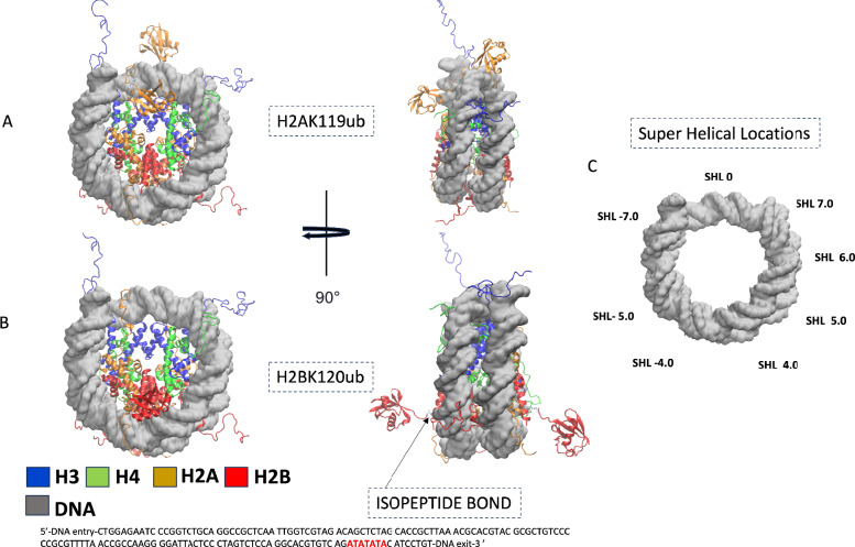
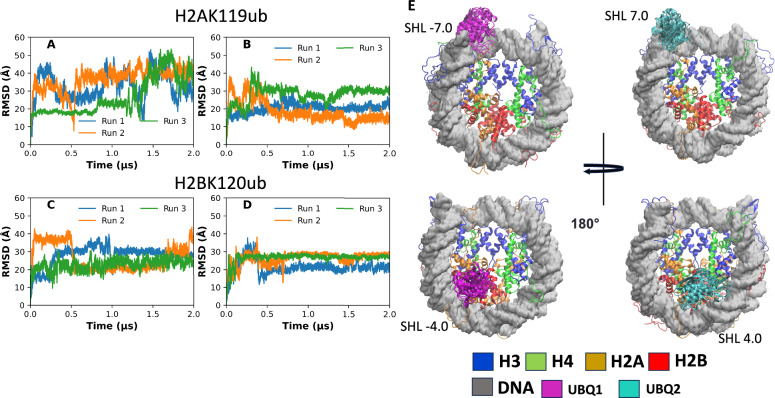
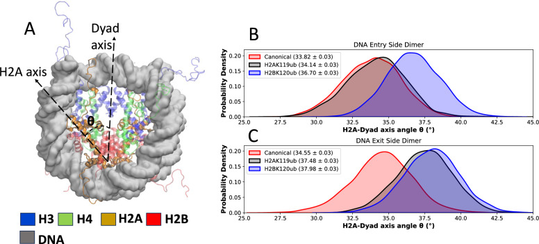
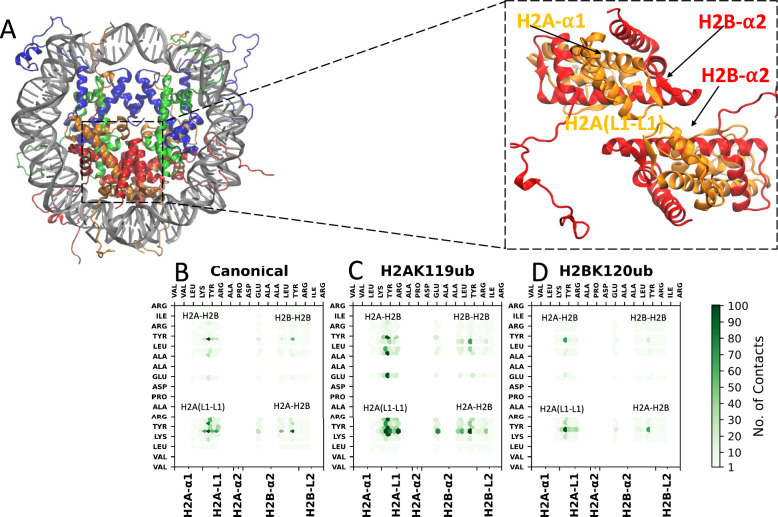
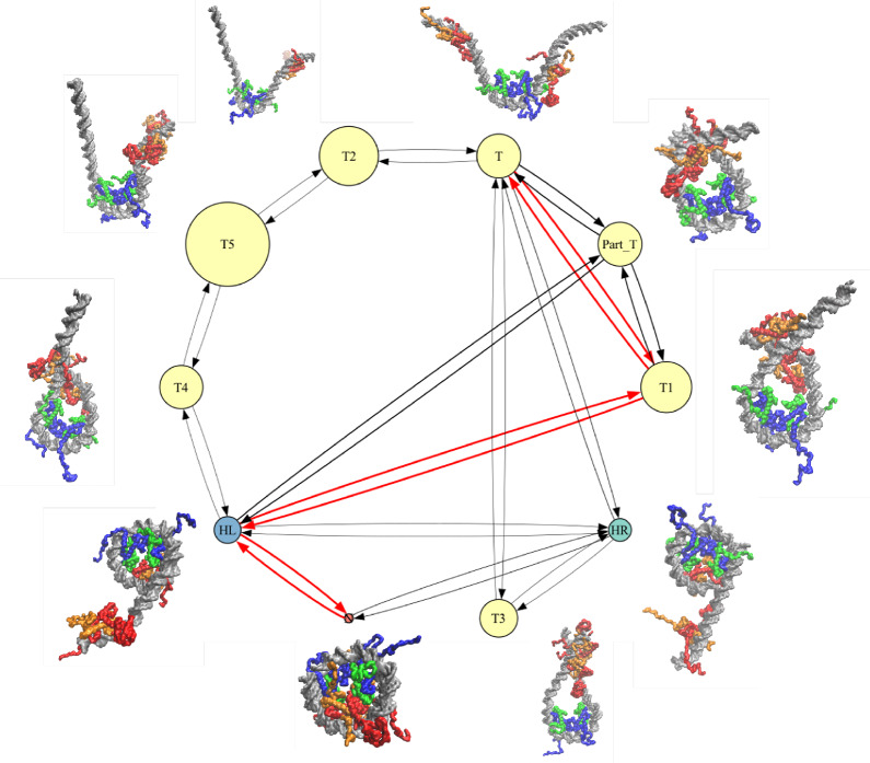
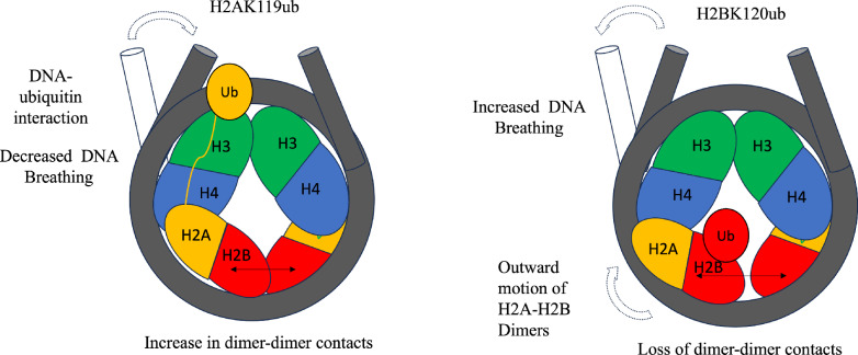
BACKGROUND: Nucleosome ubiquitination at lysine 119 of histone H2A (H2AK119ub) and lysine 120 of histone H2B (H2BK120ub) are prominent post-translational modifications with opposing roles in chromatin regulation. Although H2AK119ub is associated with transcriptional repression and H2BK120ub with activation, the molecular basis for these contrasting effects has remained unclear. RESULTS: Here, we use microsecond all-atom and millisecond coarse-grained molecular dynamics simulations to reveal how the position of ubiquitin reshapes nucleosome structure and assembly. H2AK119ub rigidifies the histone core by indirectly reinforcing the L1-L1 interface between H2A histones, strengthening both tetramer-dimer and dimer-dimer interactions, and slowing complete nucleosome assembly. In contrast, H2BK120ub disrupts these interfaces, weakens the histone core, and favors partially assembled hexasome and tetrasome states. Both modifications cause dramatic slowdowns in nucleosome folding, with H2BK120ub producing an order-of-magnitude greater effect. These simulations establish clear molecular mechanisms by which site-specific ubiquitination alters nucleosome stability and assembly kinetics. CONCLUSION: Our findings quantitatively explain how H2A and H2B ubiquitination exert opposing effects on chromatin regulation. This mechanism is directly relevant to the opposing roles of these marks in transcriptional activation and repression, and may represent one way that combinations of histone modifications modulate chromatin function in vivo.
Overview
The claims and statistics of this paper haven’t been checked yet.
Run the check — free, no account needed. It reads the open-access text and checks the sources it cites.
Integrity signals
Registry
Cites
Statistics
Figures
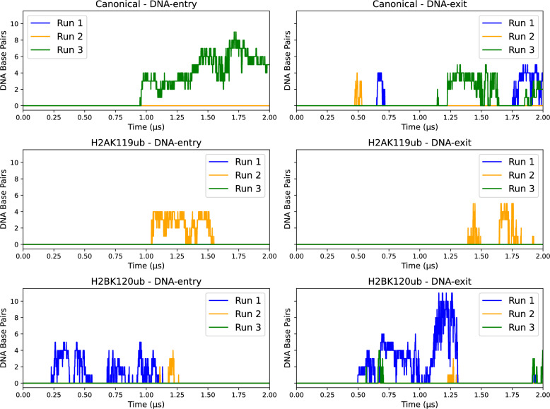
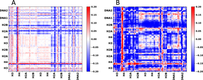
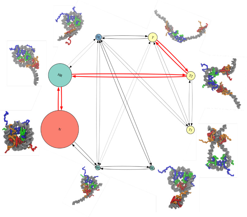
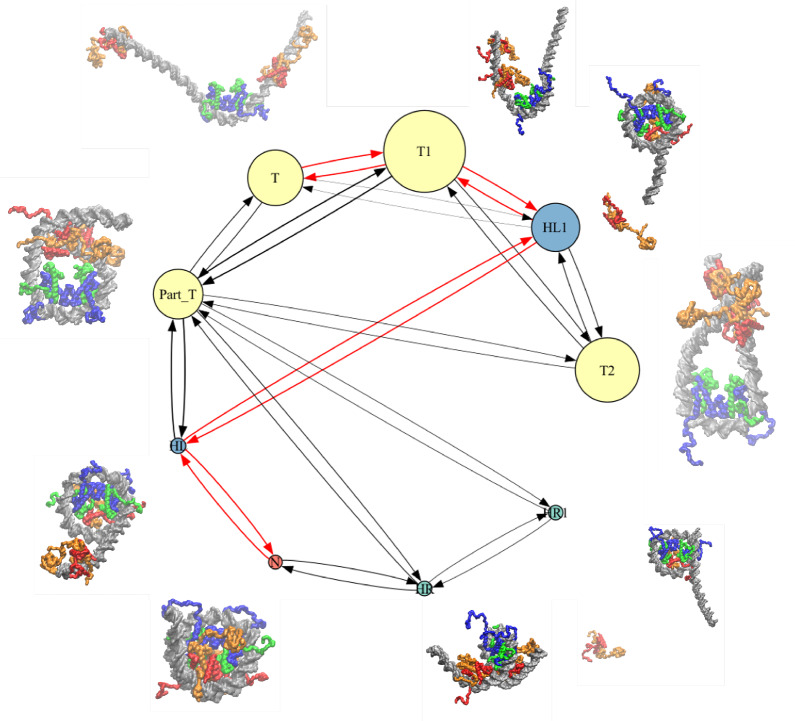
Verification
Claims the paper makes
The claims and statistics of this paper haven’t been checked yet.
Run the check — free, no account needed. It reads the open-access text and checks the sources it cites.
Self-check
Percentages, sums and reported differences re-computed from the paper’s own numbers.
The self-check needs the paper’s full text.
Citations
Cites
Histone post-translational modifications - cause and consequence of genome function
Millán-Zambrano, G., Burton, A., Bannister, A. J., & Schneider, R. (2022) · Nature Reviews Genetics · doi:10.1038/s41576-022-00468-7 · Cited by 1,116
Markov state models of biomolecular conformational dynamics
Chodera, J. D., & Noé, F. (2014) · Current Opinion in Structural Biology · doi:10.1016/j.sbi.2014.04.002 · Cited by 776
Nucleosome structure and function
McGinty, R. K., & Tan, S. (2015) · Chemical Reviews · doi:10.1021/cr500373h · Cited by 530
Research works printed but not linked: 78
- Luger K, Mäder AW, Richmond RK, Sargent DF, Richmond TJ. Crystal structure of the nucleosome core particle at 2.8 Å resolution. Nature. 1997;389:251–60. · DOI · Paper page
- Bilokapic S, Strauss M, Halic M. Histone octamer rearranges to adapt to DNA unwrapping. Nature Struct Mol Biol. 2018;25:101–8. · DOI
- Mariño-Ramírez L, Kann MG, Shoemaker BA, Landsman D. Histone structure and nucleosome stability. Expert Rev Proteom. 2005;2:719–29. · DOI
- Peng Y, Li S, Landsman D, Panchenko AR. Histone tails as signaling antennas of chromatin. Curr Opin Struct Biol. 2021;67:153–60. · DOI
- Karantza V, Freire E, Moudrianakis EN. Thermodynamic studies of the core histones: pH and ionic strength effects on the stability of the (H3–H4)/(H3–H4) 2 system. Biochemistry. 1996;35:2037–46. · DOI
- Hazan NP, Tomov TE, Tsukanov R, Liber M, Berger Y, Masoud R, et al. Nucleosome core particle disassembly and assembly kinetics studied using single-molecule fluorescence. Biophys J. 2015;109:1676–85. · DOI
- Weaver TM, Morrison EA, Musselman CA. Reading more than histones: the prevalence of nucleic acid binding among reader domains. Molecules. 2018;23:2614. · DOI
- Isaac RS, Jiang F, Doudna JA, Lim WA, Narlikar GJ, Almeida R. Nucleosome breathing and remodeling constrain CRISPR-Cas9 function. Elife. 2016;5:e13450. · DOI · Paper page
- Widom J. Structure, dynamics, and function of chromatin in vitro. Annu Rev Biophys Biomol Struct. 1998;27:285–327. · DOI
- Huertas D, Sendra R, Muñoz P. Chromatin dynamics coupled to DNA repair. Epigenetics. 2009;4:31–42. · DOI
- Bowman GD, Poirier MG. Post-translational modifications of histones that influence nucleosome dynamics. Chem Rev. 2014;115:2274–95. · DOI
- Chen JJ, Stermer D, Tanny JC. Decoding histone ubiquitylation. Front Cell Dev Biol. 2022;10:968398. · DOI
- Zhang Y. Transcriptional regulation by histone ubiquitination and deubiquitination. Genes Dev. 2003;17:2733–40. · DOI
- Goldknopf IL, Busch H. Isopeptide linkage between nonhistone and histone 2A polypeptides of chromosomal conjugate-protein A24. Proc Natl Acad Sci. 1977;74:864–8. · DOI
- Weake VM. Histone ubiquitylation control of gene expression. Fund Chrom. 2013;257–307. · DOI
- Meas R, Mao P. Histone ubiquitylation and its roles in transcription and DNA damage response. DNA Repair. 2015;36:36–42. · DOI
- Nathan D, Ingvarsdottir K, Sterner DE, Bylebyl GR, Dokmanovic M, Dorsey JA, et al. Others histone sumoylation is a negative regulator in saccharomyces cerevisiae and shows dynamic interplay with positive-acting histone modifications. Genes Dev. 2006;20:966–76. · DOI
- Wang H, Zhai L, Xu J, Joo H-Y, Jackson S, Erdjument-Bromage H, et al. Histone H3 and H4 ubiquitylation by the CUL4-DDB-ROC1 ubiquitin ligase facilitates cellular response to DNA damage. Mol Cell. 2006;22:383–94. · DOI
- Shafiq TA, Yu J, Feng W, Zhang Y, Zhou H, Paulo JA, et al. Genomic context-and H2AK119 ubiquitination-dependent inheritance of human Polycomb silencing. Sci Adv. 2024;10:eadl4529. · DOI
- Oss-Ronen L, Sarusi T, Cohen I. Histone mono-ubiquitination in transcriptional regulation and its mark on life: emerging roles in tissue development and disease. Cells. 2022;11:2404. · DOI
- Tamburri S, Lavarone E, Fernández-Pérez D, Conway E, Zanotti M, Manganaro D, et al. Histone H2AK119 mono-ubiquitination is essential for polycomb-mediated transcriptional repression. Mol Cell. 2020;77:840–56. · DOI
- Ciapponi M, Karlukova E, Schkölziger S, Benda C, Müller J. Structural basis of the histone ubiquitination read-write mechanism of RYBP-PRC1. Nature Struct Mol Biol. 2024;31:1–5. · DOI
- Xiao X, Liu C, Pei Y, Wang Y-Z, Kong J, Lu K, et al. others Histone H2A ubiquitination reinforces mechanical stability and asymmetry at the single-nucleosome level. J Am Chem Soc. 2020;142:3340–5. · DOI
- Luo A, Kong J, Chen J, Xiao X, Lan J, Li X, et al. H2B ubiquitination recruits FACT to maintain a stable altered nucleosome state for transcriptional activation. Nat Commun. 2023;14:741. · DOI
- Woods DC, Wereszczynski J. Elucidating the influence of linker histone variants on chromatosome dynamics and energetics. Nucleic Acids Res. 2020;48:3591–604. · DOI
- Armeev GA, Kniazeva AS, Komarova GA, Kirpichnikov MP, Shaytan AK. Histone dynamics mediate DNA unwrapping and sliding in nucleosomes. Nat Commun. 2021;12:2387. · DOI
- Khatua P, Tang PK, Moulick A, Patel R, Manandhar A, Loverde S. Sequence dependence in nucleosome dynamics. J Phys Chem B. 2024;128:3090–101 (PMID: 38530903). · DOI
- Portillo-Ledesma S, Tsao LH, Wagley M, Lakadamyali M, Cosma MP, Schlick T. Nucleosome clutches are regulated by chromatin internal parameters. J Mol Biol. 2021;433:166701. · DOI
- Li S, Wei T, Panchenko AR. Histone variant H2A. Z modulates nucleosome dynamics to promote DNA accessibility. Nat Commun. 2023;14:218. · DOI
- Patel R, Onyema A, Tang PK, Loverde SM. Conformational dynamics of the nucleosomal histone H2B tails revealed by molecular dynamics simulations. J Chem Inform Mod. 2024;64(12):4709–26. · DOI
- Fedulova AS, Armeev GA, Romanova TA, Singh-Palchevskaia L, Kosarim NA, Motorin NA, et al. Molecular dynamics simulations of nucleosomes are coming of age. WIREs Comput Mol Sci. 2024;14:e1728. · DOI
- Kariyawasam NL, Wereszczynski J. The influence of ionic environment on nucleosome-mica interactions revealed via molecular dynamics simulations. J Phys Chem B. 2024;128:12038–49. · DOI
- Brandani GB, Tan C, Takada S. The kinetic landscape of nucleosome assembly: a coarse-grained molecular dynamics study. PLoS Comput Biol. 2021;17:e1009253. · DOI
- Husic BE, Pande VS. Markov state models: from an art to a science. J Am Chem Soc. 2018;140:2386–96. · DOI
- Davey CA, Sargent DF, Luger K, Maeder AW, Richmond TJ. Solvent mediated interactions in the structure of the nucleosome core particle at 1.9 Å resolution. J Molecular Biol. 2002;319:1097–113. · DOI
- Pettersen EF, Goddard TD, Huang CC, Couch GS, Greenblatt DM, Meng EC, et al. UCSF Chimera-a visualization system for exploratory research and analysis. J Comput Chem. 2004;25:1605–12. · DOI
- Widom J. Role of DNA sequence in nucleosome stability and dynamics. Q Rev Biophys. 2001;34:269–324. · DOI · Paper page
- Case DA, Cheatham TE III, Darden T, Gohlke H, Luo R, Merz KM Jr, et al. The Amber biomolecular simulation programs. J Comput Chem. 2005;26:1668–88. · DOI · Paper page
- Frisch A. Others gaussian 09W Reference. vol. 470. Wallingford; 2009. p. 25.
- Tian C, Kasavajhala K, Belfon KA, Raguette L, Huang H, Migues AN, et al. others ff19SB: amino-acid-specific protein backbone parameters trained against quantum mechanics energy surfaces in solution. J Chem Theory Comput. 2019;16:528–52. · DOI
- Ivani I, Dans PD, Noy A, Pérez A, Faustino I, Hospital A, et al. others Parmbsc1: a refined force field for DNA simulations. Nat Methods. 2016;13:55–8. · DOI
- Izadi S, Anandakrishnan R, Onufriev AV. Building water models: a different approach. J Phys Chem Lett. 2014;5:3863–71. · DOI
- Hopkins CW, Le Grand S, Walker RC, Roitberg AE. Long-time-step molecular dynamics through hydrogen mass repartitioning. J Chem Theory Comput. 2015;11:1864–74. · DOI
- Ryckaert J-P, Ciccotti G, Berendsen HJ. Numerical integration of the cartesian equations of motion of a system with constraints: molecular dynamics of n-alkanes. J Comput Phys. 1977;23:327–41. · DOI
- Humphrey W, Dalke A, Schulten K. VMD: visual molecular dynamics. J Mol Graph. 1996;14:33–8. · DOI · Paper page
- DeLano WL. Others Pymol: an open-source molecular graphics tool. CCP4 Newsl Protein Crystallogr. 2002;40:82–92.
- Roe DR, Cheatham TE III. PTRAJ and CPPTRAJ: software for processing and analysis of molecular dynamics trajectory data. J Chem Theory Comput. 2013;9:3084–95. · DOI
- Li W, Wang W, Takada S. Energy landscape views for interplays among folding, binding, and allostery of calmodulin domains. Proc Natl Acad Sci. 2014;111:10550–5. · DOI
- Freeman GS, Hinckley DM, Lequieu JP, Whitmer JK, De Pablo JJ. Coarse-grained modeling of DNA curvature. J Chem Phys. 2014;141:165103. · DOI
- Niina T, Brandani GB, Tan C, Takada S. Sequence-dependent nucleosome sliding in rotation-coupled and uncoupled modes revealed by molecular simulations. PLoS Comput Biol. 2017;13:e1005880. · DOI
- Manning GS. Limiting laws and counterion condensation in polyelectrolyte solutions II. Self-diffusion of the small ions. J Chem Phys. 1969;51:934–8. · DOI
- Manning GS. The molecular theory of polyelectrolyte solutions with applications to the electrostatic properties of polynucleotides. Q Rev Biophys. 1978;11:179–246. · DOI
- Honig B, Nicholls A. Classical electrostatics in biology and chemistry. Science. 1995;268:1144–9. · DOI
- Terakawa T, Takada S. RESPAC: method to determine partial charges in coarse-grained protein model and its application to DNA-binding proteins. J Chem Theory Comput. 2014;10:711–21. · DOI
- Weeks SD, Grasty KC, Hernandez-Cuebas L, Loll PJ. Crystal structures of Lys-63-linked tri-and di-ubiquitin reveal a highly extended chain architecture. Proteins: Structure. Funct Bioinform. 2009;77:753–9. · DOI
- Terakawa T, Takada S. Multiscale ensemble modeling of intrinsically disordered proteins: p53 N-terminal domain. Biophys J. 2011;101:1450–8. · DOI
- Morrison EA, Baweja L, Poirier MG, Wereszczynski J, Musselman CA. Nucleosome composition regulates the histone H3 tail conformational ensemble and accessibility. Nucleic Acids Res. 2021;49:4750–67. · DOI
- Kenzaki H, Koga N, Hori N, Kanada R, Li W, Okazaki K-I, et al. CafeMol: a coarse-grained biomolecular simulator for simulating proteins at work. J Chem Theory Comput. 2011;7:1979–89. · DOI
- Fierz B, Kilic S, Hieb AR, Luger K, Muir TW. Stability of nucleosomes containing homogenously ubiquitylated H2A and H2B prepared using semisynthesis. J Am Chem Soc. 2012;134:19548–51. · DOI
- Schultze S, Grubmüller H. Time-lagged independent component analysis of random walks and protein dynamics. J Chem Theory Comput. 2021;17:5766–76. · DOI
- Scherer MK, Trendelkamp-Schroer B, Paul F, Pérez-Hernández G, Hoffmann M, Plattner N, et al. PyEMMA 2: a software package for estimation, validation, and analysis of Markov models. J Chem Theory Comput. 2015;11:5525–42. · DOI
- Likas A, Vlassis N, Verbeek JJ. The global k-means clustering algorithm. Pattern Recogn. 2003;36:451–61. · DOI
- Michaud-Agrawal N, Denning EJ, Woolf TB, Beckstein O. MDAnalysis: a toolkit for the analysis of molecular dynamics simulations. J Comput Chem. 2011;32:2319–27. · DOI
- Hunter JD. Matplotlib: a 2D graphics environment. Comput Sci Eng. 2007;9:90–5. · DOI · Paper page
- Miller BR III, McGee TD Jr, Swails JM, Homeyer N, Gohlke H, Roitberg AE. MMPBSA. py: an efficient program for end-state free energy calculations. J Chem Theory Comput. 2012;8:3314–21. · DOI
- Shirts MR, Chodera JD. Statistically optimal analysis of samples from multiple equilibrium states. J Chem Phys. 2008;129:124105. · DOI
- Tekpinar M, Neron B, Delarue M. Extracting dynamical correlations and identifying key residues for allosteric communication in proteins by correlationplus. J Chem Inf Model. 2021;61:4832–8. · DOI
- Hicks CW, Rahman S, Gloor SL, Fields JK, Husby NL, Vaidya A, et al. Ubiquitinated histone H2B as gatekeeper of the nucleosome acidic patch. Nucleic Acids Res. 2024;52:9978–95. · DOI
- Landré V, Revi B, Mir MG, Verma C, Hupp TR, Gilbert N, et al. Regulation of transcriptional activators by DNA-binding domain ubiquitination. Cell Death Differ. 2017;24:903–16. · DOI
- Hou T, Wang J, Li Y, Wang W. Assessing the performance of the MM/PBSA and MM/GBSA methods. 1. the accuracy of binding free energy calculations based on molecular dynamics simulations. J Chem Inf Modeling. 2011;51:69–82. · DOI
- Onufriev AV, Case DA. Generalized born implicit solvent models for biomolecules. Annu Rev Biophys. 2019;48:275–96. · DOI
- Brandani GB, Niina T, Tan C, Takada S. DNA sliding in nucleosomes via twist defect propagation revealed by molecular simulations. Nucleic Acids Res. 2018;46:2788–801. · DOI
- Mattiroli F, Penengo L. Histone ubiquitination: an integrative signaling platform in genome stability. Trends Genet. 2021;37:566–81. · DOI
- Bonisch C, Hake SB. Histone H2A variants in nucleosomes and chromatin: more or less stable? Nucleic Acids Res. 2012;40:10719–41. · DOI
- Bowerman S, Hickok RJ, Wereszczynski J. Unique Dynamics in Asymmetric macroH2A-H2A Hybrid Nucleosomes Result in Increased Complex Stability. J Phys Chem B. 2018;123:419–27. · DOI
- Chakravarthy S, Luger K. The histone variant macro-H2A preferentially forms hybrid nucleosomes. J Biol Chem. 2006;281:25522–31. · DOI
- Ullner M, Woodward CE, Jönsson B. A Debye-Hückel theory for electrostatic interactions in proteins. J Chem Phys. 1996;105:2056–65. · DOI
- Pan AC, Roux B. Building Markov state models along pathways to determine free energies and rates of transitions. J Chem Phys. 2008;129:6. · DOI
Metadata
- Authors
- Baweja, L., & Wereszczynski, J.
- Affiliations
- Illinois Institute of Technology (US)
- Corresponding author
- Wereszczynski, Jeff
- Venue
- Epigenetics & Chromatin
- Published
- 2025-11-19
- Volume
- 18
- Issue
- 1
- Pages
- 74-74
- ISSN
- 1756-8935 (ISSN-L)
- PMC
- PMC12628893
- OpenAlex
- W4416392468
- Cited by
- 3
- References
- 3
- Citations by year
- 2026: 2 · 2025: 1
- FWCI
- 0.90
- Type
- journal article
- Language
- en
- Open access
- gold
- doi.org
- Licence
- Locations
- Epigenetics & Chromatin (publishedVersion, cc-by-nc-nd)
- bioRxiv (Cold Spring Harbor Laboratory) (acceptedVersion, cc-by)
- Europe PMC (PubMed Central) (publishedVersion, cc-by-nc-nd)
- PubMed (publishedVersion)
- PubMed Central (submittedVersion, cc-by-nc)
- DOAJ (DOAJ: Directory of Open Access Journals) (submittedVersion, cc-by-sa)
- Funders
- National Institute of General Medical Sciences (R35GM119647)
- Topics
- Life Sciences › Biochemistry, Genetics and Molecular Biology › Molecular Biology › Genomics and Chromatin Dynamics
- Life Sciences › Biochemistry, Genetics and Molecular Biology › Molecular Biology › Protein Structure and Dynamics
- Life Sciences › Biochemistry, Genetics and Molecular Biology › Molecular Biology › Nuclear Structure and Function
- Keywords
- histone H2A ubiquitination, histone H2B ubiquitination, H2AK119ub, nucleosome stability, hexasome, chromatin regulation, transcriptional repression, transcriptional activation, histone modifications
About these checks
Registry, retraction, Cites and statistics results are computed, not judged, with one exception: a printed reference that no index lists is read once by a model before it counts as a citation error. Claims are extracted by a model and kept only when their quote appears verbatim in the text; each cited source is then checked, with a model’s help, for whether it says what the claim relies on. Those verdicts can be wrong — read the claim’s quote and the cited source before relying on one.
Statistics and arithmetic are recomputed, not judged.
Confidence bands are shown only for fields calibrated on our annotated test set. None is calibrated yet, so no band is shown.
What counts as a claim ›How we measure confidence ›How we check this ›
Are you an author, or do you see a problem with this page? Request a review
