

The DOI matches its Crossref record; 70 of the 70 references it cites were found in a registry or an index; 31 of the 40 sources its claims cite were checked for support.
Selective suppression of the α isoform of p38 MAPK rescues late-stage tau pathology
Open accessAbstract
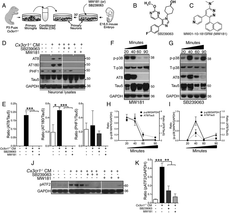
Background: Hyperphosphorylation and aggregation of tau protein are the pathological hallmarks of Alzheimer’s disease and related tauopathies. We previously demonstrated that the microglial activation induces tau hyperphosphorylation and cognitive impairment via activation of p38 mitogen-activated protein kinase (p38 MAPK) in the hTau mouse model of tauopathy that was deficient for microglial fractalkine receptor CX3CR1. Method: We report an isoform-selective, brain-permeable, and orally bioavailable small molecule inhibitor of p38α MAPK (MW181) and its effects on tau phosphorylation in vitro and in hTau mice. Results: First, pretreatment of mouse primary cortical neurons with MW181 completely blocked inflammation-induced p38α MAPK activation and AT8 (pS199/pS202) site tau phosphorylation, with the maximum effect peaking at 60–90 min after stimulation. Second, treatment of old (~20 months of age) hTau mice with MW181 (1 mg/kg body weight; 14 days via oral gavage) significantly reduced p38α MAPK activation compared with vehicle-administered hTau mice. This also resulted in a significant reduction in AT180 (pT231) site tau phosphorylation and Sarkosyl-insoluble tau aggregates. Third, MW181 treatment significantly increased synaptophysin protein expression and resulted in improved working memory. Fourth, MW181 administration reduced phosphorylated MAPK-activated protein kinase 2 (pMK2) and phosphorylated activating transcription factor 2 (pATF2), which are known substrates of p38α MAPK. Finally, MW181 reduced the expression of interferon-γ and interleukin-1β. Conclusions: Taken together, these studies support p38α MAPK as a valid therapeutic target for the treatment of tauopathies.
Overview
AI snapshot
Written from the paper’s full text.
TL;DR
MW181, an isoform-selective p38α MAPK inhibitor, reduced inflammation-induced tau phosphorylation in primary neurons and, after 14 days of oral treatment, reduced tau phosphorylation/aggregation and improved working memory in aged hTau mice. The authors conclude that p38α MAPK is a valid therapeutic target for tauopathies.
Key findings
- In primary cortical neurons, pretreatment with MW181 (2 μM) or SB239063 (100 μM) blocked Cx3cr1−/− microglial conditioned media-induced AT8 (pS199/pS202) and AT180 (pT231) tau phosphorylation; MW181’s maximum reduction of p38α MAPK (pT180/pY182) and AT8 tau phosphorylation occurred at 60–90 min after stimulation.
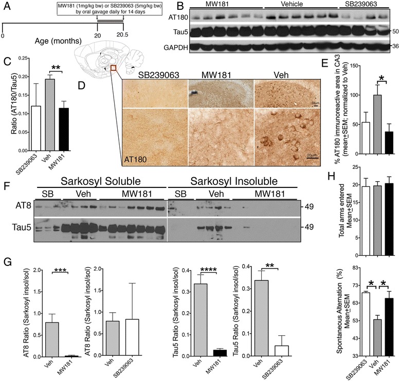
- In 20-month-old hTau mice, MW181 (1 mg/kg body weight, oral gavage, 14 days) significantly reduced AT180/Tau5 ratio (n = 6 MW181, n = 6 vehicle, n = 4 SB239063; p < 0.01), reduced Sarkosyl-insoluble tau (Tau5 ratio; n = 7 MW181, n = 5 vehicle, n = 3 SB239063; p < 0.01, *p < 0.001, p < 0.0001), increased synaptophysin, and improved Y-maze spontaneous alternation versus vehicle (n = 6 MW181, n = 5 vehicle, n = 4 SB239063; *p < 0.05).
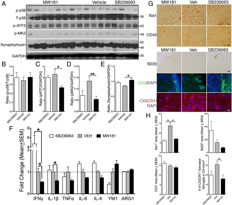
- MW181 significantly reduced hippocampal pATF2 (pT71) and pMK2 (pT334) and reduced IFNγ and IL-1β expression in aged hTau mice, while in vivo p-p38α MAPK (pT180/pY182) levels did not significantly decrease.
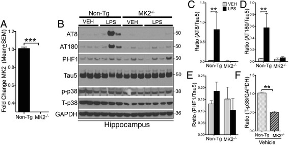
- MK2−/− mice were resistant to LPS-induced AT8 and AT180 tau phosphorylation and showed significantly reduced total p38α MAPK levels compared with nontransgenic mice.
Methods
- In vitro: primary cortical neurons from E16.5 C57BL/6J embryos grown 21 DIV; microglia from P3 Cx3cr1−/− pups; neurons pretreated 30 min with MW181 (2 μM), SB239063 (100 μM), or vehicle, then stimulated with 25% Cx3cr1−/− microglial conditioned media; lysates analyzed by western blot.
- In vivo: 20-month-old hTau mice received MW181 (1 mg/kg), SB239063 (5 mg/kg), or saline daily by oral gavage for 14 days; hippocampi analyzed by western blot, Sarkosyl-insoluble assay, qPCR, immunohistochemistry/immunofluorescence, and Y-maze. Separate 2-month-old nontransgenic and MK2−/− mice received LPS (10 mg/kg i.p.) or vehicle.
- Statistics: unpaired t tests and one-way or two-way ANOVA with indicated post-hoc tests using GraphPad Prism.
Limitations
- Stated: the authors note this is the first study targeting p38α MAPK in a 20-month-old tauopathy model and should be validated by a separate group; they did not compare MW181 effects in nontransgenic mice; MW181 did not completely abolish activated p38α MAPK; and only one MW181 dose (1 mg/kg) was tested.
- Stated: the authors note that different dosing and treatment durations are key considerations for pharmacological safety.
- Evident from the text: some treatment groups had small sample sizes (e.g., SB239063 n = 3–4 in several assays), mixed genders were used, and no dose-response or long-term safety/toxicity assessment beyond weight, grooming, and posture monitoring is reported.
Model-written summary. Check it against the paper before relying on it.
Integrity signals
Registry
Cites
Statistics
Figures
Verification
Claims the paper makes
52 Own result · 19 Own interpretation · 13 Partly supported · 5 Supported · 4 Own method · 3 No full text of the sources · 3 No basis given · accuracy not yet measured
Every source the paper cites for this claim was checked and supports it.
In the paper
“Signaling through p38 MAPK, especially the p38α isoform, has also been discovered recently to be a critical component for cytokine production in activated microglia [25, 26].”
§ Selective suppression of the α isoform of p38 MAPK rescues late-stage tau pathology > Background
What the sources say
- Supportedcites [25] Roy SM, Grum-Tokars VL, Schavocky JP, Saeed F, Staniszewski A, Teich AF, Arancio O, Bachstetter AD, Webster SJ, Van Eldik LJ, et al. Targeting human central nervous system protein kinases: An isoform selective p38alphaMAPK inhibitor that attenuates disease progression in Alzheimer’s disease mouse models. ACS Chem Neurosci. 2015;6(4):666–80. source
The Results and Discussion (Kinome Target Selectivity; page unavailable) identify the p38αMAPK–MK2 cascade as mediating LPS-induced proinflammatory cytokine production and report that a selective inhibitor reduced IL-1β production. The Methods (Cell Based Activity; page unavailable) identify the tested activated cells as BV-2 microglia. (LLM, conf=0.92)
- Supportedcites [26] Watterson DM, Grum-Tokars VL, Roy SM, Schavocky JP, Bradaric BD, Bachstetter AD, Xing B, Dimayuga E, Saeed F, Zhang H, et al. Development of novel chemical probes to address CNS protein kinase involvement in synaptic dysfunction. PLoS One. 2013;8(6), e66226. source
The Results section (page unavailable) states that LPS activation of the p38αMAPK pathway in microglial cells increases proinflammatory cytokine production. It also reports that a selective p38αMAPK inhibitor reduced IL-1β production in activated microglia, supporting the isoform-specific claim. (LLM, conf=0.92)
boosteddirect- Asserted: passes — The authors put it forward as true.
- Checkable: passes — It can be checked against evidence.
- Load-bearing: passes — The paper’s conclusions depend on it.
- Based: passes — The paper gives a reason why it holds.
- Supported
The cited sources support only part of this claim — read each source’s verdict below.
In the paper
“The p38α-mediated cytokine overproduction in microglia has been linked to inflammation-induced neurotoxicity in a variety of mouse models [22, 27–33].”
§ Selective suppression of the α isoform of p38 MAPK rescues late-stage tau pathology > Background
What the sources say
- Partly supportedcites [22] Bhaskar K, Konerth M, Kokiko-Cochran ON, Cardona A, Ransohoff RM, Lamb BT. Regulation of tau pathology by the microglial fractalkine receptor. Neuron. 2010;68(1):19–31. sourceExperimental — accuracy not yet measured
The Results section on TLR4 and IL1 signaling links microglial IL1 to neuronal p38 MAPK activation and tau phosphorylation; it also reports earlier findings linking IL1β from LPS-stimulated microglia to neurotoxicity. The paper does not establish that p38α in microglia causes cytokine overproduction or show this neurotoxic mechanism across a variety of mouse models. Page numbers are not available in our copy. (LLM, conf=0.84)
- Partly supportedcites [27] Xing B, Bachstetter AD, Van Eldik LJ. Microglial p38alpha MAPK is critical for LPS-induced neuron degeneration, through a mechanism involving TNFalpha. Mol Neurodegener. 2011;6:84. sourceExperimental — accuracy not yet measured
The Discussion reports that p38α-dependent TNFα production in mouse microglia–neuron co-cultures contributes to LPS-induced neuronal damage. The Background also mentions an earlier AD-relevant mouse model, but this paper does not itself establish the link across a variety of mouse models. (LLM, conf=0.85)
- Partly supportedcites [28] Bachstetter AD, Watterson DM, Van Eldik LJ. Target engagement analysis and link to pharmacodynamic endpoint for a novel class of CNS-penetrant and efficacious p38alpha MAPK inhibitors. J Neuroimmune Pharmacol. 2014;9(4):454–60. sourceExperimental — accuracy not yet measured
The Introduction explicitly links microglial p38α-mediated cytokine overproduction to inflammation-induced neurotoxicity, citing earlier studies. It also describes protection in a mouse brain-injury model, but does not establish this neurotoxicity link across a variety of mouse models. (LLM, conf=0.84)
- Partly supportedcites [29] Bachstetter AD, Xing B, de Almeida L, Dimayuga ER, Watterson DM, Van Eldik LJ. Microglial p38alpha MAPK is a key regulator of proinflammatory cytokine up-regulation induced by toll-like receptor (TLR) ligands or beta-amyloid (Abeta). J Neuroinflammation. 2011;8:79. sourceExperimental — accuracy not yet measured
The Background links microglial cytokine overproduction to neuronal death and reports improved neurological outcomes after p38α inhibition in an Alzheimer’s-relevant mouse model. This paper also demonstrates p38α-dependent cytokine production, but its own in vivo experiment measures cytokines in one LPS-induced mouse model, not neurotoxicity across a variety of mouse models. (LLM, conf=0.85)
- Not checked — no full text of the sourcecites [30] Barone FC, Irving EA, Ray AM, Lee JC, Kassis S, Kumar S, Badger AM, Legos JJ, Erhardt JA, Ohlstein EH, et al. Inhibition of p38 mitogen-activated protein kinase provides neuroprotection in cerebral focal ischemia. Med Res Rev. 2001;21(2):129–45. source
- Partly supportedcites [31] Correa SA, Eales KL. The role of p38 MAPK and its substrates in neuronal plasticity and neurodegenerative disease. J Signal Transduct. 2012;2012:649079. sourceExperimental — accuracy not yet measured
Section 3.2 links p38 MAPK activation in glial cells to increased inflammatory cytokines and potential neuronal toxicity; it also reports that a selective p38α inhibitor blocks increased inflammatory-molecule production. The neurotoxicity examples described there involve glial cultures and rat brains, not a variety of mouse models. No page numbers are provided in our copy. (LLM, conf=0.87)
- Not checked — no full text of the sourcecites [32] Dewil M, dela Cruz VF, Van Den Bosch L, Robberecht W. Inhibition of p38 mitogen activated protein kinase activation and mutant SOD1(G93A)-induced motor neuron death. Neurobiol Dis. 2007;26(2):332–41. source
- Not checked — no full text of the sourcecites [33] Stirling DP, Liu J, Plunet W, Steeves JD, Tetzlaff W. SB203580, a p38 mitogen-activated protein kinase inhibitor, fails to improve functional outcome following a moderate spinal cord injury in rat. Neuroscience. 2008;155(1):128–37. source
plaincorrelational- Asserted: passes — The authors put it forward as true.
- Checkable: passes — It can be checked against evidence.
- Load-bearing: passes — The paper’s conclusions depend on it.
- Based: passes — The paper gives a reason why it holds.
- Partly supported
The cited sources support only part of this claim — read each source’s verdict below.
In the paper
“MW01-10-181SRM (or MW181), a highly selective p38α MAPK inhibitor [26]”
§ Selective suppression of the α isoform of p38 MAPK rescues late-stage tau pathology > Background
What the sources say
- Partly supportedcites [26] Watterson DM, Grum-Tokars VL, Roy SM, Schavocky JP, Bradaric BD, Bachstetter AD, Xing B, Dimayuga E, Saeed F, Zhang H, et al. Development of novel chemical probes to address CNS protein kinase involvement in synaptic dysfunction. PLoS One. 2013;8(6), e66226. sourceExperimental — accuracy not yet measured
The Results section identifies MW181 as a p38αMAPK inhibitor, but describes it as a p38MAPK subfamily inhibitor rather than an isoform-selective one. Kinase profiling found similar inhibitory affinities for p38αMAPK, p38βMAPK, and NLK; the paper identifies MW108, not MW181, as more selective for p38αMAPK. (LLM, conf=0.90)
plain- Asserted: passes — The authors put it forward as true.
- Checkable: passes — It can be checked against evidence.
- Load-bearing: passes — The paper’s conclusions depend on it.
- Based: passes — The paper gives a reason why it holds.
- Partly supported
The cited sources support only part of this claim — read each source’s verdict below.
In the paper
“First, p38α MAPK has been shown to mediate Aβ-induced expression of various proinflammatory cytokines/chemokines from microglial cells that causes neuroinflammation [25, 26, 29, 35].”
§ Selective suppression of the α isoform of p38 MAPK rescues late-stage tau pathology > Discussion
What the sources say
- Partly supportedcites [25] Roy SM, Grum-Tokars VL, Schavocky JP, Saeed F, Staniszewski A, Teich AF, Arancio O, Bachstetter AD, Webster SJ, Van Eldik LJ, et al. Targeting human central nervous system protein kinases: An isoform selective p38alphaMAPK inhibitor that attenuates disease progression in Alzheimer’s disease mouse models. ACS Chem Neurosci. 2015;6(4):666–80. sourceExperimental — accuracy not yet measured
The Methods (Cell Based Activity) and Results and Discussion (Kinome Target Selectivity; page unavailable) show that inhibiting p38αMAPK signaling reduced proinflammatory IL-1β production in LPS-stimulated microglial cells. They do not show Aβ-induced production of various cytokines or chemokines by microglia, or establish that this production causes neuroinflammation. (LLM, conf=0.88)
- Partly supportedcites [26] Watterson DM, Grum-Tokars VL, Roy SM, Schavocky JP, Bradaric BD, Bachstetter AD, Xing B, Dimayuga E, Saeed F, Zhang H, et al. Development of novel chemical probes to address CNS protein kinase involvement in synaptic dysfunction. PLoS One. 2013;8(6), e66226. sourceExperimental — accuracy not yet measured
The Results section on CNS mechanism of action links microglial p38αMAPK to increased proinflammatory cytokine production, but the experiments described there use LPS or TLR ligands and measure IL-1β. The Results section on amyloid-beta elevation reports rescue of Aβ-induced synaptic deficits, not Aβ-induced microglial expression of various cytokines or chemokines. The References identify a separate study on Aβ-induced microglial cytokine up-regulation; this paper does not itself establish the full claim. (LLM, conf=0.86)
- Partly supportedcites [29] Bachstetter AD, Xing B, de Almeida L, Dimayuga ER, Watterson DM, Van Eldik LJ. Microglial p38alpha MAPK is a key regulator of proinflammatory cytokine up-regulation induced by toll-like receptor (TLR) ligands or beta-amyloid (Abeta). J Neuroinflammation. 2011;8:79. sourceExperimental — accuracy not yet measured
The Figure 4 caption reports that inhibiting p38α MAPK suppressed Aβ1-42-induced increases in IL-1β and TNFα in primary microglia. The Methods and Results address those two cytokines, not Aβ-induced chemokine expression; the in vivo neuroinflammation model used LPS rather than Aβ, so the claimed Aβ-driven neuroinflammation was not directly shown. (LLM, conf=0.86)
- Partly supportedcites [35] Munoz L, Ralay Ranaivo H, Roy SM, Hu W, Craft JM, McNamara LK, Chico LW, Van Eldik LJ, Watterson DM. A novel p38 alpha MAPK inhibitor suppresses brain proinflammatory cytokine up-regulation and attenuates synaptic dysfunction and behavioral deficits in an Alzheimer’s disease mouse model. J Neuroinflammation. 2007;4:21. sourceExperimental — accuracy not yet measured
The Background reports that Aβ activates p38 MAPK in glial cultures, followed by increased proinflammatory cytokines that p38 inhibitors block. The Results and Discussion report that a p38α inhibitor reduced Aβ-induced hippocampal cytokines in mice. The paper does not establish that those mouse cytokines came specifically from microglia, and the cited passages do not support the claim about chemokines. (LLM, conf=0.86)
plaindirect- Asserted: passes — The authors put it forward as true.
- Checkable: passes — It can be checked against evidence.
- Load-bearing: passes — The paper’s conclusions depend on it.
- Based: passes — The paper gives a reason why it holds.
- Partly supported
The cited sources support only part of this claim — read each source’s verdict below.
In the paper
“As reported previously [22], exchange of 25% of the neuronal supernatant with Cx3cr1 –/– microglia CM led to an increase in tau phosphorylation on S199/S202 (AT8) and T231 (AT180) sites (Fig. 1d, e).”
§ Selective suppression of the α isoform of p38 MAPK rescues late-stage tau pathology > Results > MW181 and SB239063 treatment rescued inflammation-induced phosphorylation of tau, p38α MAPK, and ATF2 in primary neurons
What the sources say
- Partly supportedcites [22] Bhaskar K, Konerth M, Kokiko-Cochran ON, Cardona A, Ransohoff RM, Lamb BT. Regulation of tau pathology by the microglial fractalkine receptor. Neuron. 2010;68(1):19–31. sourceExperimental — accuracy not yet measured
The Experimental Procedures describe replacing 25% of neuronal medium with microglial conditioned medium. The Results report that medium from Cx3cr1−/− microglia enhanced neuronal tau phosphorylation at AT8 and PHF1, not AT180. The paper reports AT180 increases in mice under other conditions, but not in this conditioned-medium experiment; page numbers are unavailable in our copy. (LLM, conf=0.88)
plaindirect- Asserted: passes — The authors put it forward as true.
- Checkable: passes — It can be checked against evidence.
- Load-bearing: passes — The paper’s conclusions depend on it.
- Based: passes — The paper gives a reason why it holds.
- Partly supported
Every source the paper cites for this claim was checked and supports it.
In the paper
“As reported previously [22], a single injection of LPS led to AT8 and AT180 site tau phosphorylation in nontransgenic mice (Fig. 4b–d).”
§ Selective suppression of the α isoform of p38 MAPK rescues late-stage tau pathology > Results > MK2–/– mice are resistant to LPS-induced tau phosphorylation and show reduced levels of total p38α MAPK
What the sources say
- Supportedcites [22] Bhaskar K, Konerth M, Kokiko-Cochran ON, Cardona A, Ransohoff RM, Lamb BT. Regulation of tau pathology by the microglial fractalkine receptor. Neuron. 2010;68(1):19–31. source
The Results section, “Enhanced MAPT phosphorylation after systemic LPS administration,” reports increased AT8 and AT180 phosphorylation in LPS-treated C57BL/6 mice compared with vehicle-treated mice. The Experimental Procedures section, “LPS administration,” specifies that the nontransgenic mice received a single intraperitoneal dose. (LLM, conf=0.94)
plaindirect- Asserted: passes — The authors put it forward as true.
- Checkable: passes — It can be checked against evidence.
- Load-bearing: passes — The paper’s conclusions depend on it.
- Based: passes — The paper gives a reason why it holds.
- Supported
The cited sources support only part of this claim — read each source’s verdict below.
In the paper
“Second, in neurons, p38α MAPK has been shown to act as an important kinase in inducing IL-1β-mediated hyperphosphorylation of tau [24, 61].”
§ Selective suppression of the α isoform of p38 MAPK rescues late-stage tau pathology > Discussion
What the sources say
- Partly supportedcites [24] Li Y, Liu L, Barger SW, Griffin WS. Interleukin-1 mediates pathological effects of microglia on tau phosphorylation and on synaptophysin synthesis in cortical neurons through a p38-MAPK pathway. J Neurosci. 2003;23(5):1605–11. sourceExperimental — accuracy not yet measured
The Results and Figure 8 report that IL-1β activated p38-MAPK in cortical neurons and that a p38-MAPK inhibitor suppressed IL-1β-induced tau phosphorylation. The paper identifies the kinase as p38-MAPK, but does not establish that the α isoform specifically mediates the effect. (LLM, conf=0.88)
- Not checked — no full text of the sourcecites [61] Sheng JG, Jones RA, Zhou XQ, McGinness JM, Van Eldik LJ, Mrak RE, Griffin WS. Interleukin-1 promotion of MAPK-p38 overexpression in experimental animals and in Alzheimer’s disease: potential significance for tau protein phosphorylation. Neurochem Int. 2001;39(5-6):341–8. source
plaindirect- Asserted: passes — The authors put it forward as true.
- Checkable: passes — It can be checked against evidence.
- Load-bearing: passes — The paper’s conclusions depend on it.
- Based: passes — The paper gives a reason why it holds.
- Partly supported
Every source the paper cites for this claim was checked and supports it.
In the paper
“Our previous study suggested that enhancement of microglia-specific neuroinflammation via genetic deficiency of Cx3cr1 resulted in accelerated tau pathology and cognitive impairment in an hTau mouse model of tauopathy [22].”
§ Selective suppression of the α isoform of p38 MAPK rescues late-stage tau pathology > Discussion
What the sources say
- Supportedcites [22] Bhaskar K, Konerth M, Kokiko-Cochran ON, Cardona A, Ransohoff RM, Lamb BT. Regulation of tau pathology by the microglial fractalkine receptor. Neuron. 2010;68(1):19–31. source
The Results sections report enhanced microglial activation and increased tau phosphorylation and aggregation in CX3CR1-deficient hTau mice. They also report significantly reduced spatial working memory at six months; the Discussion describes these findings as enhanced tau pathology and memory disturbances in the hTau model. (LLM, conf=0.94)
hedgeddirect- Asserted: passes — The authors put it forward as true.
- Checkable: passes — It can be checked against evidence.
- Load-bearing: passes — The paper’s conclusions depend on it.
- Based: passes — The paper gives a reason why it holds.
- Supported
A result the paper reports from its own data — it cites no other source for it, so there is none to check. The quote shows it is in the paper.
In the paper
“Fourth, MW181 administration reduced phosphorylated MAPK-activated protein kinase 2 (pMK2) and phosphorylated activating transcription factor 2 (pATF2), which are known substrates of p38α MAPK.”
- Also stated in: § In-vivo administration of MW181 reduces the level of pMK2 and pATF2, and increases synaptophysin in 20-month-old hTau mice · plain
“MW181-treated aged hTau mice showed a significant reduction in both pATF2 and pMK2 compared with vehicle-treated groups (Fig. 3a, c, d).”
§ Selective suppression of the α isoform of p38 MAPK rescues late-stage tau pathology > Abstract > Results
plaindirect- Asserted: passes — The authors put it forward as true.
- Checkable: passes — It can be checked against evidence.
- Load-bearing: passes — The paper’s conclusions depend on it.
- Based: passes — The paper gives a reason why it holds.
- Also stated in: § In-vivo administration of MW181 reduces the level of pMK2 and pATF2, and increases synaptophysin in 20-month-old hTau mice · plain
A result the paper reports from its own data — it cites no other source for it, so there is none to check. The quote shows it is in the paper.
In the paper
“Finally, MW181 reduced the expression of interferon-γ and interleukin-1β.”
- Also stated in: § In-vivo administration of MW181 reduces the level of pMK2 and pATF2, and increases synaptophysin in 20-month-old hTau mice · plain
“f Quantitative real-time PCR analysis for various markers of classical activation or M1 state (IFNγ, IL-1β, TNFα, and IL-6) and alternative activation or M2 state (IL-4, YM1, and ARG1) shows a significant reduction (n = 3 for SB239063 and n = 6 for MW181 and vehicle; mean ± SEM of IDV ratios; *p < 0.05; two-way ANOVA with Tukey’s multiple comparison test) in the levels of IFNγ and IL-1β in the MW1”
§ Selective suppression of the α isoform of p38 MAPK rescues late-stage tau pathology > Abstract > Results
plaindirectn = 6- Asserted: passes — The authors put it forward as true.
- Checkable: passes — It can be checked against evidence.
- Load-bearing: unclear — It is unclear whether the paper’s conclusions depend on it.
- Based: passes — The paper gives a reason why it holds.
- Also stated in: § In-vivo administration of MW181 reduces the level of pMK2 and pATF2, and increases synaptophysin in 20-month-old hTau mice · plain
- Probably not a substantial claim. 14
- It may state two separate claims as one. 29
- The quote only partly states this claim. 1
Every source the paper cites for this claim was checked and supports it.
In the paper
“This is consistent with a previous in vitro study in which neocortical neurons pretreated with the popular-mixed kinase inhibitor SB203580 and then treated with interleukin-1β (IL-1β) showed significantly reduced tau phosphorylation and restored synaptophysin levels [24].”
§ Selective suppression of the α isoform of p38 MAPK rescues late-stage tau pathology > Background
What the sources say
- Supportedcites [24] Li Y, Liu L, Barger SW, Griffin WS. Interleukin-1 mediates pathological effects of microglia on tau phosphorylation and on synaptophysin synthesis in cortical neurons through a p38-MAPK pathway. J Neurosci. 2003;23(5):1605–11. source
The Results state that pretreating neocortical neurons with SB203580 significantly suppressed IL-1β-induced changes in tau and synaptophysin. The Figure 8 caption identifies those changes as increased tau phosphorylation and decreased synaptophysin, supporting the reported reduction and restoration. (LLM, conf=0.91)
plaindirect- Asserted: passes — The authors put it forward as true.
- Checkable: passes — It can be checked against evidence.
- Load-bearing: passes — The paper’s conclusions depend on it.
- Based: passes — The paper gives a reason why it holds.
Probably not a substantial claim. It may state two separate claims as one.
- Supported
The cited sources support only part of this claim — read each source’s verdict below.
In the paper
“Recent studies suggest that NFT pathology closely correlates with neurodegeneration and cognitive decline [1, 2].”
§ Selective suppression of the α isoform of p38 MAPK rescues late-stage tau pathology > Background
What the sources say
- Partly supportedcites [1] Murray ME, Lowe VJ, Graff-Radford NR, Liesinger AM, Cannon A, Przybelski SA, Rawal B, Parisi JE, Petersen RC, Kantarci K, et al. Clinicopathologic and 11C-Pittsburgh compound B implications of Thal amyloid phase across the Alzheimer’s disease spectrum. Brain. 2015;138(Pt 5):1370–81. sourceExperimental — accuracy not yet measured
The Discussion reports a strong association between NFT accumulation and cognitive decline, and finds that higher Braak NFT stage predicts lower MMSE scores. It describes cortical NFTs as an example of significant neurodegeneration, but does not report a correlation between NFT pathology and an independent measure of neurodegeneration. Page numbers were not available in our copy. (LLM, conf=0.82)
- Supportedcites [2] Nelson PT, Alafuzoff I, Bigio EH, Bouras C, Braak H, Cairns NJ, Castellani RJ, Crain BJ, Davies P, Del Tredici K, et al. Correlation of Alzheimer disease neuropathologic changes with cognitive status: a review of the literature. J Neuropathol Exp Neurol. 2012;71(5):362–81. source
In “Correlation Between NFTs and Cognitive Status,” the review reports a strong association between neocortical NFTs and antemortem cognitive status. It also says that, in more advanced disease, NFT distribution correlates with where neurons die and with the cognitive domains affected. The review qualifies the relationship by NFT location and disease stage. (LLM, conf=0.90)
hedgedcorrelational- Asserted: passes — The authors put it forward as true.
- Checkable: passes — It can be checked against evidence.
- Load-bearing: fails — The paper’s conclusions may not depend on it.
- Based: passes — The paper gives a reason why it holds.
Probably not a substantial claim. The paper’s conclusions may not depend on it.
- Partly supported
The cited sources support only part of this claim — read each source’s verdict below.
In the paper
“Notably, we observed elevated levels of active (phosphorylated) p38α MAPK, but not GSK3β or regulators (p35/p25) of Cdk5, correlating with increased tau phosphorylation in the hippocampus of hTau mice deficient for the chemokine receptor CX3CR1 [22].”
§ Selective suppression of the α isoform of p38 MAPK rescues late-stage tau pathology > Background
What the sources say
- Partly supportedcites [22] Bhaskar K, Konerth M, Kokiko-Cochran ON, Cardona A, Ransohoff RM, Lamb BT. Regulation of tau pathology by the microglial fractalkine receptor. Neuron. 2010;68(1):19–31. sourceExperimental — accuracy not yet measured
The Results section, “Elevated p38 MAPK signaling in hTau-Cx3cr1−/− transgenic mice” (page unavailable), reports increased hippocampal phospho-p38 MAPK in six-month-old CX3CR1-deficient hTau mice, alongside increased tau phosphorylation; p35/p25 and phospho-ser9 GSK3β were unchanged. It does not identify the elevated p38 signal specifically as p38α, and the unchanged GSK3β measurement is limited to its serine-9-phosphorylated form. (LLM, conf=0.88)
plaincorrelational- Asserted: passes — The authors put it forward as true.
- Checkable: passes — It can be checked against evidence.
- Load-bearing: passes — The paper’s conclusions depend on it.
- Based: passes — The paper gives a reason why it holds.
It may state two separate claims as one.
- Partly supported
The cited sources support only part of this claim — read each source’s verdict below.
In the paper
“We have demonstrated recently that activated microglia induced tau hyperphosphorylation via activation of the p38α MAPK signaling pathway in the hTau mouse model of tauopathy [22, 23].”
§ Selective suppression of the α isoform of p38 MAPK rescues late-stage tau pathology > Background
What the sources say
- Partly supportedcites [22] Bhaskar K, Konerth M, Kokiko-Cochran ON, Cardona A, Ransohoff RM, Lamb BT. Regulation of tau pathology by the microglial fractalkine receptor. Neuron. 2010;68(1):19–31. sourceExperimental — accuracy not yet measured
The Results section on microglial factors shows that blocking p38 MAPK prevents the tau phosphorylation induced by factors from CX3CR1-deficient microglia. The Results section on worsened disease reports impaired spatial working memory in CX3CR1-deficient hTau mice, but does not establish that p38 MAPK caused that impairment. The paper identifies p38 MAPK, not specifically the p38α isoform. (LLM, conf=0.86)
- Partly supportedcites [23] Maphis N, Xu G, Kokiko-Cochran ON, Jiang S, Cardona A, Ransohoff RM, Lamb BT, Bhaskar K. Reactive microglia drive tau pathology and contribute to the spreading of pathological tau in the brain. Brain. 2015;138(Pt 6):1738–55. sourceExperimental — accuracy not yet measured
The Introduction reports that CX3CR1 deficiency in hTau mice increased microglial activation, accelerated tau pathology, and impaired working memory; it says mechanistic studies suggested involvement of IL-1R–p38 MAPK signaling. The Results also report that blocking IL-1 signaling reduced microglia-induced tau hyperphosphorylation alongside reduced p38 MAPK activation. The paper does not establish the cited sentence’s p38α-specific mechanism or show that cognitive impairment occurs via that pathway. (LLM, conf=0.84)
boosteddirect- Asserted: passes — The authors put it forward as true.
- Checkable: passes — It can be checked against evidence.
- Load-bearing: passes — The paper’s conclusions depend on it.
- Based: passes — The paper gives a reason why it holds.
It may state two separate claims as one.
- Partly supported
The cited sources support only part of this claim — read each source’s verdict below.
In the paper
“In fact, there are currently more than 20 serine/threonine kinases reported to phosphorylate tau in vitro: glycogen synthase kinase 3 beta (GSK-3β), cyclin-dependent kinase 5 (Cdk5), microtubule affinity regulating kinase 1–4 (MARK1–MAPK4), extracellular signal-related kinase (ERK), p38 mitogen-associated protein kinase alpha (p38α MAPK), and c-Jun N-terminal kinase (JNK) are the best examples [19”
§ Selective suppression of the α isoform of p38 MAPK rescues late-stage tau pathology > Background
What the sources say
- Not checked — no full text of the sourcecites [19] Pei JJ, Braak E, Braak H, Grundke-Iqbal I, Iqbal K, Winblad B, Cowburn RF. Distribution of active glycogen synthase kinase 3beta (GSK-3beta) in brains staged for Alzheimer disease neurofibrillary changes. J Neuropathol Exp Neurol. 1999;58(9):1010–9. source
- Not checked — no full text of the sourcecites [20] Pei JJ, Grundke-Iqbal I, Iqbal K, Bogdanovic N, Winblad B, Cowburn RF. Accumulation of cyclin-dependent kinase 5 (cdk5) in neurons with early stages of Alzheimer’s disease neurofibrillary degeneration. Brain Res. 1998;797(2):267–77. source
- Partly supportedcites [21] Kim EK, Choi EJ. Pathological roles of MAPK signaling pathways in human diseases. Biochim Biophys Acta. 2010;1802(4):396–405. sourceExperimental — accuracy not yet measured
Section 2.1 (Alzheimer’s disease; page not provided) says that JNK, p38, and ERK mediate tau phosphorylation. It does not give a count of more than 20 kinases, specify in-vitro evidence, or identify GSK-3β, Cdk5, MARK1–4, or specifically p38α as tau kinases. (LLM, conf=0.88)
plain- Asserted: passes — The authors put it forward as true.
- Checkable: passes — It can be checked against evidence.
- Load-bearing: fails — The paper’s conclusions may not depend on it.
- Based: passes — The paper gives a reason why it holds.
Probably not a substantial claim. The paper’s conclusions may not depend on it.
The cited sources support only part of this claim — read each source’s verdict below.
In the paper
“Targeted inactivation of p38α MAPK with small molecule inhibitors prevents the production of proinflammatory cytokines, reduces neurodegeneration, and improves neuronal and cognitive functions in rodent models of amyloid pathology [25, 26, 29, 35].”
§ Selective suppression of the α isoform of p38 MAPK rescues late-stage tau pathology > Discussion
What the sources say
- Partly supportedcites [25] Roy SM, Grum-Tokars VL, Schavocky JP, Saeed F, Staniszewski A, Teich AF, Arancio O, Bachstetter AD, Webster SJ, Van Eldik LJ, et al. Targeting human central nervous system protein kinases: An isoform selective p38alphaMAPK inhibitor that attenuates disease progression in Alzheimer’s disease mouse models. ACS Chem Neurosci. 2015;6(4):666–80. sourceExperimental — accuracy not yet measured
In Results and Discussion (page unavailable), the paper reports that MW150 attenuated proinflammatory IL-1β production in cultured microglia and improved memory performance in two amyloid-related mouse models. It does not show that cytokine production was prevented in those mice or that neurodegeneration was reduced; improvement in neuronal function is discussed through earlier work rather than demonstrated here. (LLM, conf=0.87)
- Partly supportedcites [26] Watterson DM, Grum-Tokars VL, Roy SM, Schavocky JP, Bradaric BD, Bachstetter AD, Xing B, Dimayuga E, Saeed F, Zhang H, et al. Development of novel chemical probes to address CNS protein kinase involvement in synaptic dysfunction. PLoS One. 2013;8(6), e66226. sourceExperimental — accuracy not yet measured
The Results sections on in vivo efficacy and CNS mechanism (page numbers unavailable) report that p38α MAPK inhibitors rescued amyloid-β-induced synaptic and memory deficits in mice and suppressed stressor-induced IL-1β increases. They do not show reduced neurodegeneration, and the cytokine experiments used LPS or other TLR stimuli rather than an amyloid-pathology model; suppression of elevated IL-1β is also narrower than preventing cytokine production. (LLM, conf=0.88)
- Partly supportedcites [29] Bachstetter AD, Xing B, de Almeida L, Dimayuga ER, Watterson DM, Van Eldik LJ. Microglial p38alpha MAPK is a key regulator of proinflammatory cytokine up-regulation induced by toll-like receptor (TLR) ligands or beta-amyloid (Abeta). J Neuroinflammation. 2011;8:79. sourceExperimental — accuracy not yet measured
The Background (page unavailable) reports that the small-molecule p38α inhibitor 069A reduced Aβ-induced cytokine up-regulation, synaptic dysfunction, and behavioral deficits in an AD-relevant mouse model. The paper does not report that the inhibitor reduced neurodegeneration in that model, and its reported cytokine effect is attenuation rather than complete prevention. Its own in vivo experiment, described in the Discussion (page unavailable), tested cytokine responses to LPS, not amyloid pathology. (LLM, conf=0.88)
- Partly supportedcites [35] Munoz L, Ralay Ranaivo H, Roy SM, Hu W, Craft JM, McNamara LK, Chico LW, Van Eldik LJ, Watterson DM. A novel p38 alpha MAPK inhibitor suppresses brain proinflammatory cytokine up-regulation and attenuates synaptic dysfunction and behavioral deficits in an Alzheimer’s disease mouse model. J Neuroinflammation. 2007;4:21. sourceExperimental — accuracy not yet measured
In Results and Discussion, ‘In vivo efficacy in AD animal model’ (page not provided), the paper reports that a p38α MAPK inhibitor reduced Aβ-induced hippocampal cytokine increases, attenuated synaptophysin loss, and improved Y-maze performance in a mouse model. It reports reduction toward baseline, not prevention of cytokine production, and does not show reduced neurodegeneration or test multiple rodent models. (LLM, conf=0.87)
plaindirect- Asserted: passes — The authors put it forward as true.
- Checkable: passes — It can be checked against evidence.
- Load-bearing: passes — The paper’s conclusions depend on it.
- Based: passes — The paper gives a reason why it holds.
It may state two separate claims as one.
- Partly supported
Every source the paper cites for this claim was checked and supports it.
In the paper
“Previous studies have suggested that hTau mice exhibit various behavioral and cognitive impairments by 20 months of age [52].”
§ Selective suppression of the α isoform of p38 MAPK rescues late-stage tau pathology > Results > Oral administration of MW181 reduces tau phosphorylation and aggregation and improves working memory in 20-month-old hTau mice with an advanced stage of tau pathology
What the sources say
- Supportedcites [52] Polydoro M, Acker CM, Duff K, Castillo PE, Davies P. Age-dependent impairment of cognitive and synaptic function in the htau mouse model of tau pathology. J Neurosci. 2009;29(34):10741–9. source
The Introduction and Results report that htau mice developed deficits in object recognition and spatial memory at 12 months of age, which is by 20 months. These are impairments measured in behavioral tasks; the Results also distinguish them from general behavioral and motor deficits, which were not observed. (LLM, conf=0.88)
hedged- Asserted: passes — The authors put it forward as true.
- Checkable: passes — It can be checked against evidence.
- Load-bearing: passes — The paper’s conclusions depend on it.
- Based: passes — The paper gives a reason why it holds.
Probably not a substantial claim.
- Supported
The cited sources support only part of this claim — read each source’s verdict below.
In the paper
“Furthermore, hyperphosphorylation of tau in this model was mediated by upregulation of p38α the MAPK pathway, but not GSK3β or CDK5 pathways [22].”
§ Selective suppression of the α isoform of p38 MAPK rescues late-stage tau pathology > Discussion
What the sources say
- Partly supportedcites [22] Bhaskar K, Konerth M, Kokiko-Cochran ON, Cardona A, Ransohoff RM, Lamb BT. Regulation of tau pathology by the microglial fractalkine receptor. Neuron. 2010;68(1):19–31. sourceExperimental — accuracy not yet measured
The Results section on elevated p38 MAPK signaling reports increased activated p38 MAPK in hTau-Cx3cr1−/− mice, while the measured GSK3β and Cdk5-regulator markers were unchanged. The cell-culture Results show p38 MAPK-dependent tau phosphorylation, but the paper does not identify the α isoform or establish that p38α mediated the effect in the mice. (LLM, conf=0.86)
plaindirect- Asserted: passes — The authors put it forward as true.
- Checkable: passes — It can be checked against evidence.
- Load-bearing: passes — The paper’s conclusions depend on it.
- Based: passes — The paper gives a reason why it holds.
It may state two separate claims as one.
- Partly supported
The cited sources support only part of this claim — read each source’s verdict below.
In the paper
“In a recent study, we demonstrated that adoptive transfer of reactive microglia from hTau-Cx3cr1 –/– mice led to induction of tau hyperphosphorylation and aggregation within the brains of recipient mice, which was also mediated via the IL-1β–p38α MAPK signaling pathway [23].”
§ Selective suppression of the α isoform of p38 MAPK rescues late-stage tau pathology > Discussion
What the sources say
- Partly supportedcites [23] Maphis N, Xu G, Kokiko-Cochran ON, Jiang S, Cardona A, Ransohoff RM, Lamb BT, Bhaskar K. Reactive microglia drive tau pathology and contribute to the spreading of pathological tau in the brain. Brain. 2015;138(Pt 6):1738–55. sourceExperimental — accuracy not yet measured
The Discussion reports that transferred hTauCx3cr1−/− microglia induced tau phosphorylation and ‘pre-tangle-type’ aggregation in recipient brains. The Results show that an IL-1 receptor antagonist reduced tau hyperphosphorylation and p38 MAPK activation, supporting the proposed pathway. The paper does not identify the p38α isoform specifically or establish that this pathway mediates the aggregation finding. (LLM, conf=0.85)
boosteddirect- Asserted: passes — The authors put it forward as true.
- Checkable: passes — It can be checked against evidence.
- Load-bearing: passes — The paper’s conclusions depend on it.
- Based: passes — The paper gives a reason why it holds.
It may state two separate claims as one.
- Partly supported
The cited sources support only part of this claim — read each source’s verdict below.
In the paper
“The recent description [25] of a drug candidate that retains all of the positive attributes of MW181, but is further optimized for bioavailability and in-vivo safety, provides the means to extend the rationale presented here for tauopathies toward clinical proof of concept.”
§ Selective suppression of the α isoform of p38 MAPK rescues late-stage tau pathology > Conclusions
What the sources say
- Partly supportedcites [25] Roy SM, Grum-Tokars VL, Schavocky JP, Saeed F, Staniszewski A, Teich AF, Arancio O, Bachstetter AD, Webster SJ, Van Eldik LJ, et al. Targeting human central nervous system protein kinases: An isoform selective p38alphaMAPK inhibitor that attenuates disease progression in Alzheimer’s disease mouse models. ACS Chem Neurosci. 2015;6(4):666–80. sourceExperimental — accuracy not yet measured
Split into 2 atoms — 1 confirmed, 1 partially supported, 0 disputed, 0 misattributed, 0 unverifiable. Expand the claim to see per-atom verdicts.
plaindirect- Asserted: passes — The authors put it forward as true.
- Checkable: passes — It can be checked against evidence.
- Load-bearing: fails — The paper’s conclusions may not depend on it.
- Based: passes — The paper gives a reason why it holds.
Probably not a substantial claim. The paper’s conclusions may not depend on it.
- Partly supported
A result the paper reports from its own data — it cites no other source for it, so there is none to check. The quote shows it is in the paper.
In the paper
“There was also no significant effect of either LPS or MK2 deficiency on PHF1 site tau phosphorylation (Fig. 4b, e).”
§ Selective suppression of the α isoform of p38 MAPK rescues late-stage tau pathology > Results > MK2–/– mice are resistant to LPS-induced tau phosphorylation and show reduced levels of total p38α MAPK
plain- Asserted: passes — The authors put it forward as true.
- Checkable: passes — It can be checked against evidence.
- Load-bearing: unclear — It is unclear whether the paper’s conclusions depend on it.
- Based: passes — The paper gives a reason why it holds.
It may state two separate claims as one.
A result the paper reports from its own data — it cites no other source for it, so there is none to check. The quote shows it is in the paper.
In the paper
“SB239063 induced a modest increase in synaptophysin levels; however, the difference (versus vehicle control) was not statistically significant when analyzed with multiple comparisons via one-way ANOVA (Fig. 3a, e).”
§ Selective suppression of the α isoform of p38 MAPK rescues late-stage tau pathology > Results > In-vivo administration of MW181 reduces the level of pMK2 and pATF2, and increases synaptophysin in 20-month-old hTau mice
plain- Asserted: passes — The authors put it forward as true.
- Checkable: passes — It can be checked against evidence.
- Load-bearing: unclear — It is unclear whether the paper’s conclusions depend on it.
- Based: passes — The paper gives a reason why it holds.
It may state two separate claims as one.
A result the paper reports from its own data — it cites no other source for it, so there is none to check. The quote shows it is in the paper.
In the paper
“Inhibition of p38α MAPK by MW181, but not by SB239063, led to a significant decrease in both IFNγ and IL-1β expression in the 20-month-old hTau mice (Fig. 3f).”
§ Selective suppression of the α isoform of p38 MAPK rescues late-stage tau pathology > Results > MW181 blocks IFNγ and IL-1β expression and promotes alternative immune activation
plaindirect- Asserted: passes — The authors put it forward as true.
- Checkable: passes — It can be checked against evidence.
- Load-bearing: passes — The paper’s conclusions depend on it.
- Based: passes — The paper gives a reason why it holds.
It may state two separate claims as one.
A result the paper reports from its own data — it cites no other source for it, so there is none to check. The quote shows it is in the paper.
In the paper
“Interestingly, quantitative morphometric scoring of the percentage of AT180+ area in two different fields covering CA3/DG with three sections per mouse and from three mice per group showed a statistically significant decrease in AT180+ neurons in MW181-treated but not in SB239063-treated hTau mice (Fig. 2e).”
§ Selective suppression of the α isoform of p38 MAPK rescues late-stage tau pathology > Results > Oral administration of MW181 reduces tau phosphorylation and aggregation and improves working memory in 20-month-old hTau mice with an advanced stage of tau pathology
plaindirectn = 3- Asserted: passes — The authors put it forward as true.
- Checkable: passes — It can be checked against evidence.
- Load-bearing: passes — The paper’s conclusions depend on it.
- Based: passes — The paper gives a reason why it holds.
It may state two separate claims as one.
A result the paper reports from its own data — it cites no other source for it, so there is none to check. The quote shows it is in the paper.
In the paper
“In contrast to MW181, SB239063 showed a biphasic effect with an initial drop in the p-p38α MAPK and AT8 site tau phosphorylation at 20 min and then again at 60 min after Cx3cr1 –/– CM treatment (Fig. 1g, i).”
- Also stated in: § MW181 and SB239063 treatment rescued inflammation-induced phosphorylation of tau, p38α MAPK, and ATF2 in primary neurons · plain
“There was a sharp spike in both p38α MAPK and AT8 site tau phosphorylation at the 40-min time point (Fig. 1g, i).”
§ Selective suppression of the α isoform of p38 MAPK rescues late-stage tau pathology > Results > MW181 and SB239063 treatment rescued inflammation-induced phosphorylation of tau, p38α MAPK, and ATF2 in primary neurons
plaindirect- Asserted: passes — The authors put it forward as true.
- Checkable: passes — It can be checked against evidence.
- Load-bearing: unclear — It is unclear whether the paper’s conclusions depend on it.
- Based: passes — The paper gives a reason why it holds.
It may state two separate claims as one.
- Also stated in: § MW181 and SB239063 treatment rescued inflammation-induced phosphorylation of tau, p38α MAPK, and ATF2 in primary neurons · plain
A result the paper reports from its own data — it cites no other source for it, so there is none to check. The quote shows it is in the paper.
In the paper
“Upon quantification, we observed statistically significant reduction in the Iba1 immunoreactivity and the number of CX3CR1+ activated microglial cells, but not in the CD3+ T cells/B220+ B cells, in MW181-treated or SB239063-treated groups compared with vehicle-treated 20-month-old hTau mice (Fig. 3h).”
§ Selective suppression of the α isoform of p38 MAPK rescues late-stage tau pathology > Results > MW181 blocks IFNγ and IL-1β expression and promotes alternative immune activation
plaindirect- Asserted: passes — The authors put it forward as true.
- Checkable: passes — It can be checked against evidence.
- Load-bearing: passes — The paper’s conclusions depend on it.
- Based: passes — The paper gives a reason why it holds.
It may state two separate claims as one.
A result the paper reports from its own data — it cites no other source for it, so there is none to check. The quote shows it is in the paper.
In the paper
“Because of the variability, the difference in the Sarkosyl insoluble/soluble ratios for AT8 showed statistically significant reduction only with MW181, but not with SB239063 treatment, compared with vehicle (Fig. 2g).”
§ Selective suppression of the α isoform of p38 MAPK rescues late-stage tau pathology > Results > Oral administration of MW181 reduces tau phosphorylation and aggregation and improves working memory in 20-month-old hTau mice with an advanced stage of tau pathology
plain- Asserted: passes — The authors put it forward as true.
- Checkable: passes — It can be checked against evidence.
- Load-bearing: passes — The paper’s conclusions depend on it.
- Based: passes — The paper gives a reason why it holds.
It may state two separate claims as one.
A result the paper reports from its own data — it cites no other source for it, so there is none to check. The quote shows it is in the paper.
In the paper
“Interestingly, both IL-6 and TNFα levels also displayed marginal reduction with MW181 treatment during this same time. However, the differences were not statistically significant (Fig. 3f).”
§ Selective suppression of the α isoform of p38 MAPK rescues late-stage tau pathology > Results > MW181 blocks IFNγ and IL-1β expression and promotes alternative immune activation
plain- Asserted: passes — The authors put it forward as true.
- Checkable: passes — It can be checked against evidence.
- Load-bearing: unclear — It is unclear whether the paper’s conclusions depend on it.
- Based: passes — The paper gives a reason why it holds.
It may state two separate claims as one.
A result the paper reports from its own data — it cites no other source for it, so there is none to check. The quote shows it is in the paper.
In the paper
“Immunohistochemical and immunofluorescence analysis shows Iba1+ microglia and CD45+ microglia/macrophages in the CA3 region of hippocampus, B220+ B cells and CD3+ T cells in the meninges, and CX3CR1+ microglia in the CA3 region of the hippocampus in MW181-treated, Veh-treated, or SB239063-treated 20-month-old hTau mice.”
§ Selective suppression of the α isoform of p38 MAPK rescues late-stage tau pathology > Results > In-vivo administration of MW181 reduces the level of pMK2 and pATF2, and increases synaptophysin in 20-month-old hTau mice
plainn = 3- Asserted: passes — The authors put it forward as true.
- Checkable: passes — It can be checked against evidence.
- Load-bearing: unclear — It is unclear whether the paper’s conclusions depend on it.
- Based: passes — The paper gives a reason why it holds.
It may state two separate claims as one.
A result the paper reports from its own data — it cites no other source for it, so there is none to check. The quote shows it is in the paper.
In the paper
“Since p38α MAPK inhibition by MW181 leads to both MK2 deactivation and reduced tau phosphorylation (Fig. 3), we tested whether MK2 deficiency would prevent LPS-induced tau phosphorylation in vivo.”
§ Selective suppression of the α isoform of p38 MAPK rescues late-stage tau pathology > Results > MK2–/– mice are resistant to LPS-induced tau phosphorylation and show reduced levels of total p38α MAPK
plaindirect- Asserted: passes — The authors put it forward as true.
- Checkable: passes — It can be checked against evidence.
- Load-bearing: passes — The paper’s conclusions depend on it.
- Based: passes — The paper gives a reason why it holds.
It may state two separate claims as one.
A result the paper reports from its own data — it cites no other source for it, so there is none to check. The quote shows it is in the paper.
In the paper
“Note that B220 and CD3 showed a reduced trend in MW181-treated groups but were not statistically significant, unlike Iba1 and CX3CR1 which were significant.”
§ Selective suppression of the α isoform of p38 MAPK rescues late-stage tau pathology > Results > In-vivo administration of MW181 reduces the level of pMK2 and pATF2, and increases synaptophysin in 20-month-old hTau mice
plainn = 3- Asserted: passes — The authors put it forward as true.
- Checkable: passes — It can be checked against evidence.
- Load-bearing: unclear — It is unclear whether the paper’s conclusions depend on it.
- Based: passes — The paper gives a reason why it holds.
It may state two separate claims as one.
A result the paper reports from its own data — it cites no other source for it, so there is none to check. The quote shows it is in the paper.
In the paper
“MW181-treated aged hTau mice showed a significant reduction in both pATF2 and pMK2 compared with vehicle-treated groups (Fig. 3a, c, d).”
§ Selective suppression of the α isoform of p38 MAPK rescues late-stage tau pathology > Results > In-vivo administration of MW181 reduces the level of pMK2 and pATF2, and increases synaptophysin in 20-month-old hTau mice
plaindirect- Asserted: passes — The authors put it forward as true.
- Checkable: passes — It can be checked against evidence.
- Load-bearing: passes — The paper’s conclusions depend on it.
- Based: passes — The paper gives a reason why it holds.
It may state two separate claims as one.
The paper states this without a source or data behind it here, so there is nothing to check it against. The quote shows it is in the paper.
In the paper
“Indeed, this is the reason why we observed no alterations in the levels of pT180/pY182 in p38α MAPK in vivo but we did in vitro (Figs. 1 and 3).”
§ Selective suppression of the α isoform of p38 MAPK rescues late-stage tau pathology > Discussion
plain- Asserted: passes — The authors put it forward as true.
- Checkable: passes — It can be checked against evidence.
- Load-bearing: passes — The paper’s conclusions depend on it.
- Based: passes — The paper gives a reason why it holds.
It may state two separate claims as one.
The paper states this without a source or data behind it here, so there is nothing to check it against. The quote shows it is in the paper.
In the paper
“Based on these studies, the current consensus in the field is that tau can contribute to the disease process either via loss of function (e.g., failure to bind to microtubules) or gain of toxic function (e.g., by affecting various types of non-microtubule binding functions).”
§ Selective suppression of the α isoform of p38 MAPK rescues late-stage tau pathology > Background
plainconditional- Asserted: passes — The authors put it forward as true.
- Checkable: passes — It can be checked against evidence.
- Load-bearing: fails — The paper’s conclusions may not depend on it.
- Based: passes — The paper gives a reason why it holds.
Probably not a substantial claim. The paper’s conclusions may not depend on it. It may state two separate claims as one.
In the paper
“These inhibitors vary in their target specificity, which raises concerns about linkage of target modulation to phenotype—a key aspect of drug discovery target validation, as documented previously [34].”
§ Selective suppression of the α isoform of p38 MAPK rescues late-stage tau pathology > Background
What the sources say
- Not checked — no full text of the sourcecites [34] Laco F, Low JL, Seow J, Woo TL, Zhong Q, Seayad J, Liu Z, Wei H, Reuveny S, Elliott DA, et al. Cardiomyocyte differentiation of pluripotent stem cells with SB203580 analogues correlates with Wnt pathway CK1 inhibition independent of p38 MAPK signaling. J Mol Cell Cardiol. 2015;80:56–70. source
plaincorrelational- Asserted: passes — The authors put it forward as true.
- Checkable: passes — It can be checked against evidence.
- Load-bearing: passes — The paper’s conclusions depend on it.
- Based: passes — The paper gives a reason why it holds.
Probably not a substantial claim. It may state two separate claims as one.
What the paper did — it cites no source for this, so there is none to check. The quote shows it is in the paper.
In the paper
“Slides were coded prior to scoring and the analysis was performed in a blinded manner.”
§ Selective suppression of the α isoform of p38 MAPK rescues late-stage tau pathology > Methods > Immunofluorescence, immunohistochemistry, and quantitative morphometry
plain- Asserted: passes — The authors put it forward as true.
- Checkable: passes — It can be checked against evidence.
- Load-bearing: fails — The paper’s conclusions may not depend on it.
- Based: passes — The paper gives a reason why it holds.
Probably not a substantial claim. The paper’s conclusions may not depend on it. It may state two separate claims as one.
A conclusion the paper draws from its own results — it cites no other source for it, so there is none to check. The quote shows it is in the paper.
In the paper
“We also show that MW181 may prevent cognitive impairment in the aged hTau mice, possibly attributed to increasing synaptophysin levels.”
§ Selective suppression of the α isoform of p38 MAPK rescues late-stage tau pathology > Background
hedgedconditional- Asserted: passes — The authors put it forward as true.
- Checkable: passes — It can be checked against evidence.
- Load-bearing: passes — The paper’s conclusions depend on it.
- Based: passes — The paper gives a reason why it holds.
It may state two separate claims as one.
What the paper did — it cites no source for this, so there is none to check. The quote shows it is in the paper.
In the paper
“All experiments were performed in triplicate with independent cultures.”
§ Selective suppression of the α isoform of p38 MAPK rescues late-stage tau pathology > Methods > Neuron-microglia CM experiments and pharmacokinetics
plain- Asserted: passes — The authors put it forward as true.
- Checkable: passes — It can be checked against evidence.
- Load-bearing: fails — The paper’s conclusions may not depend on it.
- Based: passes — The paper gives a reason why it holds.
Probably not a substantial claim. The paper’s conclusions may not depend on it. The quote only partly states this claim.
A conclusion the paper draws from its own results — it cites no other source for it, so there is none to check. The quote shows it is in the paper.
In the paper
“These results indicate that MW181 can significantly affect the in-vivo catalytic activity of p38α MAPK with a resultant reduction in the site-specific phosphorylation of known substrates (T71 and T334 sites in ATF2 and MK2, respectively).”
§ Selective suppression of the α isoform of p38 MAPK rescues late-stage tau pathology > Results > In-vivo administration of MW181 reduces the level of pMK2 and pATF2, and increases synaptophysin in 20-month-old hTau mice
plaindirect- Asserted: passes — The authors put it forward as true.
- Checkable: passes — It can be checked against evidence.
- Load-bearing: passes — The paper’s conclusions depend on it.
- Based: passes — The paper gives a reason why it holds.
It may state two separate claims as one.
What the paper did — it cites no source for this, so there is none to check. The quote shows it is in the paper.
In the paper
“Three different fields (covering the dorsolateral hippocampal area) per section and three random sections per mouse brain were scored by an individual blinded to the treatment/genotype.”
§ Selective suppression of the α isoform of p38 MAPK rescues late-stage tau pathology > Methods > Immunofluorescence, immunohistochemistry, and quantitative morphometry
plain- Asserted: passes — The authors put it forward as true.
- Checkable: passes — It can be checked against evidence.
- Load-bearing: passes — The paper’s conclusions depend on it.
- Based: passes — The paper gives a reason why it holds.
Probably not a substantial claim.
What the paper did — it cites no source for this, so there is none to check. The quote shows it is in the paper.
In the paper
“All animals receiving treatment were monitored for weight loss/gain, grooming changes and posture over 14 days; no differences were noted.”
§ Selective suppression of the α isoform of p38 MAPK rescues late-stage tau pathology > Methods > Oral Gavage (p.o.) experiments
plain- Asserted: passes — The authors put it forward as true.
- Checkable: passes — It can be checked against evidence.
- Load-bearing: fails — The paper’s conclusions may not depend on it.
- Based: passes — The paper gives a reason why it holds.
Probably not a substantial claim. The paper’s conclusions may not depend on it. It may state two separate claims as one.
A conclusion the paper draws from its own results — it cites no other source for it, so there is none to check. The quote shows it is in the paper.
In the paper
“Together, these results suggest that primarily MW181, but to a lesser extent SB239063, are both capable of suppressing proinflammatory responses, while inducing alternative, more neuroprotective, anti-inflammatory responses in the brain.”
§ Selective suppression of the α isoform of p38 MAPK rescues late-stage tau pathology > Results > MW181 blocks IFNγ and IL-1β expression and promotes alternative immune activation
hedgeddirect- Asserted: passes — The authors put it forward as true.
- Checkable: passes — It can be checked against evidence.
- Load-bearing: passes — The paper’s conclusions depend on it.
- Based: passes — The paper gives a reason why it holds.
It may state two separate claims as one.
In the paper
“A symmetrical Y maze was used to evaluate spatial working memory [49].”
§ Selective suppression of the α isoform of p38 MAPK rescues late-stage tau pathology > Methods > Y-maze behavioral analysis
What the sources say
- Not checked — no full text of the sourcecites [49] Hughes RN. The value of spontaneous alternation behavior (SAB) as a test of retention in pharmacological investigations of memory. Neurosci Biobehav Rev. 2004;28(5):497–505. source
plain- Asserted: passes — The authors put it forward as true.
- Checkable: passes — It can be checked against evidence.
- Load-bearing: fails — The paper’s conclusions may not depend on it.
- Based: passes — The paper gives a reason why it holds.
Probably not a substantial claim. The paper’s conclusions may not depend on it.
A conclusion the paper draws from its own results — it cites no other source for it, so there is none to check. The quote shows it is in the paper.
In the paper
“These studies suggest that targeting the p38α MAPK signaling pathway should be tested as a potential strategy to suppress microglial innate immune responses and prevent inflammation-mediated tau pathology.”
§ Selective suppression of the α isoform of p38 MAPK rescues late-stage tau pathology > Background
hedgedconditional- Asserted: passes — The authors put it forward as true.
- Checkable: fails — It may be too vague to check against evidence.
- Load-bearing: passes — The paper’s conclusions depend on it.
- Based: passes — The paper gives a reason why it holds.
Probably not a substantial claim. It may be too vague to check against evidence.
A conclusion the paper draws from its own results — it cites no other source for it, so there is none to check. The quote shows it is in the paper.
In the paper
“Together, these results suggest that SB239063 and MW181 could reduce tau hyperphosphorylation and aggregation as well as improve working memory in 20-month-old hTau mice.”
§ Selective suppression of the α isoform of p38 MAPK rescues late-stage tau pathology > Results > Oral administration of MW181 reduces tau phosphorylation and aggregation and improves working memory in 20-month-old hTau mice with an advanced stage of tau pathology
hedgedconditional- Asserted: passes — The authors put it forward as true.
- Checkable: passes — It can be checked against evidence.
- Load-bearing: passes — The paper’s conclusions depend on it.
- Based: passes — The paper gives a reason why it holds.
It may state two separate claims as one.
A conclusion the paper draws from its own results — it cites no other source for it, so there is none to check. The quote shows it is in the paper.
In the paper
“These results identify p38α MAPK as a druggable target that can block inflammation-induced tau pathology and improve cognitive function in tauopathies.”
§ Selective suppression of the α isoform of p38 MAPK rescues late-stage tau pathology > Discussion
plaindirect- Asserted: passes — The authors put it forward as true.
- Checkable: passes — It can be checked against evidence.
- Load-bearing: passes — The paper’s conclusions depend on it.
- Based: passes — The paper gives a reason why it holds.
It may state two separate claims as one.
A conclusion the paper draws from its own results — it cites no other source for it, so there is none to check. The quote shows it is in the paper.
In the paper
“Therefore, based on current evidence, it is conceivable that reduced LPS-induced tau phosphorylation in MK2 deficient mice is due to the feed-forward abrogation of the MK2 > IL-1β > IL-1R > p38α MAPK > tau axis.”
§ Selective suppression of the α isoform of p38 MAPK rescues late-stage tau pathology > Discussion
hedgedconditional- Asserted: passes — The authors put it forward as true.
- Checkable: passes — It can be checked against evidence.
- Load-bearing: passes — The paper’s conclusions depend on it.
- Based: passes — The paper gives a reason why it holds.
Probably not a substantial claim.
A conclusion the paper draws from its own results — it cites no other source for it, so there is none to check. The quote shows it is in the paper.
In the paper
“To our knowledge, this report provides the most conclusive demonstration that selective inhibition of p38α MAPK alone can significantly reduce Sarkosyl-insoluble NFTs, increase synaptophysin, and reduce the expression of proinflammatory cytokines in a pure tauopathy model with advanced stages of disease.”
§ Selective suppression of the α isoform of p38 MAPK rescues late-stage tau pathology > Discussion
hedgeddirect- Asserted: passes — The authors put it forward as true.
- Checkable: passes — It can be checked against evidence.
- Load-bearing: passes — The paper’s conclusions depend on it.
- Based: passes — The paper gives a reason why it holds.
It may state two separate claims as one.
The paper relies on this without support.
Hyperphosphorylation and aggregation of tau protein are pathological hallmarks of Alzheimer’s disease and related tauopathies.
“Hyperphosphorylation and aggregation of tau protein are the pathological hallmarks of Alzheimer’s disease and related tauopathies.”
The paper relies on this without support.
MW181 is brain-permeable and orally bioavailable.
“We report an isoform-selective, brain-permeable, and orally bioavailable small molecule inhibitor of p38α MAPK (MW181) and its effects on tau phosphorylation in vitro and in hTau mice.”
The paper relies on this without support.
Activation of both ATF2 and MK2 may lead to the expression of various proinflammatory cytokines/chemokines including IL-1β.
“Activation of both ATF2 and MK2 may lead to the expression of various proinflammatory cytokines/chemokines including IL-1β.”
The paper relies on this without support.
IFNγ is primarily secreted by T cells.
“IFNγ is primarily secreted by T cells”
The paper relies on this without support.
IL-1β is one of the key proinflammatory cytokines important for LPS-induced p38α MAPK activation.
“IL-1β is one of the key proinflammatory cytokines important for LPS-induced p38α MAPK activation”
Claims other papers make about this one
Each is checked against this paper’s own text: does it say what the citing paper says it does?
In the citing paper
“Selective suppression of p38α MAPK significantly reduces tau hyperphosphorylation and improves working memory in hTau mice [164].”
From Tau and neuroinflammation in Alzheimer’s disease: interplay mechanisms and clinical translation (2023) · cites this paper as [164]
Self-check
Percentages, sums and reported differences re-computed from the paper’s own numbers.
Each contradiction between two passages listed here was confirmed by an independent check.
Rated major: it would touch a result — a value a result rests on, reported and recomputed p-values on opposite sides of the significance level, two passages that conflict with no sign which is right, or a cited work retracted before this paper was published.
In the paper
§ Selective suppression of the α isoform of p38 MAPK rescues late-stage tau pathology > Abstract > Results
“treatment of old (~20 months of age) hTau mice with MW181 (1 mg/kg body weight; 14 days via oral gavage) significantly reduced p38α MAPK activation compared with vehicle-administered hTau mice.”
§ Selective suppression of the α isoform of p38 MAPK rescues late-stage tau pathology > Results > In-vivo administration of MW181 reduces the level of pMK2 and pATF2, and increases synaptophysin in 20-month-old hTau mice
“Unlike cell culture studies, the levels of p-p38/T-p38α MAPK in MW181-treated or SB239063-treated mice did not significantly decrease compared with vehicle-treated groups (Fig. 3a, b).”
Rated minor: it would not touch a result — a value no result rests on, a p-value that stays on the same side of the significance level, or a conflict that is clerical, resolved elsewhere in the paper, or outside its own findings.
In the paper
§ Selective suppression of the α isoform of p38 MAPK rescues late-stage tau pathology > Methods > Antibodies and reagents
“phosphorylated MK2 (pT233) antibodies were from Cell Signaling”
§ Selective suppression of the α isoform of p38 MAPK rescues late-stage tau pathology > Results > In-vivo administration of MW181 reduces the level of pMK2 and pATF2, and increases synaptophysin in 20-month-old hTau mice
“we assessed the phosphorylation of downstream pathway targets: pATF2 (T71) and pMK2 (pT334).”
Rated minor: it would not touch a result — a value no result rests on, a p-value that stays on the same side of the significance level, or a conflict that is clerical, resolved elsewhere in the paper, or outside its own findings.
In the paper
§ Selective suppression of the α isoform of p38 MAPK rescues late-stage tau pathology > Methods > Immunofluorescence, immunohistochemistry, and quantitative morphometry
“Three different fields (covering the dorsolateral hippocampal area) per section and three random sections per mouse brain were scored by an individual blinded to the treatment/genotype.”
§ Selective suppression of the α isoform of p38 MAPK rescues late-stage tau pathology > Results > Oral administration of MW181 reduces tau phosphorylation and aggregation and improves working memory in 20-month-old hTau mice with an advanced stage of tau pathology
“two random fields in three sections per brain were quantified”
Citations
Cites
Amyloid oligomers exacerbate tau pathology in a mouse model of tauopathy
Selenica, M.-L. B., Brownlow, M., Jimenez, J. P., Lee, D. C., Pena, G., Dickey, C. A., Gordon, M. N., & Morgan, D. (2013) · Neurodegenerative Diseases · doi:10.1159/000337230 · Cited by 10,359
Grundke-Iqbal, I., Iqbal, K., Tung, Y. C., Quinlan, M., Wisniewski, H. M., & Binder, L. I. (1986) · Proceedings of the National Academy of Sciences · doi:10.1073/pnas.83.13.4913 · Cited by 3,676 · cited 1×
“contribute to neurodegeneration in AD [14, 15] (reviewed in [16, 17]).”
Jung, S., Aliberti, J., Graemmel, P., Sunshine, M. J., Kreutzberg, G. W., Sher, A., & Littman, D. R. (2000) · Molecular and Cellular Biology · doi:10.1128/mcb.20.11.4106-4114.2000 · Cited by 2,632
Pathological roles of MAPK signaling pathways in human diseases
Kim, E. K., & Choi, E.-J. (2010) · Biochimica et Biophysica Acta (BBA) - Molecular Basis of Disease · doi:10.1016/j.bbadis.2009.12.009 · Cited by 2,472 · cited 1×
“…synthase kinase 3 beta (GSK-3 β ), cyclin-dependent kinase 5 (Cdk5), microtubule affinity regulating kinase 1 – 4 (MARK1 – MAPK4), extracellular signal-related kinase (ERK), p38 mitogen-associated protein kinase alpha (p38 α MAPK), and c-Jun N-terminal kinase (JNK) are the best examples [19 – 21].”
Tau in physiology and pathology
Wang, Y., & Mandelkow, E. (2016) · Nature Reviews Neuroscience · doi:10.1038/nrn.2015.1 · Cited by 2,251 · cited 1×
“contribute to neurodegeneration in AD [14, 15] (reviewed in [16, 17]).”
Nelson, P. T., Alafuzoff, I., Bigio, E. H., Bouras, C., Braak, H., Cairns, N. J., Castellani, R. J., Crain, B. J., Davies, P., Tredici, K. D., et al. (2012) · Journal of Neuropathology & Experimental Neurology · doi:10.1097/nen.0b013e31825018f7 · Cited by 2,050 · cited 1×
“Recent studies suggest that NFT pathology closely correlates with neurodegeneration and cognitive decline [1, 2].”
Mitogen-activated protein kinases in innate immunity
Arthur, J. S. C., & Ley, S. C. (2013) · Nature reviews. Immunology · doi:10.1038/nri3495 · Cited by 1,898 · cited 1×
“Mitogen-activated protein kinase (p38α MAPK is one of the members of this large family of kinases) is associated with various cellular processes including cell division [53], immunity [54], differentiation, oncogenesis [55], neurogenesis, and apoptosis [56] as well as learning and memory [57] (reviewed in [58, 59]).”
Dendritic Function of Tau Mediates Amyloid-β Toxicity in Alzheimer's Disease Mouse Models
Ittner, L. M., Ke, Y. D., Delerue, F., Bi, M., Gladbach, A., van Eersel, J., Wölfing, H., Chieng, B. C., Christie, M. J., Napier, I. A., et al. (2010) · Cell · doi:10.1016/j.cell.2010.06.036 · Cited by 1,821
Tau in Alzheimer Disease and Related Tauopathies
Iqbal, K., Liu, F., Gong, C., & Grundke, I. (2010) · Current Alzheimer Research · doi:10.2174/156720510793611592 · Cited by 1,163
The structural and mechanical complexity of cell-growth control
Huang, S., & Ingber, D. E. (1999) · Nature Cell Biology · doi:10.1038/13043 · Cited by 803 · cited 1×
“…kinase (p38 α MAPK is one of the members of this large family of kinases) is associated with various cellular processes including cell division [53], immunity [54], differentiation, oncogenesis [55], neurogenesis, and apoptosis [56] as well as learning and memory [57] (reviewed in [58, 59]).”
Altered microtubule organization in small-calibre axons of mice lacking tau protein
Harada, A., Oguchi, K., Okabe, S., Kuno, J., Terada, S., Ohshima, T., Sato-Yoshitake, R., Takei, Y., Noda, T., & Hirokawa, N. (1994) · Nature · doi:10.1038/369488a0 · Cited by 802
MAPKAP kinase 2 is essential for LPS-induced TNF-α biosynthesis
Kotlyarov, A., Neininger, A., Schubert, C., Eckert, R., Birchmeier, C., Volk, H.-D., & Gaestel, M. (1999) · Nature Cell Biology · doi:10.1038/10061 · Cited by 763
Greenberg, S. G., & Davies, P. J. (1990) · Proceedings of the National Academy of Sciences · doi:10.1073/pnas.87.15.5827 · Cited by 735 · cited 1×
“The Sarkosyl-insoluble fraction of MAPT was isolated from hippocampal tissues as described before [48], with minor modifications described previously [22].”
Hyperphosphorylation and aggregation of tau in mice expressing normal human tau isoforms
Andorfer, C., Kress, Y., Espinoza, M., De Silva, R., Tucker, K. L., Barde, Y., Duff, K., & Davies, P. (2003) · Journal of Neurochemistry · doi:10.1046/j.1471-4159.2003.01879.x · Cited by 722 · cited 2×
“Notably, original reports on hTau mice suggested formation of mature tangles and neurodegeneration by 20 months of age [45, 62, 63].”
Hughes, R. N. (2004) · Neuroscience & Biobehavioral Reviews · doi:10.1016/j.neubiorev.2004.06.006 · Cited by 641 · cited 1×
“A symmetrical Y maze was used to evaluate spatial working memory [49].”
Regulation of Tau Pathology by the Microglial Fractalkine Receptor
Bhaskar, K., Konerth, M., Kokiko-Cochran, O. N., Cardona, A., Ransohoff, R. M., & Lamb, B. T. (2010) · Neuron · doi:10.1016/j.neuron.2010.08.023 · Cited by 639 · cited 21×
“Furthermore, hyperphosphorylation of tau in this model was mediated by upregulation of p38α the MAPK pathway, but not GSK3β or CDK5 pathways [22].”
InfluentialSelf-citationMaphis, N., Xu, G., Kokiko-Cochran, O. N., Jiang, S., Cardona, A., Ransohoff, R. M., Lamb, B. T., & Bhaskar, K. (2015) · Brain · doi:10.1093/brain/awv081 · Cited by 595 · cited 3×
“However, a recent study by our group has suggested that deficiency of microglial Cx3cr1 can enhance neuroinflammation and lead to the formation of Sarkosyl-insoluble tau in hTau mice as early as 2 months of age [23].”
Self-citationHigh‐yield isolation of murine microglia by mild trypsinization
Saura, J., Tusell, J. M., & Serratosa, J. (2003) · Glia · doi:10.1002/glia.10274 · Cited by 592 · cited 2×
“After 14 DIV, a differential trypsinization [43] protocol was utilized to remove the astrocytes in the flasks and the pure population of Fig.”
Li, Y., Liu, L., Barger, S. W., & Griffin, W. S. T. (2003) · The Journal of Neuroscience · doi:10.1523/jneurosci.23-05-01605.2003 · Cited by 563 · cited 2×
“This is consistent with a previous in vitro study in which neocortical neurons pretreated with the popular-mixed kinase inhibitor SB203580 and then treated with interleukin-1β (IL-1β) showed significantly reduced tau phosphorylation and restored synaptophysin levels [24].”
Cell-cycle reentry and cell death in transgenic mice expressing nonmutant human tau isoforms
Andorfer, C., Acker, C. M., Kress, Y., Hof, P. R., Duff, K., & Davies, P. (2005) · The Journal of Neuroscience · doi:10.1523/jneurosci.4637-04.2005 · Cited by 511 · cited 1×
“Notably, original reports on hTau mice suggested formation of mature tangles and neurodegeneration by 20 months of age [45, 62, 63].”
Tau interacts with src-family non-receptor tyrosine kinases
Lee, G. S., Newman, S. T., Gard, D. L., Band, H., & Panchamoorthy, G. (1998) · Journal of Cell Science · doi:10.1242/jcs.111.21.3167 · Cited by 471
Pei, J.-J., Braak, E., Braak, H., Grundke-Iqbal, I., Iqbal, K., Winblad, B., & Cowburn, R. F. (1999) · Journal of Neuropathology & Experimental Neurology · doi:10.1097/00005072-199909000-00011 · Cited by 459
Wang, Q., Walsh, D. M., Rowan, M. J., Selkoe, D. J., & Anwyl, R. (2004) · The Journal of Neuroscience · doi:10.1523/jneurosci.1633-03.2004 · Cited by 437
Phosphorylation of Tau by Fyn: Implications for Alzheimer's Disease
Lee, G. S., Thangavel, R., Sharma, V. M., Litersky, J. M., Bhaskar, K., Fang, S. M., Lana, H., Andreadis, A., Hoesen, G. W. V., & Ksiȩżak-Reding, H. (2004) · Journal of Neuroscience · doi:10.1523/jneurosci.4162-03.2004 · Cited by 435
Self-citationSpecification of synaptic connectivity by cell surface interactions
de Wit, J., & Ghosh, A. (2015) · Nature reviews. Neuroscience · doi:10.1038/nrn.2015.3 · Cited by 415
The Role of Mitogen-Activated Protein Kinase Pathways in Alzheimer’s Disease
Zhu, X., Lee, H., Raina, A. K., Perry, G. L., & Smith, M. A. (2002) · Neurosignals · doi:10.1159/000067426 · Cited by 392 · cited 1×
“In AD/tauopathies, MAPKs play various roles at different stages of disease [60].”
Age-Dependent Impairment of Cognitive and Synaptic Function in the htau Mouse Model of Tau Pathology
Polydoro, M., Acker, C. M., Duff, K., Castillo, P. E., & Davies, P. (2009) · The Journal of Neuroscience · doi:10.1523/jneurosci.1065-09.2009 · Cited by 329 · cited 1×
“Previous studies have suggested that hTau mice exhibit various behavioral and cognitive impairments by 20 months of age [52].”
Murray, M. E., Lowe, V. J., Graff-Radford, N. R., Liesinger, A. M., Cannon, A., Przybelski, S. A., Rawal, B., Parisi, J. E., Petersen, R. C., Kantarci, K., et al. (2015) · Brain · doi:10.1093/brain/awv050 · Cited by 324 · cited 1×
“…of the major pathological hallmarks of AD and related tauopathies, occurs when microtubule-associated protein tau (MAPT or tau) undergoes hyper-phosphorylation and aggregates as NFTs. Recent studies suggest that NFT pathology closely correlates with neurodegeneration and cognitive decline [1, 2].”
Molecular basis of MAP kinase regulation
Peti, W., & Page, R. (2013) · Protein Science · doi:10.1002/pro.2374 · Cited by 302 · cited 1×
“Mitogen-activated protein kinase (p38α MAPK is one of the members of this large family of kinases) is associated with various cellular processes including cell division [53], immunity [54], differentiation, oncogenesis [55], neurogenesis, and apoptosis [56] as well as learning and memory [57] (reviewed in [58, 59]).”
Underwood, D. C., Osborn, R. R., Bochnowicz, S., Webb, E. F., Rieman, D. J., Lee, J. C., Romanic, A. M., Adams, J. L., Hay, D. W. P., & Griswold, D. E. (2000) · American Journal of Physiology-Lung Cellular and Molecular Physiology · doi:10.1152/ajplung.2000.279.5.l895 · Cited by 297 · cited 1×
“1% dimethyl sulfoxide (DMSO) in H2O, with 50% polyethylene glycol (PEG) 400 and carboxymethyl cellulose as described previously [47] and at a final concentration of 5 mg/kg, body weight (b.”
Bachstetter, A. D., Xing, B., de Almeida, L., Dimayuga, E. R., Watterson, D. M., & Van Eldik, L. J. (2011) · Journal of Neuroinflammation · doi:10.1186/1742-2094-8-79 · Cited by 282
Self-citationDuff, K., Knight, H., Refolo, L. M., Sanders, S., Yu, X., Picciano, M., Malester, B., Hutton, M., Adamson, J., Goedert, M., et al. (2000) · Neurobiology of Disease · doi:10.1006/nbdi.1999.0279 · Cited by 269 · cited 1×
“Notably, original reports on hTau mice suggested formation of mature tangles and neurodegeneration by 20 months of age [45, 62, 63].”
The Role of p38 MAPK and Its Substrates in Neuronal Plasticity and Neurodegenerative Disease
Corrêa, S. A. L., & Eales, K. L. (2012) · Journal of Signal Transduction · doi:10.1155/2012/649079 · Cited by 258
Protein phosphorylation in neurodegeneration: friend or foe?
Tenreiro, S., Eckermann, K., & Outeiro, T. F. (2014) · Frontiers in Molecular Neuroscience · doi:10.3389/fnmol.2014.00042 · Cited by 252 · cited 1×
“Most of these tau phosphorylation sites are considered to be targeted by more than one kinase [18].”
Underwood, D. C., Osborn, R. R., Kotzer, C. J., Adams, J. L., Lee, J. C., Webb, E. F., Carpenter, D. C., Bochnowicz, S., Thomas, H. C., Hay, D. W. P., et al. (2000) · Journal of Pharmacology and Experimental Therapeutics · doi:10.1016/s0022-3565(24)39230-4 · Cited by 241 · cited 1×
“b, c Structural formulae of SB239063 (adapted from [68]) and MW181 (adapted from [26]).”
Generation and Characterization of p38β (MAPK11) Gene-Targeted Mice
Beardmore, V. A., Hinton, H. J., Eftychi, C., Apostolaki, M., Armaka, M., Darragh, J., McIlrath, J. M., Carr, J. M., Armit, L. J., Clacher, C., et al. (2005) · Molecular and Cellular Biology · doi:10.1128/mcb.25.23.10454-10464.2005 · Cited by 236
Disease-related Modifications in Tau Affect the Interaction between Fyn and Tau
Bhaskar, K., Yen, S.-H. C., & Lee, G. S. (2005) · Journal of Biological Chemistry · doi:10.1074/jbc.m505895200 · Cited by 228 · cited 1×
“disease-related point mutations in tau affect tau–Fyn interactions and may contribute to pathogenesis [12, 13].”
Self-citationPei, J.-J., Grundke-Iqbal, I., Iqbal, K., Bogdanovic, N., Winblad, B., & Cowburn, R. F. (1998) · Brain Research · doi:10.1016/s0006-8993(98)00296-0 · Cited by 226
Munoz, L., Ranaivo, H. R., Roy, S. M., Hu, W., Craft, J. M., McNamara, L. K., Chico, L. W., Van Eldik, L. J., & Watterson, D. M. (2007) · Journal of Neuroinflammation · doi:10.1186/1742-2094-4-21 · Cited by 224
Self-citationBarone, F. C., Irving, E. A., Ray, A. M., Lee, J., Kassis, S., Kumar, S., Badger, A. M., White, R. F., McVey, M. J., Legos, J. J., et al. (2001) · Journal of Pharmacology and Experimental Therapeutics · doi:10.1016/s0022-3565(24)38747-6 · Cited by 214 · cited 1×
“…contusion model of spinal cord injury [33], controlled cortical impact model of traumatic brain injury [36], cerebral ischemia [37], neuropathic pain in a rat model of lumbar disc herniation [38], cerebral ischemia in a rat model of stroke [39], and Parkinson ’ s disease in MPTP-treated mice [40].”
p38 MAPK in Development and Cancer
Bradham, C. A., & McClay, D. R. (2006) · Cell Cycle · doi:10.4161/cc.5.8.2685 · Cited by 214 · cited 1×
“Mitogen-activated protein kinase (p38α MAPK is one of the members of this large family of kinases) is associated with various cellular processes including cell division [53], immunity [54], differentiation, oncogenesis [55], neurogenesis, and apoptosis [56] as well as learning and memory [57] (reviewed in [58, 59]).”
Bikkavilli, R. K., Feigin, M. E., & Malbon, C. C. (2008) · Journal of Cell Science · doi:10.1242/jcs.032854 · Cited by 193
Janning, D., Igaev, M., Sündermann, F., Brühmann, J., Beutel, O., Heinisch, J. J., Bakota, L., Piehler, J., Junge, W., & Brandt, R. (2014) · Molecular Biology of the Cell · doi:10.1091/mbc.e14-06-1099 · Cited by 181 · cited 1×
“The non-microtubule binding functions of tau are equally important because a recent study demonstrated that tau has a very short interaction time with microtubules in physiological conditions [11].”
The PI3K-Akt-mTOR pathway regulates Aβ oligomer induced neuronal cell cycle events
Bhaskar, K., Miller, M., Chludzinski, A., Herrup, K., Zagorski, M., & Lamb, B. T. (2009) · Molecular Neurodegeneration · doi:10.1186/1750-1326-4-14 · Cited by 177
Self-citationSheng, J. G., Jones, R. A., Zhou, X. Q., McGinness, J. M., Van Eldik, L. J., Mrak, R. E., & Griffin, W. S. T. (2001) · Neurochemistry International · doi:10.1016/s0197-0186(01)00041-9 · Cited by 176 · cited 1×
“Second, in neurons, p38 α MAPK has been shown to act as an important kinase in inducing IL-1 β -mediated hyperphosphorylation of tau [24, 61].”
Mitogen-Activated Protein Kinase Inhibition in Traumatic Brain Injury: In Vitro and In Vivo Effects
Mori, T., Wang, X., Jung, J., Sumii, T., Singhal, A. B., Fini, M. E., Dixon, C. E., Alessandrini, A., & Lo, E. H. (2002) · Journal of Cerebral Blood Flow & Metabolism · doi:10.1097/00004647-200204000-00008 · Cited by 168 · cited 1×
“[36], cerebral ischemia [37], neuropathic pain in a rat model of lumbar disc herniation [38], cerebral ischemia in a rat model of stroke [39], and Parkinson’s disease in MPTP-treated mice [40].”
MAPKAP Kinase 2-Deficient Mice Are Resistant to Collagen-Induced Arthritis
Hegen, M., Gaestel, M., Nickerson‐Nutter, C. L., Lin, L., & Telliez, J. (2006) · The Journal of Immunology · doi:10.4049/jimmunol.177.3.1913 · Cited by 147 · cited 1×
“MK2 mice [46] were a kind gift from Dr Matthias Gaestel, Medical School Hannover, provided by Dr Ellen Beswick at the University of New Mexico.”
Normal and Pathological Tau Proteins as Factors for Microtubule Assembly
Delacourte, A., & Buée, L. (1997) · International review of cytology · doi:10.1016/s0074-7696(08)62588-7 · Cited by 145 · cited 1×
“These tau–fyn [8] and tau–actin [9] (also reviewed in [10]) interactions were also demonstrated in earlier studies.”
Karunakaran, S., Saeed, U., Mishra, M., Valli, R. K., Joshi, S. D., Meka, D. P., Seth, P., & Ravindranath, V. (2008) · Journal of Neuroscience · doi:10.1523/jneurosci.4511-08.2008 · Cited by 134 · cited 1×
“[36], cerebral ischemia [37], neuropathic pain in a rat model of lumbar disc herniation [38], cerebral ischemia in a rat model of stroke [39], and Parkinson’s disease in MPTP-treated mice [40].”
Dewil, M., dela Cruz, V. F., Van Den Bosch, L., & Robberecht, W. (2007) · Neurobiology of Disease · doi:10.1016/j.nbd.2006.12.023 · Cited by 133 · cited 1×
“sclerosis [32], contusion model of spinal cord injury [33], controlled cortical impact model of traumatic brain injury”
Piao, C. S., Kim, J., Han, P., & Lee, J. (2003) · Journal of Neuroscience Research · doi:10.1002/jnr.10671 · Cited by 123 · cited 1×
“[36], cerebral ischemia [37], neuropathic pain in a rat model of lumbar disc herniation [38], cerebral ischemia in a rat model of stroke [39], and Parkinson’s disease in MPTP-treated mice [40].”
p38 MAPK Autophosphorylation Drives Macrophage IL-12 Production during Intracellular Infection
Kim, L., Rı́o, L. D., Butcher, B. A., Mogensen, T. H., Paludan, S. R., Flavell, R. A., & Denkers, E. (2005) · The Journal of Immunology · doi:10.4049/jimmunol.174.7.4178 · Cited by 118 · cited 1×
“To some extent, the levels of pT180/pY182 in p38α MAPK could also be attributed to the extent of autophosphorylation by itself [64].”
Xing, B., Bachstetter, A. D., & Van Eldik, L. J. (2011) · Molecular Neurodegeneration · doi:10.1186/1750-1326-6-84 · Cited by 101
Roy, S. M., Grum-Tokars, V. L., Schavocky, J. P., Saeed, F., Staniszewski, A., Teich, A. F., Arancio, O., Bachstetter, A. D., Webster, S. J., Van Eldik, L. J., et al. (2015) · ACS Chemical Neuroscience · doi:10.1021/acschemneuro.5b00002 · Cited by 99
Self-citationTint, I. S., Slaughter, T., Fischer, I., & Black, M. M. (1998) · Journal of Neuroscience · doi:10.1523/jneurosci.18-21-08660.1998 · Cited by 84 · cited 1×
“However, immune-mediated or genetic-mediated depletion of tau did not seem to affect microtubule stability in vitro and in vivo, suggesting the possibility of non-microtubule binding functions of tau [3, 4].”
Watterson, D. M., Grum-Tokars, V. L., Roy, S. M., Schavocky, J. P., Bradaric, B. D., Bachstetter, A. D., Xing, B., Dimayuga, E., Saeed, F., Zhang, H., et al. (2013) · PLoS ONE · doi:10.1371/journal.pone.0066226 · Cited by 80
Self-citationBhaskar, K., Hobbs, G. A., Yen, S.-H., & Lee, G. S. (2010) · Neuropathology and Applied Neurobiology · doi:10.1111/j.1365-2990.2010.01103.x · Cited by 79 · cited 1×
“disease-related point mutations in tau affect tau–Fyn interactions and may contribute to pathogenesis [12, 13].”
Self-citationTau impacts on growth-factor-stimulated actin remodeling
Sharma, V. M., Litersky, J. M., Bhaskar, K., & Lee, G. S. (2007) · Journal of Cell Science · doi:10.1242/jcs.03378 · Cited by 75 · cited 1×
“Indeed, our previous studies have suggested that the PXXP motif of tau can interact with SH3 domains of the Src family kinases (such as Fyn and Src) [5 – 7], which in turn affect actin remodeling in growth cones [7].”
Self-citationMicrofilament‐associated growth cone component depends upon Tau for its intracellular localization
Ditella, M. C., Feiguin, F. M., Morfini, G. A., & Cáceres, A. (1994) · Cell Motility and the Cytoskeleton · doi:10.1002/cm.970290204 · Cited by 73 · cited 1×
“These tau–fyn [8] and tau–actin [9] (also reviewed in [10]) interactions were also demonstrated in earlier studies.”
Ito, T., Ohtori, S., Inoue, G., et al. (2007) · Spine · doi:10.1097/01.brs.0000251437.10545.e9 · Cited by 67 · cited 1×
“[36], cerebral ischemia [37], neuropathic pain in a rat model of lumbar disc herniation [38], cerebral ischemia in a rat model of stroke [39], and Parkinson’s disease in MPTP-treated mice [40].”
Straßburger, M., Braun, H., & Reymann, K. G. (2008) · European Journal of Pharmacology · doi:10.1016/j.ejphar.2008.06.099 · Cited by 61 · cited 1×
“Similarly, 100 μM of SB239063 was selected based on a previous study where 84% downregulation of IL-1β mRNA was observed in microglial cells in an organotypic hippocampal slice culture model [44].”
Verkaar, F., van der Doelen, A. A., Smits, J. F. M., Blankesteijn, W. M., & Zaman, G. J. R. (2011) · Chemistry & Biology · doi:10.1016/j.chembiol.2011.01.015 · Cited by 42
2-Deoxy-d-ribose inhibits hypoxia-induced apoptosis by suppressing the phosphorylation of p38 MAPK
Ikeda, R., Che, X., Ushiyama, M., et al. (2006) · Biochemical and Biophysical Research Communications · doi:10.1016/j.bbrc.2006.01.142 · Cited by 40 · cited 1×
“Mitogen-activated protein kinase (p38α MAPK is one of the members of this large family of kinases) is associated with various cellular processes including cell division [53], immunity [54], differentiation, oncogenesis [55], neurogenesis, and apoptosis [56] as well as learning and memory [57] (reviewed in [58, 59]).”
Li, S.-Z., Zhang, H.-H., Liang, J.-B., Song, Y., Jin, B.-X., Xing, N.-N., Fan, G.-C., Du, R.-L., & Zhang, X.-D. (2014) · Biochimica et Biophysica Acta (BBA) - Molecular Cell Research · doi:10.1016/j.bbamcr.2014.03.028 · Cited by 35
Gaundar, S. S., & Bendall, L. J. (2010) · Current Drug Targets · doi:10.2174/138945010791320854 · Cited by 26 · cited 1×
“Mitogen-activated protein kinase (p38α MAPK is one of the members of this large family of kinases) is associated with various cellular processes including cell division [53], immunity [54], differentiation, oncogenesis [55], neurogenesis, and apoptosis [56] as well as learning and memory [57] (reviewed in [58, 59]).”
Laco, F., Low, J.-L., Seow, J., Woo, T. L., Zhong, Q., Seayad, J., Liu, Z., Wei, H., Reuveny, S., Elliott, D. A., et al. (2015) · Journal of Molecular and Cellular Cardiology · doi:10.1016/j.yjmcc.2014.12.003 · Cited by 23 · cited 2×
“, [34, 51]), where each kinase target may have a role that varies quantitatively during the phenotypic change.”
Stirling, D. P., Liu, J., Plunet, W., Steeves, J. D., & Tetzlaff, W. (2008) · Neuroscience · doi:10.1016/j.neuroscience.2008.05.007 · Cited by 22
Bachstetter, A. D., Watterson, D. M., & Van Eldik, L. J. (2014) · Journal of Neuroimmune Pharmacology · doi:10.1007/s11481-014-9543-3 · Cited by 13
Self-citationTau Protein: Function and Pathology
Rosenmann, H., Blum, D. L., Kayed, R., & Ittner, L. M. (2012) · International Journal of Alzheimer s Disease · doi:10.1155/2012/707482 · Cited by 12 · cited 1×
“contribute to neurodegeneration in AD [14, 15] (reviewed in [16, 17]).”
Cited by
Sorted by citations
Tau and neuroinflammation in Alzheimer’s disease: interplay mechanisms and clinical translation
Chen, Y., & Yu, Y. (2023) · Journal of Neuroinflammation · doi:10.1186/s12974-023-02853-3 · Cited by 516
Pathogenesis, diagnostics, and therapeutics for Alzheimer's disease: Breaking the memory barrier
Tryphena, K. P., Shukla, R., Khatri, D. K., & Vora, L. K. (2024) · Ageing Research Reviews · doi:10.1016/j.arr.2024.102481 · Cited by 438
“In aged hTau mice, MW181 reversed tau phosphorylation and prevented cognitive deterioration (Maphis et al., 2016).”
BackgroundLee, J. K., & Kim, N. (2017) · Molecules · doi:10.3390/molecules22081287 · Cited by 326
“In addition, p38α-specific inhibition was effective in memory recovery and ameliorating AD symptoms in in vitro/vivo models [163].”
BackgroundChallenges and hopes for Alzheimer’s disease
Tatulian, S. A. (2022) · Drug Discovery Today · doi:10.1016/j.drudis.2022.01.016 · Cited by 291
Randomized crossover trial of a modified ketogenic diet in Alzheimer’s disease
Phillips, M. C. L., Deprez, L., Mortimer, G. M. N., Murtagh, D. K. J., McCoy, S. L., Mylchreest, R., Gilbertson, L. J., Clark, K. M., Simpson, P. V., McManus, E. J., et al. (2021) · Alzheimer s Research & Therapy · doi:10.1186/s13195-021-00783-x · Cited by 254
Metadata
- Authors
- Maphis, N., Jiang, S., Xu, G., Kokiko-Cochran, O. N., Roy, S. M., Van Eldik, L. J., Watterson, D. M., Lamb, B. T., & Bhaskar, K.
- Affiliations
- University of New Mexico (US)
- Indiana University Indianapolis (US)
- Northwestern University (US)
- Corresponding author
- Bhaskar, Kiran
- Venue
- Alzheimer's Research & Therapy
- Published
- 2016-12
- Volume
- 8
- Issue
- 1
- Pages
- 54-54
- ISSN
- 1758-9193 (ISSN-L)
- PMC
- PMC5157054
- OpenAlex
- W2558063058
- Cited by
- 147
- References
- 70
- Citations by year
- 2026: 11 · 2025: 15 · 2024: 14 · 2023: 15 · 2022: 18 · 2021: 23 · 2020: 15 · 2019: 15 · 2018: 12 · 2017: 9
- FWCI
- 4.88
- Type
- journal article
- Language
- en
- Open access
- gold
- doi.org
- Licence
- Locations
- Alzheimer s Research & Therapy (publishedVersion, cc-by)
- PubMed Central (publishedVersion, cc-by)
- PubMed (publishedVersion)
- UKnowledge (University of Kentucky) (submittedVersion, cc-by)
- IUScholarWorks (Indiana University) (submittedVersion, cc-by-nc-nd)
- PMC
- Funders
- BrightFocus Foundation (AHAF0311KB)
- National Institute of Neurological Disorders and Stroke (NS074804, NS093920)
- Alzheimer's Association (NIRG-11-204995)
- U.S. Department of Defense (ERMS#12109018)
- National Institute on Aging (AG023012)
- Topics
- Life Sciences › Neuroscience › Cellular and Molecular Neuroscience › Alzheimer's disease research and treatments
- Life Sciences › Neuroscience › Neurology › Neuroinflammation and Neurodegeneration Mechanisms
- Health Sciences › Medicine › Psychiatry and Mental health › Dementia and Cognitive Impairment Research
- Keywords
- p38 MAPK, tau hyperphosphorylation, tauopathy, Alzheimer's disease, microglial activation, CX3CR1, small molecule inhibitors, MAPKAPK2, ATF2, neuroinflammation, synaptophysin, working memory, interferon-gamma, IL-1β, tau aggregation
- MeSH terms
- MAP-Kinase-Activated Kinase 2, Animals, Behavior, Animal, Cerebral Cortex, Disease Models, Animal, Interferon-gamma, Memory, Short-Term, Mice, Inbred C57BL, Neurons, Pyridazines, Pyridines, tau Proteins, Protein Serine-Threonine Kinases, Microglia, Mice, Knockout, Tauopathies, Protein Kinase Inhibitors, Intracellular Signaling Peptides and Proteins, p38 Mitogen-Activated Protein Kinases, Mice, Activating Transcription Factor 2, Interleukin-1beta
About these checks
Registry, retraction, Cites and statistics results are computed, not judged, with one exception: a printed reference that no index lists is read once by a model before it counts as a citation error. Claims are extracted by a model and kept only when their quote appears verbatim in the text; each cited source is then checked, with a model’s help, for whether it says what the claim relies on. Those verdicts can be wrong — read the claim’s quote and the cited source before relying on one.
A language model drew the claims from the paper on 2026-10-18, under version 3 of the claim definition (model deepseek-v4.1-flash, prompt extract-v3).
A second model labelled each claim on 2026-10-18 (model jev-1.13-20260917, definition revision gold-guideline-v0.2).
Cited sources are checked with a language model’s help (gpt-6-sol).
Statistics and arithmetic are recomputed, not judged.
Confidence bands are shown only for fields calibrated on our annotated test set. None is calibrated yet, so no band is shown.
What counts as a claim ›How we measure confidence ›How we check this ›
Are you an author, or do you see a problem with this page? Request a review
