

The DOI matches its Crossref record; 54 of the 54 references it cites were found in a registry or an index.
White matter hyperintensities across the adult lifespan: relation to age, Aβ load, and cognition
Open accessAbstract
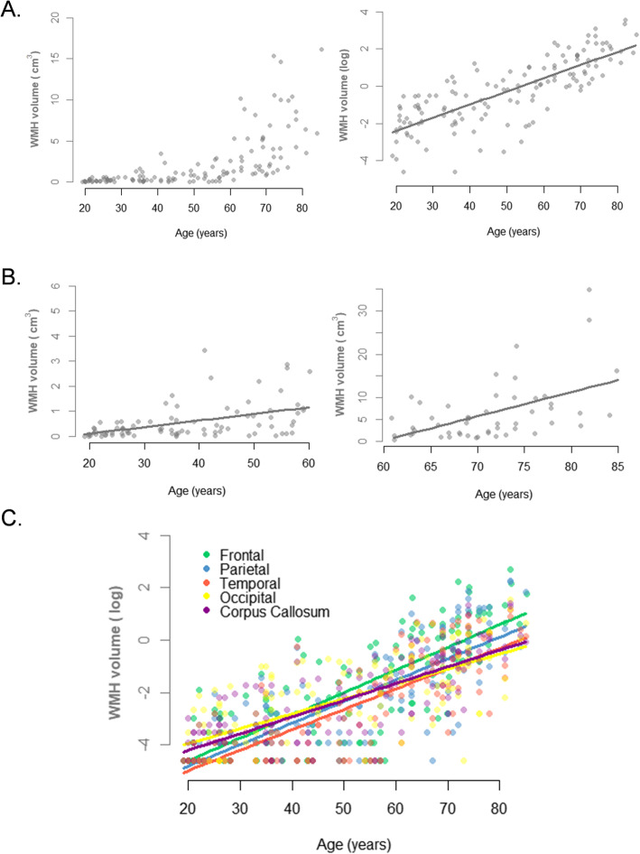
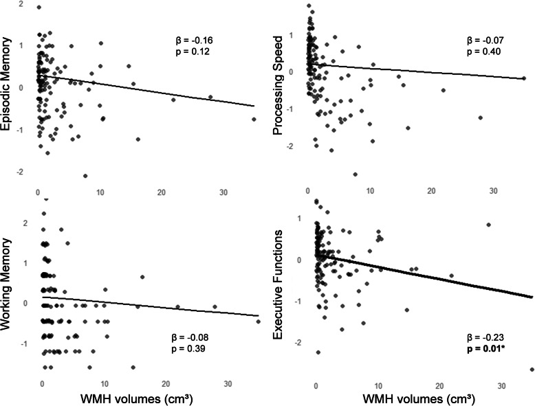
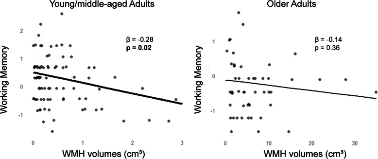
BACKGROUND: White matter hyperintensities (WMH) are very frequent in older adults and associated with worse cognitive performance. Little is known about the links between WMH and vascular risk factors, cortical β-amyloid (Aβ) load, and cognition in cognitively unimpaired adults across the entire lifespan, especially in young and middle-aged adults. METHODS: One hundred and thirty-seven cognitively unimpaired adults from the community were enrolled (IMAP cohort). Participants underwent (i) a comprehensive neuropsychological assessment of episodic memory, processing speed, working memory, and executive functions; (ii) brain structural T1 and FLAIR MRI scans used for the automatic segmentation of total and regional (frontal, parietal, temporal, occipital, and corpus callosum) WMH; and (iii) a Florbetapir-PET scan to measure cortical Aβ. The relationships of total and regional WMH to age, vascular risk factors, cortical Aβ, and cognition were assessed within the whole sample, but also splitting the sample in two age groups (≤ or > 60 years old). RESULTS: WMH increased with age across the adult lifespan, i.e., even in young and middle-aged adults. Systolic blood pressure, diastolic blood pressure, and glycated hemoglobin were all associated with higher WMH before, but not after, adjusting for age and the other vascular risk factors. Higher frontal, temporal, and occipital WMH were associated with greater Aβ, but this association was no longer significant when adjusting for age and vascular risk factors. Higher total and frontal WMH were associated with worse performance in executive functions, with no interactive effect of the age group. In contrast, there was a significant interaction of the age group on the link between WMH and working memory, which was significant within the subgroup of young/middle-aged adults only. Adding cortical Aβ load in the models did not alter the results, and there was no interaction between WMH and Aβ on cognition. CONCLUSION: WMH increased with age and were associated with worse executive functions across the adult lifespan and with worse working memory in young/middle-aged adults. Aβ load was weakly associated with WMH and did not change the relationship found between WMH and executive functions. This study argues for the clinical relevance of WMH across the adult lifespan, even in young and middle-aged adults with low WMH.
Overview
The claims and statistics of this paper haven’t been checked yet.
Run the check — free, no account needed. It reads the open-access text and checks the sources it cites.
Integrity signals
Registry
Cites
Statistics
Figures
Verification
Claims the paper makes
The claims and statistics of this paper haven’t been checked yet.
Run the check — free, no account needed. It reads the open-access text and checks the sources it cites.
Claims other papers make about this one
Each is checked against this paper’s own text: does it say what the citing paper says it does?
The source backs part of the claim, not all of it — or backs it for a different population, condition or comparison.
In the citing paper
“Previous studies investigating the impact of regional distribution of WMHs on executive function (EF) found that WMHs in all cortical regions (frontal areas, occipital, parietal and temporal) are associated with deficits in EF scores [15,16,17]”
From Amyloid-PET and White Matter Hyperintensities Have Independent Effects on Baseline Cognitive Function and Synergistic Effects on Longitudinal Executive Function (2023) · cites this paper as [16]
Experimental — accuracy not yet measured
Self-check
Percentages, sums and reported differences re-computed from the paper’s own numbers.
The self-check needs the paper’s full text.
Citations
Cites
Dementia prevention, intervention, and care
Livingston, G., Sommerlad, A., Orgeta, V., Costafreda, S. G., Huntley, J., Ames, D., Ballard, C., Banerjee, S., Burns, A., Cohen-Mansfield, J., et al. (2017) · The Lancet · doi:10.1016/s0140-6736(17)31363-6 · Cited by 6,072
Pantoni, L. (2010) · The Lancet Neurology · doi:10.1016/s1474-4422(10)70104-6 · Cited by 3,405
Wechsler Adult Intelligence Scale--Fourth Edition
Wechsler, D. (2012) · PsycTESTS Dataset · doi:10.1037/t15169-000 · Cited by 2,232
Reproducibility of quantitative tractography methods applied to cerebral white matter
Wakana, S., Caprihan, A., Panzenboeck, M. M., Fallon, J. H., Perry, M., Gollub, R. L., Hua, K., Zhang, J., Jiang, H., Dubey, P., et al. (2007) · NeuroImage · doi:10.1016/j.neuroimage.2007.02.049 · Cited by 1,686
Hua, K., Zhang, J., Wakana, S., Jiang, H., Li, X., Reich, D. S., Calabresi, P. A., Pekar, J. J., van Zijl, P. C. M., & Mori, S. (2007) · NeuroImage · doi:10.1016/j.neuroimage.2007.07.053 · Cited by 1,507
de Leeuw, F.-E. (2001) · Journal of Neurology, Neurosurgery & Psychiatry · doi:10.1136/jnnp.70.1.9 · Cited by 1,291
White matter hyperintensities, cognitive impairment and dementia: an update
Prins, N. D., & Scheltens, P. (2015) · Nature Reviews Neurology · doi:10.1038/nrneurol.2015.10 · Cited by 1,284
Hammers, A., Allom, R. J., Koepp, M. J., Free, S. L., Myers, R., Lemieux, L., Mitchell, T. N., Brooks, D. J., & Duncan, J. S. (2003) · Human Brain Mapping · doi:10.1002/hbm.10123 · Cited by 1,270
Screening for dementia by memory testing
Grober, E. H., Herman Buschke, L., Crystal, H., Bang, S., & Dresner, R. (1988) · Neurology · doi:10.1212/wnl.38.6.900 · Cited by 956
What are White Matter Hyperintensities Made of?
Wardlaw, J. M., Hernández, M. C. V., & Maniega, S. M. (2015) · Journal of the American Heart Association · doi:10.1161/jaha.114.001140 · Cited by 948
Defining imaging biomarker cut points for brain aging and Alzheimer's disease
Jack, C. R., Wiste, H. J., Weigand, S. D., Therneau, T. M., Lowe, V. J., Knopman, D. S., Gunter, J. L., Senjem, M. L., Jones, D. T., Kantarci, K., et al. (2017) · Alzheimer's & Dementia · doi:10.1016/j.jalz.2016.08.005 · Cited by 931
Midlife vascular risk factor exposure accelerates structural brain aging and cognitive decline
Debette, S., Seshadri, S., Beiser, A., Au, R., Himali, J. J., Palumbo, C., Wolf, P. A., & DeCarli, C. (2011) · Neurology · doi:10.1212/wnl.0b013e318227b227 · Cited by 772
Vascular dysfunction—The disregarded partner of Alzheimer's disease
Sweeney, M. D., Montagne, A., Sagare, A. P., Nation, D. A., Schneider, L. S., Chui, H. C., Harrington, M. G., Pa, J., Law, M., Wang, D. J. J., et al. (2019) · Alzheimer's & Dementia · doi:10.1016/j.jalz.2018.07.222 · Cited by 711
White matter lesions impair frontal lobe function regardless of their location
Tullberg, M., Fletcher, E., DeCarli, C., Mungas, D., Reed, B. R., Harvey, D. J., Weiner, M. W., Chui, H. C., & Jagust, W. J. (2004) · Neurology · doi:10.1212/01.wnl.0000130530.55104.b5 · Cited by 530
Age, Sex, andAPOEε4 Effects on Memory, Brain Structure, and β-Amyloid Across the Adult Life Span
Jack, C. R., Wiste, H. J., Weigand, S. D., Knopman, D. S., Vemuri, P., Mielke, M. M., Lowe, V. J., Senjem, M. L., Gunter, J. L., Machulda, M. M., et al. (2015) · JAMA Neurology · doi:10.1001/jamaneurol.2014.4821 · Cited by 412
La Joie, R., Perrotin, A., Barré, L., Hommet, C., Mézenge, F., Ibazizene, M., Camus, V., Abbas, A., Landeau, B., Guilloteau, D., et al. (2012) · The Journal of Neuroscience · doi:10.1523/jneurosci.2170-12.2012 · Cited by 374
Self-citationPresence and progression of white matter hyperintensities and cognition: A meta-analysis
Kloppenborg, R. P., Nederkoorn, P. J., Geerlings, M. I., & van den Berg, E. (2014) · Neurology · doi:10.1212/wnl.0000000000000505 · Cited by 338
Associations between vascular risk factors and brain MRI indices in UK Biobank
Cox, S. R., Lyall, D. M., Ritchie, S. J., Bastin, M. E., Harris, M. A., Buchanan, C. R., Fawns‐Ritchie, C., Barbu, M. C., de Nooij, L., Reus, L. M., et al. (2019) · European Heart Journal · doi:10.1093/eurheartj/ehz100 · Cited by 332
Vascular and amyloid pathologies are independent predictors of cognitive decline in normal elderly
Vemuri, P., Lesnick, T. G., Przybelski, S. A., Knopman, D. S., Preboske, G. M., Kantarci, K., Raman, M. R., Machulda, M. M., Mielke, M. M., Lowe, V. J., et al. (2015) · Brain · doi:10.1093/brain/awu393 · Cited by 299
White matter hyperintensities: relationship to amyloid and tau burden
Graff-Radford, J., Arenaza-Urquijo, E. M., Knopman, D. S., Schwarz, C. G., Brown, R. D., Rabinstein, A. A., Gunter, J. L., Senjem, M. L., Przybelski, S. A., Lesnick, T., et al. (2019) · Brain · doi:10.1093/brain/awz162 · Cited by 253
Bolandzadeh, N., Davis, J. C., Tam, R. C., Handy, T. C., & Liu-Ambrose, T. Y. L. (2012) · BMC Neurology · doi:10.1186/1471-2377-12-126 · Cited by 221
Rabin, J. S., Schultz, A. P., Hedden, T., Viswanathan, A., Marshall, G. A., Kilpatrick, E., Klein, H., Buckley, R. F., Yang, H.-S., Properzi, M., et al. (2018) · JAMA Neurology · doi:10.1001/jamaneurol.2018.1123 · Cited by 221
Wen, W., Sachdev, P. S., Li, J. J., Chen, X., & Anstey, K. J. (2009) · Human Brain Mapping · doi:10.1002/hbm.20586 · Cited by 210
Early cerebral small vessel disease and brain volume, cognition, and gait
Smith, E. E., O'Donnell, M., Dagenais, G., Lear, S. A., Wielgosz, A., Sharma, M., Poirier, P., Stotts, G., Black, S. E., Strother, S., et al. (2015) · Annals of Neurology · doi:10.1002/ana.24320 · Cited by 207
Hedden, T., Mormino, E. C., Amariglio, R. E., Younger, A. P., Schultz, A. P., Becker, J. A., Buckner, R. L., Johnson, K. A., Sperling, R. A., & Rentz, D. M. (2012) · The Journal of Neuroscience · doi:10.1523/jneurosci.2462-12.2012 · Cited by 182
Prevalence of White Matter Hyperintensities in a Young Healthy Population
Hopkins, R. O., Beck, C. J., Burnett, D. L., Weaver, L. K., Victoroff, J., & Bigler, E. D. (2006) · Journal of Neuroimaging · doi:10.1111/j.1552-6569.2006.00047.x · Cited by 163
Habes, M., Sotiras, A., Erus, G., Toledo, J. B., Janowitz, D., Wolk, D. A., Shou, H., Bryan, R. N., Doshi, J. J., Völzke, H., et al. (2018) · Neurology · doi:10.1212/wnl.0000000000006116 · Cited by 147
Besson, F. L., La Joie, R., Doeuvre, L., Gaubert, M., Mezenge, F., Egret, S., Landeau, B., Barre, L., Abbas, A., Ibazizene, M., et al. (2015) · Journal of Neuroscience · doi:10.1523/jneurosci.0150-15.2015 · Cited by 141
Self-citationAssociations between amyloid β and white matter hyperintensities: A systematic review
Roseborough, A. D., Ramirez, J., Black, S. E., & Edwards, J. D. (2017) · Alzheimer s & Dementia · doi:10.1016/j.jalz.2017.01.026 · Cited by 139
Cerebrovascular disease, beta-amyloid, and cognition in aging
Marchant, N. L., Reed, B. R., DeCarli, C. S., Madison, C. M., Weiner, M. W., Chui, H. C., & Jagust, W. J. (2012) · Neurobiology of Aging · doi:10.1016/j.neurobiolaging.2011.10.001 · Cited by 127
Epidemiology of White Matter Lesions
Launer, L. J. (2004) · Topics in Magnetic Resonance Imaging · doi:10.1097/01.rmr.0000168216.98338.8d · Cited by 125
White matter hyperintensities are common in midlife and already associated with cognitive decline
d’Arbeloff, T. C., Elliott, M. L., Knodt, A. R., Melzer, T. R., Keenan, R. J., Ireland, D. R., Ramrakha, S., Poulton, R., Anderson, T., Caspi, A., et al. (2019) · Brain Communications · doi:10.1093/braincomms/fcz041 · Cited by 108
Lee, M. J., Seo, S. W., Na, D. L., Kim, C., Park, J. H., Kim, G. H., Kim, C. H., Noh, Y., Cho, H., Kim, H. J., et al. (2014) · JAMA Psychiatry · doi:10.1001/jamapsychiatry.2013.4506 · Cited by 105
Periventricular hyperintensities are associated with elevated cerebral amyloid
Marnane, M., Al-Jawadi, O., Mortazavi, S. A., Pogorzelec, K. J., Wang, B. W., Feldman, H., Hsiung, G. R., Initiative, A. D. N., Initiative, A. D. N., Weiner, M. W., et al. (2016) · Neurology · doi:10.1212/wnl.0000000000002352 · Cited by 91
André, C., Tomadesso, C., de Florès, R., Branger, P., Rehel, S., Mézenge, F., Landeau, B., Sayette, V. D. L., Eustache, F., Chételat, G., et al. (2019) · Alzheimer s & Dementia Diagnosis Assessment & Disease Monitoring · doi:10.1016/j.dadm.2018.12.009 · Cited by 71
Self-citationBunce, D. J., Anstey, K. J., Cherbuin, N., Burns, R. A., Christensen, H. M., Wen, W. X., & Sachdev, P. S. (2010) · PLoS ONE · doi:10.1371/journal.pone.0013567 · Cited by 68
Wechsler Adult Intelligence Scale–Fourth Edition
Dumont, R., Willis, J. O., Veizel, K., & Zibulsky, J. (2014) · Encyclopedia of Special Education · doi:10.1002/9781118660584.ese2520 · Cited by 66
White Matter Abnormalities and Cognition in a Community Sample
Vannorsdall, T. D., Waldstein, S. R., Kraut, M. A., Pearlson, G. D., & Schretlen, D. J. (2009) · Archives of Clinical Neuropsychology · doi:10.1093/arclin/acp037 · Cited by 66
Dissociable Effects of Alzheimer Disease and White Matter Hyperintensities on Brain Metabolism
Haight, T. J. (2013) · JAMA Neurology · doi:10.1001/jamaneurol.2013.1878 · Cited by 65
King, K. S., Peshock, R. M., Rossetti, H. C., McColl, R. W., Ayers, C. R., Hulsey, K., & Das, S. R. (2013) · Stroke · doi:10.1161/strokeaha.113.003602 · Cited by 65
Study, F. T. A., Brugulat-Serrat, A., Salvadó, G., Sudre, C. H., Grau-Rivera, O., Suárez-Calvet, M., Falcon, C., Sánchez-Benavides, G., Gramunt, N., Fauria, K., et al. (2020) · Brain Imaging and Behavior · doi:10.1007/s11682-019-00151-2 · Cited by 64
The effects of white matter hyperintensities and amyloid deposition on Alzheimer dementia
Gordon, B. A., Najmi, S., Hsu, P. J., Roe, C. M., Morris, J. C., & Benzinger, T. L. S. (2015) · NeuroImage Clinical · doi:10.1016/j.nicl.2015.04.017 · Cited by 62
Soriano-Raya, J. J., Miralbell, J., Elena López-Cancio, M., Bargalló, N., Arenillas, J. F., Barrios, M., Cáceres, C. M., Torán‐Monserrat, P., Alzamora, M. T., Dávalos, A., et al. (2012) · Journal of the International Neuropsychological Society · doi:10.1017/s1355617712000677 · Cited by 60
Vascular and neurodegenerative pathways—will they meet?
Kalaria, R. N., & Ihara, M. (2013) · Nature Reviews Neurology · doi:10.1038/nrneurol.2013.164 · Cited by 60
Increased florbetapir binding in the temporal neocortex from age 20 to 60 years
Gonneaud, J., Arenaza‐Urquijo, E. M., Mézenge, F., Landeau, B., Gaubert, M., Bejanin, A., de Florès, R., Wirth, M., Tomadesso, C., Poisnel, G., et al. (2017) · Neurology · doi:10.1212/wnl.0000000000004733 · Cited by 49
Self-citationWalsh, P., Sudre, C. H., Fiford, C. M., Ryan, N. S., Lashley, T., Frost, C. D., & Barnes, J. (2020) · Neurobiology of Aging · doi:10.1016/j.neurobiolaging.2020.03.008 · Cited by 49
Jiang, J., Paradise, M., Liu, T., Armstrong, N. J., Zhu, W., Kochan, N. A., Brodaty, H., Sachdev, P. S., & Wen, W. X. (2018) · NeuroImage Clinical · doi:10.1016/j.nicl.2018.03.035 · Cited by 48
Moura, A. R., Lee, S., Habeck, C. G., Razlighi, Q. R., & Stern, Y. (2019) · Neurobiology of Aging · doi:10.1016/j.neurobiolaging.2019.08.024 · Cited by 36
Amyloid burden and white matter hyperintensities mediate age-related cognitive differences
Dupont, P. S., Bocti, C., Joannette, M., Lavallée, M. M., Nikelski, J., Vallet, G. T., Chertkow, H., & Joubert, S. (2020) · Neurobiology of Aging · doi:10.1016/j.neurobiolaging.2019.08.025 · Cited by 25
Fonctions exécutives et pathologies neurologiques et psychiatriques
Godefroy, O. (2012) · HAL (Le Centre pour la Communication Scientifique Directe) · Cited by 25
Relevance of brain lesion location for cognition in vascular mild cognitive impairment
Giorgio, A., Donato, I. D., Leucio, A. D., Zhang, J., Salvadori, E., Poggesi, A., Diciotti, S., Cosottini, M., Ciulli, S., Inzitari, D., et al. (2019) · NeuroImage Clinical · doi:10.1016/j.nicl.2019.101789 · Cited by 18
Eustache, F., Laisney, M., Lalevée, C., Pèlerin, A., Perrotin, A., Egret, S., Chételat, G., & Desgranges, B. (2015) · Revue de neuropsychologie · doi:10.1684/nrp.2015.0351 · Cited by 14
Self-citationWirth, A., Johannesen, S. W., Khomenko, A., Baldaranov, D., Bruun, T., Wendl, C. M., Schuierer, G. R., Greenlee, M. W., & Bogdahn, U. (2018) · Journal of Magnetic Resonance Imaging · doi:10.1002/jmri.26577 · Cited by 11
Eustache, F., Laisney, M., Lalevée, C., Pèlerin, A., Perrotin, A., Egret, S., Chételat, G., & Desgranges, B. (2015) · Revue de neuropsychologie · doi:10.3917/rne.073.0217 · Cited by 8
Self-citation
Cited by
Sorted by citations
White matter hyperintensity topography in Alzheimer's disease and links to cognition
Garnier‐Crussard, A., Bougacha, S., Wirth, M., Dautricourt, S., Sherif, S., Landeau, B., Gonneaud, J., De Flores, R., de la Sayette, V., Vivien, D., et al. (2022) · Alzheimer's & Dementia · doi:10.1002/alz.12410 · Cited by 170
Self-citationKellar, D., Lockhart, S. N., Aisen, P. S. S., Raman, R., Rissman, R. A., Brewer, J. B., & Craft, S. (2021) · The Journal of Prevention of Alzheimer s Disease · doi:10.14283/jpad.2021.14 · Cited by 92
Gaubert, M., Lange, C., Garnier‐Crussard, A., Köbe, T., Bougacha, S., Gonneaud, J., de Florès, R., Tomadesso, C., Mézenge, F., Landeau, B., et al. (2021) · Alzheimer s Research & Therapy · doi:10.1186/s13195-020-00759-3 · Cited by 67
Self-citationVascular burden and cognition: Mediating roles of neurodegeneration and amyloid PET
Ottoy, J., Ozzoude, M., Zukotynski, K., Adamo, S. M., Scott, C. J. M., Gaudet, V., Ramirez, J., Swardfager, W., Cogo‐Moreira, H., Lam, B., et al. (2022) · Alzheimer s & Dementia · doi:10.1002/alz.12750 · Cited by 48
White matter hyperintensities in former American football players
Alosco, M. L., Tripodis, Y., Baucom, Z., Adler, C. H., Balcer, L. J., Bernick, C. B., Mariani, M. L., Au, R., Banks, S. J., Barr, W., et al. (2022) · Alzheimer s & Dementia · doi:10.1002/alz.12779 · Cited by 35
Metadata
- Authors
- Garnier-Crussard, A., Bougacha, S., Wirth, M., André, C., Delarue, M., Landeau, B., Mézenge, F., Kuhn, E., Gonneaud, J., Chocat, A., Quillard, A., Ferrand-Devouge, E., de La Sayette, V., Vivien, D., Krolak-Salmon, P., & Chételat, G.
- Affiliations
- Inserm (FR)
- German Center for Neurodegenerative Diseases (DZNE) Dresden (DE)
- Corresponding author
- Chételat, Gaël
- ORCID
- Venue
- Alzheimer's Research & Therapy
- Published
- 2020-10-08
- Volume
- 12
- Issue
- 1
- Pages
- 127-127
- ISSN
- 1758-9193 (ISSN-L)
- PMC
- PMC7545576
- OpenAlex
- W3092538613
- Cited by
- 130
- References
- 54
- Citations by year
- 2026: 18 · 2025: 33 · 2024: 22 · 2023: 25 · 2022: 20 · 2021: 12
- FWCI
- 6.17
- Type
- journal article
- Language
- en
- Open access
- gold
- doi.org
- Locations
- Alzheimer s Research & Therapy (publishedVersion, cc-by)
- PubMed Central (publishedVersion, cc-by)
- PubMed (publishedVersion)
- HAL (submittedVersion)
- DOAJ (DOAJ: Directory of Open Access Journals) (submittedVersion, cc-by-sa)
- DZNE Pub (submittedVersion, other-oa)
- Funders
- Institut National de la Santé et de la Recherche Medicale
- Programme Hospitalier de Recherche Clinique (PHRCN 2011-A01493-38, 2012 12-006-0347)
- Agence Nationale de la Recherche
- Fondation Plan Alzheimer
- Fondation LECMA-Vaincre Alzheimer (13732)
- Association France Alzheimer et maladies apparentées, the Région Basse-Normandie
- Agence régionale de santé Auvergne-Rhône-Alpes
- Horizon 2020 (667696)
- Topics
- Health Sciences › Medicine › Clinical Neurology › Cerebrovascular and genetic disorders
- Health Sciences › Medicine › Psychiatry and Mental health › Dementia and Cognitive Impairment Research
- Life Sciences › Neuroscience › Neurology › Neurological Disease Mechanisms and Treatments
- Keywords
- white matter hyperintensities, vascular risk factors, aging, executive function, working memory, episodic memory, processing speed, young and middle-aged adults, systolic blood pressure, HbA1c, cognitive aging
- MeSH terms
- Aged, Brain, Cognition, Humans, Longevity, Magnetic Resonance Imaging, Middle Aged, Neuropsychological Tests, White Matter
About these checks
Registry, retraction, Cites and statistics results are computed, not judged, with one exception: a printed reference that no index lists is read once by a model before it counts as a citation error. Claims are extracted by a model and kept only when their quote appears verbatim in the text; each cited source is then checked, with a model’s help, for whether it says what the claim relies on. Those verdicts can be wrong — read the claim’s quote and the cited source before relying on one.
Statistics and arithmetic are recomputed, not judged.
Confidence bands are shown only for fields calibrated on our annotated test set. None is calibrated yet, so no band is shown.
What counts as a claim ›How we measure confidence ›How we check this ›
Are you an author, or do you see a problem with this page? Request a review
