

The DOI matches its Crossref record; 4 of the 4 references it cites were found in a registry or an index.
Human umbilical endothelial cells (HUVECs) have a sex: characterisation of the phenotype of male and female cells
Overview
The claims and statistics of this paper haven’t been checked yet.
Run the check — free, no account needed. It reads the open-access text and checks the sources it cites.
Integrity signals
Registry
Cites
Statistics
Figures
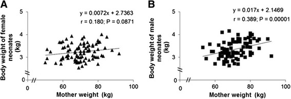
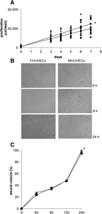
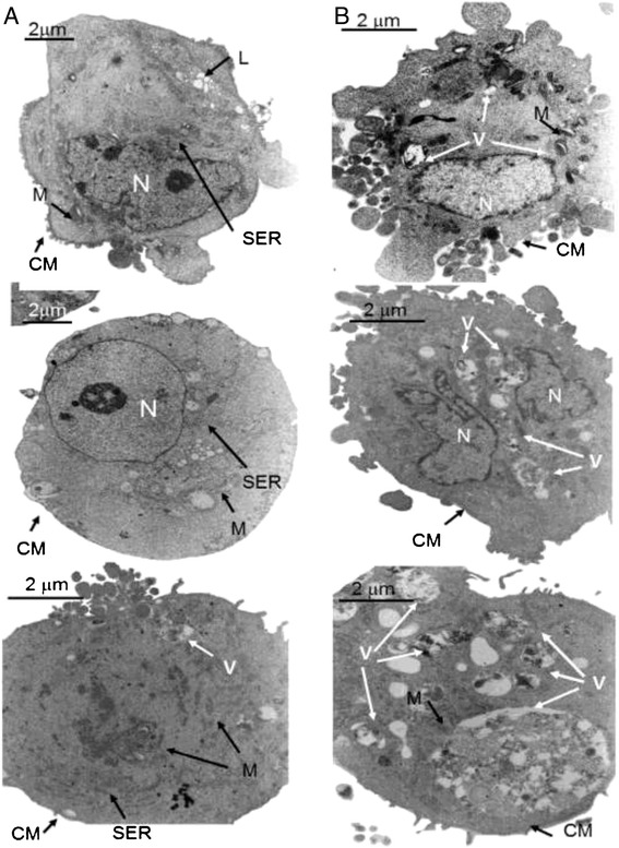
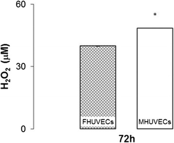
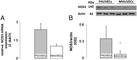
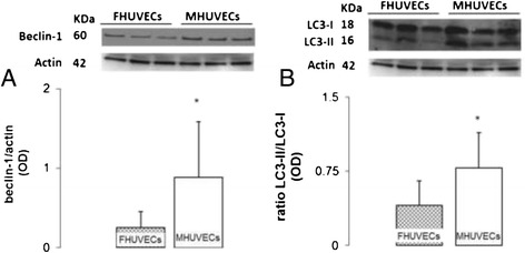
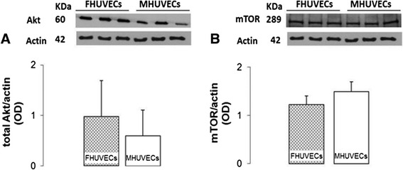
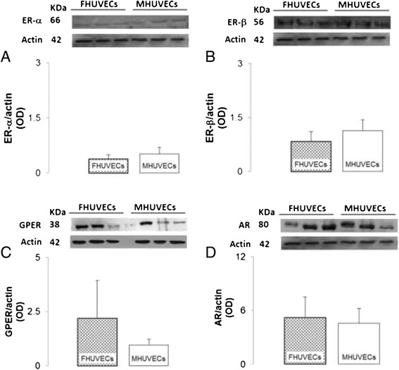
Verification
Claims the paper makes
The claims and statistics of this paper haven’t been checked yet.
Run the check — free, no account needed. It reads the open-access text and checks the sources it cites.
Claims other papers make about this one
Each is checked against this paper’s own text: does it say what the citing paper says it does?
In the citing paper
“For example, as human umbilical vein endothelial cells (HUVECs) are obtained from the umbilical cord, they are not only influenced by maternal hormones but also affected by embryonic gonads.”
From Cell Immortality: In Vitro Effective Techniques to Achieve and Investigate Its Applications and Challenges (2024) · cites this paper as [9]
Self-check
Percentages, sums and reported differences re-computed from the paper’s own numbers.
The self-check needs the paper’s full text.
Citations
Cites
Rabinowitz, J. D., & White, E. (2010) · Science · doi:10.1126/science.1193497 · Cited by 1,982
Autophagy, mitochondria and oxidative stress: cross-talk and redox signalling.
Lee, J., Giordano, S., & Zhang, J. (2012) · Biochemical Journal · doi:10.1042/bj20111451 · Cited by 1,438
Starving Neurons Show Sex Difference in Autophagy
Du, L., Hickey, R. W., Bayir, H., Watkins, S. C., Tyurin, V. A., Guo, F., Kochanek, P. M., Jenkins, L. W., Ren, J., Gibson, G., et al. (2009) · Journal of Biological Chemistry · doi:10.1074/jbc.m804396200 · Cited by 204
Molecular Links Between Endothelial Dysfunction and Neurodegeneration in Alzheimer's Disease
Lyros, E., Bakogiannis, C., Liu, Y., & Fassbender, K. (2014) · Current Alzheimer Research · doi:10.2174/1567205010666131119235254 · Cited by 70 · cited 2×
“The awareness of such sex differences could help researchers understand the sexual differences observed in the incidence, presentation and outcome of cardiovascular diseases [5] and in other pathologies such as immune disorders and neurodegeneration [3,4].”
Background
Research works printed but not linked: 57
- Sena CM, Pereira AM, Seica R: Endothelial dysfunction - a major mediator of diabetic vascular disease. Biochim Biophys Acta (BBA) - Mol Basis Dis 2013, 1832: 2216–2231. 10.1016/j.bbadis.2013.08.006 · DOI
- Xiao L, Liu Y, Wang N: New paradigms in inflammatory signaling in vascular endothelial cells. Am J Physiol Heart Circ Physiol 2014, 306: H317-H325. 10.1152/ajpheart.00182.2013 · DOI
- Dai R, Ahmed S: Sexual dimorphism of miRNA expression: a new perspective in understanding the sex bias of autoimmune diseases. Ther Clin Risk Manag 2014, 10: 151–163.
- Legato MJ: Principles of Gender-Specific Medicine: 2. 2nd edition. Academic Press, San Diego; 2009.
- Papakonstantinou NA, Stamou MI, Baikoussis NG, Goudevenos J, Apostolakis E: Sex differentiation with regard to coronary artery disease. J Cardiol 2013, 62: 4–11. 10.1016/j.jjcc.2013.03.001 · DOI
- Kublickiene K, Luksha L: Gender and the endothelium. Pharmacol Rep 2008, 60: 49–60.
- Shah K, McCormack CE, Bradbury NA: Do you know the sex of your cells? Am J Physiol Cell Physiol 2014, 306: C3-C18. 10.1152/ajpcell.00281.2013 · DOI · Paper page
- Taylor KE, Vallejo-Giraldo C, Schaible N, Zakeri R, Miller V: Reporting of sex as a variable in cardiovascular studies using cultured cells. Biol Sex Differ 2011, 2: 11. 10.1186/2042-6410-2-11 · DOI
- Wang M, Wang Y, Weil B, Abarbanell A, Herrmann J, Tan J, Kelly M, Meldrum DR: Estrogen receptor beta mediates increased activation of PI3K/Akt signaling and improved myocardial function in female hearts following acute ischemia. Am J Physiol Regul Integr Comp Physiol 2009, 296: R972-R978. 10.1152/ajpregu.00045.2009 · DOI
- Oviedo PJ, Sobrino A, Laguna-Fernandez A, Novella S, Tarìn JJ, Garcia-Pérez MA, Sanchìs J, Cano A, Hermenegildo C: Estradiol induces endothelial cell migration and proliferation through estrogen receptor-enhanced RhoA/ROCK pathway. Mol Cell Endocrinol 2011, 335: 96. 10.1016/j.mce.2010.06.020 · DOI
- Kublickiene K, Fu XD, Svedas E, Landgren BM, Genazzani AR, Simoncini T: Effects in postmenopausal women of estradiol and medroxyprogesterone alone and combined on resistance artery function and endothelial morphology and movement. J Clin Endocrinol Metabol 2008, 93: 1874–1883. 10.1210/jc.2007-2651 · DOI
- Reckelhoff JF: Sex and sex steroids in cardiovascular-renal physiology and pathophysiology. Gender Med 2008, 1(Supplement 1):S1-S2. 10.1016/j.genm.2008.03.001 · DOI
- Regitz-Zagrosek V, Wintermantel TM, Schubert C: Estrogens and SERMs in coronary heart disease. Curr Opin Pharmacol 2007, 7: 130–139. 10.1016/j.coph.2006.10.009 · DOI
- Kypreos KE, Zafirovic S, Petropoulou PI, Bjelogrlic P, Resanovic I, Traish A, Isenovic ER: Regulation of endothelial nitric oxide synthase and high-density lipoprotein quality by estradiol in cardiovascular pathology. J Cardiovasc Pharmacol Ther 2014, 19: 256–268. 10.1177/1074248413513499 · DOI
- Wu Q, Chambliss K, Umetani M, Mineo C, Shaul PW: Non-nuclear estrogen receptor signaling in the endothelium. J Biol Chem 2011, 286: 14737–14743. 10.1074/jbc.R110.191791 · DOI
- Onat D, Brillon D, Colombo P, Schmidt A: Human vascular endothelial cells: a model system for studying vascular inflammation in diabetes and atherosclerosis. Current Diabetes Reports 2011, 11: 193–202. 10.1007/s11892-011-0182-2 · DOI
- Cines DB, Pollak ES, Buck CA, Loscalzo J, Zimmerman GA, McEver RP, Pober JS, Wick TM, Konkle BA, Schwartz BS, Barnathan ES, McCrae KR, Hug BA, Schmidt AM, Stern DM: Endothelial cells in physiology and in the pathophysiology of vascular disorders. Blood 1998, 91: 3527–3561.
- Batres RO, Dupont J: Gender differences in prostacyclin and prostaglandin E2 synthesis by human endothelial cells. Prostaglandins Leukot Med 1986, 22: 159–171. 10.1016/0262-1746(86)90085-5 · DOI
- Matarrese P, Colasanti T, Ascione B, Margutti P, Franconi F, Alessandri C, Conti F, Riccieri V, Rosano G, Ortona E, Malorni W: Gender disparity in susceptibility to oxidative stress and apoptosis induced by autoantibodies specific to RLIP76 in vascular cells. Antioxid Redox Signal 2011, 15: 2825–2836. 10.1089/ars.2011.3942 · DOI
- Villar IC, Scotland RS, Khambata RS, Chan M, Duchene J, Sampaio AL, Perretti M, Hobbs AJ, Ahluwalia A: Suppression of endothelial P-selectin expression contributes to reduced cell trafficking in females. Arterioscler Thromb Vasc Biol 2011, 31: 1075–1083. 10.1161/ATVBAHA.111.223545 · DOI
- Gupta NC, Davis CM, Nelson JW, Young JM, Alkayed NJ: Soluble epoxide hydrolase: sex differences and role in endothelial cell survival. Arterioscler Thromb Vasc Biol 2012, 32: 1936–1942. 10.1161/ATVBAHA.112.251520 · DOI
- Straface E, Vona R, Gambardella L, Ascione B, Marino M, Bulzomi P, Canu S, Coinu R, Rosano G, Malorni W, Franconi F: Cell sex determines anoikis resistance in vascular smooth muscle cells. FEBS Lett 2009, 583: 3448–3454. 10.1016/j.febslet.2009.09.052 · DOI
- Campesi I, Straface E, Occhioni S, Montella A, Franconi F: Protein oxidation seems to be linked to constitutive autophagy: a sex study. Life Sci 2013, 93: 145–152. 10.1016/j.lfs.2013.06.001 · DOI
- Chen C, Hu LX, Dong T, Wang GQ, Wang LH, Zhou XP, Jiang Y, Murao K, Lu SQ, Chen JW, Zhang GX: Apoptosis and autophagy contribute to gender difference in cardiac ischemia-reperfusion induced injury in rats. Life Sci 2013, 93: 265–270. 10.1016/j.lfs.2013.06.019 · DOI
- Balconi G, Pietra A, Busacca M, de Gaetano G, Dejana E: Success rate of primary human endothelial cell culture from umbilical cords is influenced by maternal and fetal factors and interval from delivery. Vitro 1983, 19: 807–810. 10.1007/BF02618159 · DOI
- de Llano JJM, Fuertes G, Torro I, Garcia Vicent C, Fayos JL, Lurbe E: Birth weight and characteristics of endothelial and smooth muscle cell cultures from human umbilical cord vessels. J Transl Med 2009, 7: 30. 10.1186/1479-5876-7-30 · DOI
- Bertino E, Gilli G, Occhi L, Giuliani F, Di Nicola P, Spada E, Fabris C: Postnatal growth of preterm infants: which reference charts? Minerva Pediatr 2010, 62(3 Suppl 1):71–74.
- Crampton SP, Davis J, Hughes CC: Isolation of human umbilical vein endothelial cells (HUVEC). J Vis Exp 2007, 3: 183.
- Schatz F, Soderland C, Hendricks-Munoz KD, Gerrets RP, Lockwood CJ: Human endometrial endothelial cells: isolation, characterization, and inflammatory-mediated expression of tissue factor and type 1 plasminogen activator inhibitor. Biol Reprod 2000, 62: 691–697. 10.1095/biolreprod62.3.691 · DOI
- Handin RI, Wagner DD: Molecular and cellular biology of von Willebrand factor. Prog Hemost Thromb 1989, 9: 233–259.
- Cattaneo MG, Cappellini E, Benfante R, Ragni M, Omodeo-Salé F, Nisoli E, Borgese N, Vicentini LM: Chronic deficiency of nitric oxide affects hypoxia inducible factor-1a (HIF-1a) stability and migration in human endothelial cells. PLoS One 2011, 6: e29680. 10.1371/journal.pone.0029680 · DOI
- Sapan CV, Lundblad RL, Price NC: Colorimetric protein assay techniques. Biotechnol Appl Biochem 1999, 29: 99–108. · DOI
- Vandesompele J, De Preter K, Pattyn F, Poppe B, Van Roy N, De Paepe A, Speleman F: Accurate normalization of real-time quantitative RT-PCR data by geometric averaging of multiple internal control genes. Genome Biol 2002, 3: research0034.0031-research0034.0011. 10.1186/gb-2002-3-7-research0034 · DOI · Paper page
- de Jonge HJM, Fehrmann RSN, de Bont ESJM, Hofstra RMW, Gerbens F, Kamps WA, de Vries EGE, van der Zee AGJ, te Meerman GJ, ter Elst A: Evidence based selection of housekeeping genes. PLoS One 2007, 2: e898. 10.1371/journal.pone.0000898 · DOI
- Pfaffl MW, Horgan GW, Dempfle L: Relative expression software tool (REST©) for group-wise comparison and statistical analysis of relative expression results in real-time PCR. Nucleic Acids Res 2002, 30: e36. 10.1093/nar/30.9.e36 · DOI
- Malorni W, Campesi I, Straface E, Vella S, Franconi F: Redox features of the cell: a gender perspective. Antioxid Redox Signal 2007, 9: 1779–1801. 10.1089/ars.2007.1596 · DOI
- Ma XH, Piao S, Wang D, McAfee QW, Nathanson KL, Lum JJ, Li LZ, Amaravadi RK: Measurements of tumor cell autophagy predict invasiveness, resistance to chemotherapy, and survival in melanoma. Clin Cancer Res 2011, 17: 3478–3489. 10.1158/1078-0432.CCR-10-2372 · DOI · Paper page
- Prossnitz ER, Barton M: Estrogen biology: new insights into GPER function and clinical opportunities. Mol Cell Endocrinol 2014, 389: 71–83. 10.1016/j.mce.2014.02.002 · DOI
- Pellegrini M, Bulzomi P, Lecis M, Leone S, Campesi I, Franconi F, Marino M: Endocrine disruptors differently influence estrogen receptor β and androgen receptor in male and female rat VSMC. J Cell Physiol 2014, 229: 1061–1068. 10.1002/jcp.24530 · DOI
- Hervé MAJ, Meduri G, Petit FG, Domet TS, Lazennec G, Mourah S, Perrot-Applanat M: Regulation of the vascular endothelial growth factor (VEGF) receptor Flk-1/KDR by estradiol through VEGF in uterus. J Endocrinol 2006, 188: 91–99. 10.1677/joe.1.06184 · DOI
- Toth B, Saadat G, Geller A, Scholz C, Schulze S, Friese K, Jeschke U: Human umbilical vascular endothelial cells express estrogen receptor beta (ERβ) and progesterone receptor A (PR-A), but not ERα and PR-B. Histochem Cell Biol 2008, 130: 399. 10.1007/s00418-008-0426-7 · DOI
- Evans MJ, Harris HA, Miller CP, Karathanasis SK, Adelman SJ: Estrogen receptors a and b have similar activities in multiple endothelial cell pathways. Endocrinology 2002, 143: 3785–3795. 10.1210/en.2002-220356 · DOI
- Laughlin MH, Welshons WV, Sturek M, Rush JW, Turk JR, Taylor JA, Judy BM, Henderson KK, Ganjam V: Gender, exercise training, and eNOS expression in porcine skeletal muscle arteries. J Appl Physiol 2003, 95: 250–264. · DOI
- Ogueta SB, Schwartz SD, Yamashita CK, Farber DB: Estrogen receptor in the human eye: influence of gender and age on gene expression. Invest Ophthalmol Vis Sci 1999, 40: 1906–1911.
- Kim-Schulze S, McGowan KA, Hubchak SC, Cid MC, Martin MB, Kleinman HK, Greene GL, Schnaper HW: Expression of an estrogen receptor by human coronary artery and umbilical vein endothelial cells. Circulation 1996, 94: 1402–1407. 10.1161/01.CIR.94.6.1402 · DOI
- Greaves E, Collins F, Critchley HOD, Saunders PTK: ERb-dependent effects on uterine endothelial cells are cell specific and mediated via Sp1. Hum Reprod 2013, 28: 2490–2501. 10.1093/humrep/det235 · DOI
- Simard M, Drolet R, Blomquist C, Tremblay Y: Human type 2 17beta-hydroxysteroid dehydrogenase in umbilical vein and artery endothelial cells: differential inactivation of sex steroids according to the vessel type. Endocrine 2011, 40: 203–211. 10.1007/s12020-011-9519-5 · DOI
- Annibalini G, Agostini D, Calcabrini C, Martinelli C, Colombo E, Guescini M, Tibollo P, Stocchi V, Sestili P: Effects of sex hormones on inflammatory response in male and female vascular endothelial cells. J Endocrinol Investig 2014, 37: 861–869. 10.1007/s40618-014-0118-1 · DOI
- Death AK, McGrath KCY, Sader MA, Nakhla S, Jessup W, Handelsman DJ, Celermajer DS: Dihydrotestosterone promotes vascular cell adhesion molecule-1 expression in male human endothelial cells via a nuclear factor-kappaB-dependent pathway. Endocrinology 2004, 145: 1889–1897. 10.1210/en.2003-0789 · DOI
- McGrath K, Hill M, Mcrobb L, Heather A: The androgen receptor drives the sex-specific expression of vascular cell adhesion molecule-1 in endothelial cells but not lipid metabolism genes in monocyte-derived macrophages. Horm Mol Biol Clin Invest 2010, 2: 203–209.
- Bae S, Zhang L: Gender differences in cardioprotection against ischemia/reperfusion injury in adult rat hearts: focus on Akt and protein kinase C signaling. J Pharmacol Exp Ther 2005, 315: 1125–1135. 10.1124/jpet.105.090803 · DOI
- Wang F, He Q, Sun Y, Dai X, Yang XP: Female adult mouse cardiomyocytes are protected against oxidative stress. Hypertension 2010, 55: 1172–1178. 10.1161/HYPERTENSIONAHA.110.150839 · DOI
- Le TYL, Ashton AW, Mardini M, Stanton PG, Funder JW, Handelsman DJ, Mihailidou AS: Role of androgens in sex differences in cardiac damage during myocardial infarction. Endocrinology 2014, 155: 568–575. 10.1210/en.2013-1755 · DOI
- Cuestas E, Darauich L, Corredera L, Costa ME: Is there any correlation between mothers birth weight with the first child birth weight? Rev Fac Cien Med Univ Nac 2007, 3: 68–72.
- Hales CN, Barker DJP: The thrifty phenotype hypothesis: type 2 diabetes. Br Med Bull 2001, 60: 5–20. 10.1093/bmb/60.1.5 · DOI
- Zhang Y, Li H, Liu S, Fu G, Zhao Y, Xie YJ, Zhang Y, Wang Y: The associations of high birth weight with blood pressure and hypertension in later life: a systematic review and meta-analysis. Hypertens Res 2013, 36: 725–735. 10.1038/hr.2013.33 · DOI
- Murrow L, Debnath J: Autophagy as a stress-response and quality-control mechanism: implications for cell injury and human disease. Annu Rev Pathol 2013, 8: 105–137. 10.1146/annurev-pathol-020712-163918 · DOI
Cited by
Guidelines for the use and interpretation of assays for monitoring autophagy (4th edition) 1
Klionsky, D. J., Abdel-Aziz, A. K., Abdelfatah, S., Abdellatif, M., Abdoli, A., Abel, S., Abeliovich, H., Abildgaard, M. H., Abudu, Y. P., Acevedo-Arozena, A., et al. (2021) · Autophagy · doi:10.1080/15548627.2020.1797280 · Cited by 2,897
Self-citationGenes that escape X chromosome inactivation modulate sex differences in valve myofibroblasts
Aguado, B. A., Walker, C. J., Grim, J. C., Schroeder, M. E., Batan, D., Vogt, B. J., Rodriguez, A. G., Schwisow, J. A., Moulton, K. S., Weiss, R. M., et al. (2022) · Circulation · doi:10.1161/circulationaha.121.054108 · Cited by 87
Chalak, M., Hesaraki, M., Mirbahari, S. N., Yeganeh, M., Abdi, S., Rajabi, S., & Hemmatzadeh, F. (2024) · Life · doi:10.3390/life14030417 · Cited by 66
Metadata
- Authors
- Addis, R., Campesi, I., Fois, M., Capobianco, G., Dessole, S., Fenu, G., Montella, A., Cattaneo, M. G., Vicentini, L. M., & Franconi, F.
- Venue
- Biology of Sex Differences
- Published
- 2014-12
- Volume
- 5
- Issue
- 1
- ISSN
- 2042-6410
- PMC
- PMC4273493
- Cited by
- 104
- References
- 4
- Language
- en
About these checks
Registry, retraction, Cites and statistics results are computed, not judged, with one exception: a printed reference that no index lists is read once by a model before it counts as a citation error. Claims are extracted by a model and kept only when their quote appears verbatim in the text; each cited source is then checked, with a model’s help, for whether it says what the claim relies on. Those verdicts can be wrong — read the claim’s quote and the cited source before relying on one.
Statistics and arithmetic are recomputed, not judged.
Confidence bands are shown only for fields calibrated on our annotated test set. None is calibrated yet, so no band is shown.
What counts as a claim ›How we measure confidence ›How we check this ›
Are you an author, or do you see a problem with this page? Request a review
