

The DOI matches its Crossref record; 38 of the 38 references it cites were found in a registry or an index.
Muscle-specific loss of Bmal1 leads to disrupted tissue glucose metabolism and systemic glucose homeostasis
Open accessAbstract
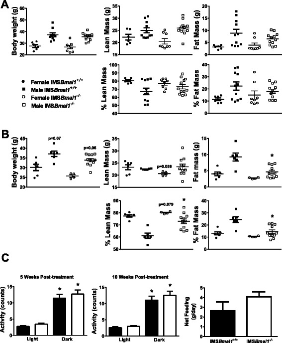
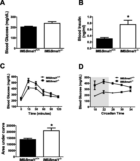
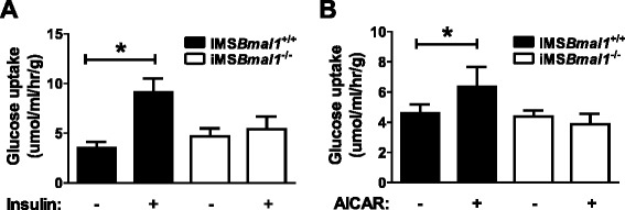
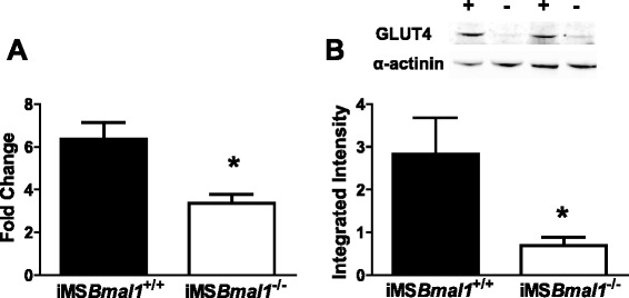
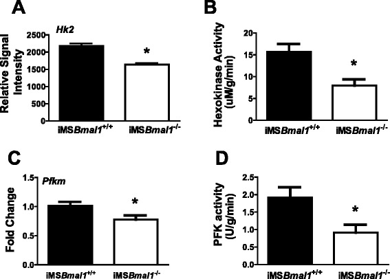
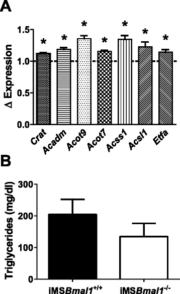
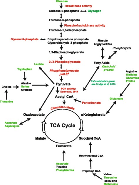
BACKGROUND: Diabetes is the seventh leading cause of death in the USA, and disruption of circadian rhythms is gaining recognition as a contributing factor to disease prevalence. This disease is characterized by hyperglycemia and glucose intolerance and symptoms caused by failure to produce and/or respond to insulin. The skeletal muscle is a key insulin-sensitive metabolic tissue, taking up ~80 % of postprandial glucose. To address the role of the skeletal muscle molecular clock to insulin sensitivity and glucose tolerance, we generated an inducible skeletal muscle-specific Bmal1 (-/-) mouse (iMSBmal1 (-/-)). RESULTS: Progressive changes in body composition (decreases in percent fat) were seen in the iMSBmal1 (-/-) mice from 3 to 12 weeks post-treatment as well as glucose intolerance and non-fasting hyperglycemia. Ex vivo analysis of glucose uptake revealed that the extensor digitorum longus (EDL) muscles did not respond to either insulin or 5-aminoimidazole-4-carboxamide ribonucleotide (AICAR) stimulation. RT-PCR and Western blot analyses demonstrated a significant decrease in mRNA expression and protein content of the muscle glucose transporter (Glut4). We also found that both mRNA expression and activity of two key rate-limiting enzymes of glycolysis, hexokinase 2 (Hk2) and phosphofructokinase 1 (Pfk1), were significantly reduced in the iMSBmal1 (-/-) muscle. Lastly, results from metabolomics analyses provided evidence of decreased glycolytic flux and uncovered decreases in some tricarboxylic acid (TCA) intermediates with increases in amino acid levels in the iMSBmal1 (-/-) muscle. These findings suggest that the muscle is relying predominantly on fat as a fuel with increased protein breakdown to support the TCA cycle. CONCLUSIONS: These data support a fundamental role for Bmal1, the endogenous circadian clock, in glucose metabolism in the skeletal muscle. Our findings have implicated altered molecular clock dictating significant changes in altered substrate metabolism in the absence of feeding or activity changes. The changes in body composition in our model also highlight the important role that changes in skeletal muscle carbohydrate, and fat metabolism can play in systemic metabolism.
Overview
The claims and statistics of this paper haven’t been checked yet.
Run the check — free, no account needed. It reads the open-access text and checks the sources it cites.
Integrity signals
Registry
Cites
Statistics
Figures
Verification
Claims the paper makes
The claims and statistics of this paper haven’t been checked yet.
Run the check — free, no account needed. It reads the open-access text and checks the sources it cites.
Claims other papers make about this one
Each is checked against this paper’s own text: does it say what the citing paper says it does?
In the citing paper
“Considering the negative metabolic consequences, such as reduction in insulin sensitivity and mitochondrial function, attributed to both circadian misalignment and sleep loss [21,67] and the potential benefits of exercise in this context [17,68], further research is warranted.”
From Exercise mitigates sleep-loss-induced changes in glucose tolerance, mitochondrial function, sarcoplasmic protein synthesis, and diurnal rhythms (2021) · cites this paper as [67]
In the citing paper
“Disruption of the muscle clock has also been shown to have significant impact on glucose intolerance and insulin resistance with very small changes in fiber types (Dyar et al., 2014; Schroder et al., 2015; Harfmann et al., 2016).”
From Myosteatosis in the Context of Skeletal Muscle Function Deficit: An Interdisciplinary Workshop at the National Institute on Aging (2020) · cites this paper as Harfmann et al., 2016
In the citing paper
“When investigating glucose transport ex vivo, it was clear that there was no increase in glucose uptake in response to insulin.”
From Myosteatosis in the Context of Skeletal Muscle Function Deficit: An Interdisciplinary Workshop at the National Institute on Aging (2020) · cites this paper as Harfmann et al., 2016
In the citing paper
“Muscles from Bmal1 knockout mice exhibit reduced glycolytic enzyme activity (e.g., 50% reduction in both hexokinase and phosphofructokinase activity) and a significant reduction in expression of the glucose transporter type 4 (GLUT-4) suggests that the molecular clock regulates muscle mechanisms that impact glucose uptake in muscle (Harfmann et al., 2016).”
From Myosteatosis in the Context of Skeletal Muscle Function Deficit: An Interdisciplinary Workshop at the National Institute on Aging (2020) · cites this paper as Harfmann et al., 2016
In the citing paper
“Using an inducible muscle specific Bmal1 knockout mouse, Harfmann et al. (2016), showed that non-fasted blood glucose (measured every 4 h for about 32 h) in the mice was always elevated compared to controls.”
From Myosteatosis in the Context of Skeletal Muscle Function Deficit: An Interdisciplinary Workshop at the National Institute on Aging (2020) · cites this paper as Harfmann et al., 2016
In the citing paper
“Further, use of 5-aminoimidazole-4-carboxamide-1-D-ribofuranoside (AICAR) to activate adenosine monophosphate (AMP)-activated protein kinase (AMPK) also did not increase glucose uptake indicating that loss of Bmal1 affects both exercise as well as insulin-induced glucose uptake.”
From Myosteatosis in the Context of Skeletal Muscle Function Deficit: An Interdisciplinary Workshop at the National Institute on Aging (2020) · cites this paper as Harfmann et al., 2016
Self-check
Percentages, sums and reported differences re-computed from the paper’s own numbers.
The self-check needs the paper’s full text.
Citations
Cites
Adverse metabolic and cardiovascular consequences of circadian misalignment
Scheer, F. A. J. L., Hilton, M. F., Mantzoros, C. S., & Shea, S. A. (2009) · Proceedings of the National Academy of Sciences · doi:10.1073/pnas.0808180106 · Cited by 2,351
Coordinated Transcription of Key Pathways in the Mouse by the Circadian Clock
Panda, S., Antoch, M. P., Miller, B. H., Su, A. I., Schook, A. C., Straume, M., Schultz, P. G., Kay, S. A., Takahashi, J. S., & Hogenesch, J. B. (2002) · Cell · doi:10.1016/s0092-8674(02)00722-5 · Cited by 2,310
mCRY1 and mCRY2 are essential components of the negative limb of the circadian clock feedback loop
Kume, K., Zylka, M. J., Sriram, S., Shearman, L. P., Weaver, D. R., Jin, X., Maywood, E. S., Hastings, M. H., & Reppert, S. M. (1999) · Cell · doi:10.1016/s0092-8674(00)81014-4 · Cited by 1,617
Extensive and divergent circadian gene expression in liver and heart
Storch, K.-F., Lipan, O., Leykin, I., Viswanathan, N., Davis, F. C., Wong, W. H., & Weitz, C. J. (2002) · Nature · doi:10.1038/nature744 · Cited by 1,509
Exercise, GLUT4, and skeletal muscle glucose uptake
Richter, E. A., & Hargreaves, M. (2013) · Physiological Reviews · doi:10.1152/physrev.00038.2012 · Cited by 1,407
Interacting molecular loops in the mammalian circadian clock
Shearman, L. P., Sriram, S., Weaver, D. R., Maywood, E. S., Chaves, I., Zheng, B., Kume, K., Lee, C. C., van der, G. T. J., Horst, et al. (2000) · Science · doi:10.1126/science.288.5468.1013 · Cited by 1,367
The key role of anaplerosis and cataplerosis for citric acid cycle function
Owen, O. E., Kalhan, S. C., & Hanson, R. W. (2002) · Journal of Biological Chemistry · doi:10.1074/jbc.r200006200 · Cited by 1,172 · cited 1×
“In healthy muscle, pyruvate generated from glycolysis may enter the TCA cycle as acetyl CoA at oxaloacetate and then the TCA cycle proceeds [23].”
BackgroundRudic, R. D., McNamara, P., Curtis, A.-M., Boston, R. C., Panda, S., Hogenesch, J. B., & FitzGerald, G. A. (2004) · PLoS Biology · doi:10.1371/journal.pbio.0020377 · Cited by 1,059
Physiological significance of a peripheral tissue circadian clock
Lamia, K. A., Storch, K.-F., & Weitz, C. J. (2008) · Proceedings of the National Academy of Sciences · doi:10.1073/pnas.0806717105 · Cited by 1,048
Goodpaster, B. H., Kelley, D. E., Thaete, F. L., He, J., & Ross, R. (2000) · Journal of Applied Physiology · doi:10.1152/jappl.2000.89.1.104 · Cited by 956
Cited by
Sorted by citations
Circadian clocks and insulin resistance
Stenvers, D. J., Scheer, F. A. J. L., Schrauwen, P., la Fleur, S. E., & Kalsbeek, A. (2019) · Nature Reviews Endocrinology · doi:10.1038/s41574-018-0122-1 · Cited by 733
Circadian clock genes and the transcriptional architecture of the clock mechanism
Cox, K. H., & Takahashi, J. S. (2019) · Journal of Molecular Endocrinology · doi:10.1530/jme-19-0153 · Cited by 514
Correa-de-Araujo, R., Addison, O., Miljkovic, I., Goodpaster, B. H., Bergman, B. C., Clark, R. V., Elena, J. W., Esser, K. A., Ferrucci, L., Harris-Love, M. O., et al. (2020) · Frontiers in Physiology · doi:10.3389/fphys.2020.00963 · Cited by 468
Self-citationCircadian rhythms and exercise — re-setting the clock in metabolic disease
Gabriel, B. M., & Zierath, J. R. (2019) · Nature Reviews Endocrinology · doi:10.1038/s41574-018-0150-x · Cited by 382
Exercise Metabolism and Adaptation in Skeletal Muscle
Smith, J. A. B., Murach, K. A., Dyar, K. A., & Zierath, J. R. (2023) · Nature Reviews Molecular Cell Biology · doi:10.1038/s41580-023-00606-x · Cited by 342
Metadata
- Authors
- Harfmann, B. D., Schroder, E. A., Kachman, M. T., Hodge, B. A., Zhang, X., & Esser, K. A.
- Affiliations
- University of Kentucky (US)
- University of Michigan (US)
- Corresponding author
- Esser, Karyn A.
- ORCID
- Venue
- Skeletal Muscle
- Published
- 2016-03-30
- Volume
- 6
- Issue
- 1
- Pages
- 12-12
- ISSN
- 2044-5040 (ISSN-L)
- PMC
- PMC4969979
- OpenAlex
- W2333878333
- Cited by
- 231
- References
- 38
- Citations by year
- 2026: 24 · 2025: 30 · 2024: 24 · 2023: 20 · 2022: 29 · 2021: 19 · 2020: 33 · 2019: 17 · 2018: 15 · 2017: 18 · 2016: 2
- FWCI
- 11.03
- Type
- journal article
- Language
- en
- Open access
- gold
- doi.org
- Locations
- Skeletal Muscle (publishedVersion, cc-by)
- PubMed Central (publishedVersion, cc-by)
- PubMed (publishedVersion)
- University of Florida Digital Collections (University of Florida) (submittedVersion, other-oa)
- UKnowledge (University of Kentucky) (submittedVersion, cc-by)
- Funders
- National Institutes of Health (R01AR066082, RC1ES018636)
- Topics
- Life Sciences › Neuroscience › Endocrine and Autonomic Systems › Circadian rhythm and melatonin
- Life Sciences › Biochemistry, Genetics and Molecular Biology › Molecular Biology › Metabolism, Diabetes, and Cancer
- Health Sciences › Medicine › Physiology (medical) › Adipose Tissue and Metabolism
- Keywords
- Bmal1, circadian clock, glucose metabolism, glucose homeostasis, impaired glucose tolerance, hyperglycemia, insulin sensitivity, GLUT4, hexokinase 2, phosphofructokinase-1, glycolysis, TCA cycle, substrate metabolism, fat metabolism, protein degradation, body composition, diabetes mellitus
- MeSH terms
- Adipose Tissue, Aminoimidazole Carboxamide, Animals, Blood Glucose, Body Weight, Circadian Rhythm, Female, Hexokinase, Homeostasis, Hypoglycemic Agents, Insulin, Male, Motor Activity, Ribonucleotides, RNA, Messenger, Mice, Knockout, Muscle, Skeletal, Phosphofructokinase-1, Muscle Type, Glucose Transporter Type 4, Mice, ARNTL Transcription Factors
About these checks
Registry, retraction, Cites and statistics results are computed, not judged, with one exception: a printed reference that no index lists is read once by a model before it counts as a citation error. Claims are extracted by a model and kept only when their quote appears verbatim in the text; each cited source is then checked, with a model’s help, for whether it says what the claim relies on. Those verdicts can be wrong — read the claim’s quote and the cited source before relying on one.
Statistics and arithmetic are recomputed, not judged.
Confidence bands are shown only for fields calibrated on our annotated test set. None is calibrated yet, so no band is shown.
What counts as a claim ›How we measure confidence ›How we check this ›
Are you an author, or do you see a problem with this page? Request a review
