

The DOI matches its Crossref record; 37 of the 38 references it cites were found in a registry or an index.
Performance evaluation of the Biograph mCT Flow PET/CT system according to the NEMA NU2-2012 standard
Open accessAbstract
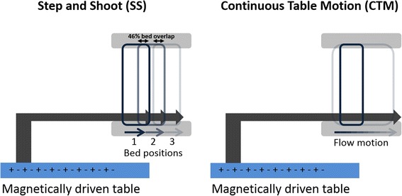
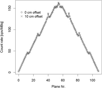
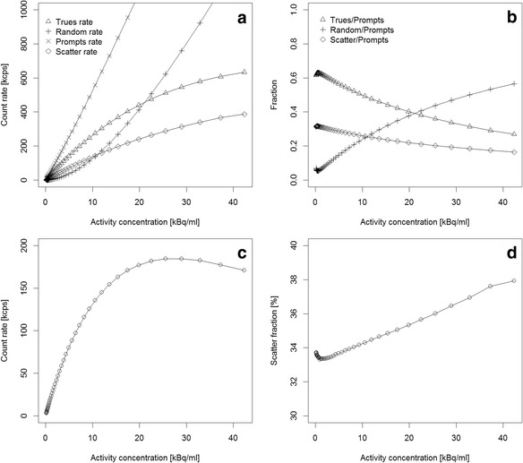
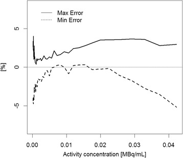
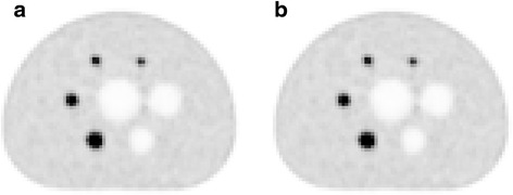
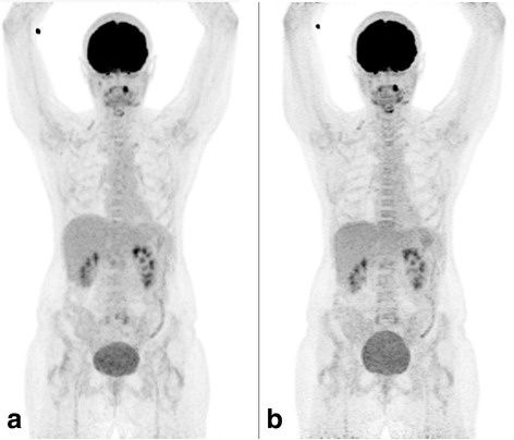
BACKGROUND: The purpose of the study is to evaluate the physical performance of a Biograph mCT Flow 64-4R PET/CT system (Siemens Healthcare, Germany) and to compare clinical image quality in step-and-shoot (SS) and continuous table motion (CTM) acquisitions. METHODS: The spatial resolution, sensitivity, count rate curves, and Image Quality (IQ) parameters following the National Electrical Manufactures Association (NEMA) NU2-2012 standard were evaluated. For resolution measurements, an (18)F point source inside a glass capillary tube was used. Sensitivity measurements were based on a 70-cm-long polyethylene tube, filled with 4.5 MBq of FDG. Scatter fraction and count rates were measured using a 70-cm-long polyethylene cylinder with a diameter of 20 cm and a line source (1.04 GBq of FDG) inserted axially into the cylinder 4.5 cm off-centered. A NEMA IQ phantom containing six spheres (10- to 37-mm diameter) was used for the evaluation of the image quality. First, a single-bed scan was acquired (NEMA standard), followed by a two-bed scan (4 min each) of the IQ phantom with the image plane containing the spheres centered in the overlap region of the two bed positions. In addition, a scan of the same region in CTM mode was performed with a table speed of 0.6 mm/s. Furthermore, two patient scans were performed in CTM and SS mode. Image contrasts and patient images were compared between SS and CTM acquisitions. RESULTS: Full Width Half Maximum (FWHM) of the spatial resolution ranged from 4.3 to 7.8 mm (radial distance 1 to 20 cm). The measured sensitivity was 9.6 kcps/MBq, both at the center of the FOV and 10 cm off-center. The measured noise equivalent count rate (NECR) peak was 185 kcps at 29.0 kBq/ml. The scatter fraction was 33.5 %. Image contrast recovery values (sphere-to-background of 8:1) were between 42 % (10-mm sphere) to 79 % (37-mm sphere). The background variability was between 2.1 and 5.3 % (SS) and between 2.4 and 6.9 % (CTM). No significant difference in image quality was observed between SS and CTM mode. CONCLUSIONS: The spatial resolution, sensitivity, scatter fraction, and count rates were in concordance with the published values for the predecessor system, the Biograph mCT. Contrast recovery values as well as image quality obtained in SS and CTM acquisition modes were similar.
Overview
The claims and statistics of this paper haven’t been checked yet.
Run the check — free, no account needed. It reads the open-access text and checks the sources it cites.
Integrity signals
Registry
Cites
Statistics
Figures
Verification
Claims the paper makes
The claims and statistics of this paper haven’t been checked yet.
Run the check — free, no account needed. It reads the open-access text and checks the sources it cites.
Claims other papers make about this one
Each is checked against this paper’s own text: does it say what the citing paper says it does?
The source backs part of the claim, not all of it — or backs it for a different population, condition or comparison.
In the citing paper
“In contrast, a SiPM system with 100% detector coverage and 214 ps timing resolution [47] can offer increased system sensitivity over a comparable PMT system with lutetium oxyorthosilicate (LSO) crystals [48] that exceeds the expected difference based on the slightly larger axial FOV (16.4 vs. 9.6 cps/kBq with a 25.6 vs. 22.1 cm FOV) [47].”
From Influences on PET Quantification and Interpretation (2022) · cites this paper as [48]
Experimental — accuracy not yet measured
Self-check
Percentages, sums and reported differences re-computed from the paper’s own numbers.
The self-check needs the paper’s full text.
Citations
Cites
A combined PET/CT scanner for clinical oncology.
Beyer, T., Townsend, D. W., Brun, T., Kinahan, P. E., Charron, M., Roddy, R., Jerin, J., Young, J., Byars, L. G., & Nutt, R. (2000) · PubMed · Cited by 1,655
Self-citationNational electrical manufacturers association
Sokole, E. B., & Cradduck, T. D. (1926) · Journal of the A.I.E.E. · doi:10.1109/jaiee.1926.6537344 · Cited by 860
Surti, S., Kuhn, A., Werner, M. E., Perkins, A. E., Kolthammer, J. A., & Karp, J. S. (2007) · PubMed · Cited by 639
Benefit of Time-of-Flight in PET: Experimental and Clinical Results
Karp, J. S., Surti, S., Daube-Witherspoon, M. E., & Muehllehner, G. (2008) · Journal of Nuclear Medicine · doi:10.2967/jnumed.107.044834 · Cited by 585
Fully 3-D PET reconstruction with system matrix derived from point source measurements
Panin, V. Y., Kehren, F., Michel, C., & Casey, M. (2006) · IEEE Transactions on Medical Imaging · doi:10.1109/tmi.2006.876171 · Cited by 536
Physical and clinical performance of the mCT time-of-flight PET/CT scanner
Jakoby, B., Bercier, Y., Conti, M., Casey, M. E., Bendriem, B., & Townsend, D. W. (2011) · Physics in Medicine and Biology · doi:10.1088/0031-9155/56/8/004 · Cited by 487
Resolution modeling in PET imaging: Theory, practice, benefits, and pitfalls
Rahmim, A., Qi, J., & Sossi, V. (2013) · Medical Physics · doi:10.1118/1.4800806 · Cited by 371
Time of flight in PET revisited
Moses, W. W. (2003) · IEEE Transactions on Nuclear Science · doi:10.1109/tns.2003.817319 · Cited by 338
An Assessment of the Impact of Incorporating Time-of-Flight Information into Clinical PET/CT Imaging
Lois, C., Jakoby, B., Long, M. J., Hübner, K. F., Barker, D. W., Casey, M., Conti, M., Panin, V., Kadrmas, D. J., & Townsend, D. W. (2010) · Journal of Nuclear Medicine · doi:10.2967/jnumed.109.068098 · Cited by 263
Karakatsanis, N. A., Lodge, M. A., Tahari, A. K., Zhou, Y., Wahl, R. L., & Rahmim, A. (2013) · Physics in Medicine and Biology · doi:10.1088/0031-9155/58/20/7391 · Cited by 247
Research works printed but not linked: 1
- Melcher CL. Scintillator crystals for PET. J Nucl Med. 2000;41:1051–5.
Cited by
Sorted by citations
Performance Characteristics of the Digital Biograph Vision PET/CT System
van Sluis, J., de Jong, J., Schaar, J., Noordzij, W., van Snick, P., Dierckx, R., Borra, R., Willemsen, A., & Boellaard, R. (2019) · Journal of Nuclear Medicine · doi:10.2967/jnumed.118.215418 · Cited by 484
History and future technical innovation in positron emission tomography
Jones, T., & Townsend, D. W. (2017) · Journal of medical imaging · doi:10.1117/1.jmi.4.1.011013 · Cited by 324
Alberts, I., Hünermund, J., Prenosil, G. A., Mingels, C., Bohn, K. P., Viscione, M., Sari, H., Vollnberg, B., Shi, K., Afshar‐Oromieh, A., et al. (2021) · European Journal of Nuclear Medicine and Molecular Imaging · doi:10.1007/s00259-021-05282-7 · Cited by 286
Grant, A. M., Deller, T. W., Khalighi, M. M., Maramraju, S. H., Delso, G., & Levin, C. S. (2016) · Medical Physics · doi:10.1118/1.4945416 · Cited by 246
Studies of a Next-Generation Silicon-Photomultiplier–Based Time-of-Flight PET/CT System
Hsu, D. F. C., Ilan, E., Peterson, W. T., Uribe, J., Lubberink, M., & Levin, C. S. (2017) · Journal of Nuclear Medicine · doi:10.2967/jnumed.117.189514 · Cited by 241
Quantification, improvement, and harmonization of small lesion detection with state-of-the-art PET
van der Vos, C. S., Koopman, D., Rijnsdorp, S., Arends, A. J., Boellaard, R., van Dalen, J. A., Lubberink, M., Willemsen, A. T. M., & Visser, E. P. (2017) · European Journal of Nuclear Medicine and Molecular Imaging · doi:10.1007/s00259-017-3727-z · Cited by 206
EANM/EARL FDG-PET/CT accreditation - summary results from the first 200 accredited imaging systems
Kaalep, A., Sera, T., Oyen, W., Krause, B. J., Chiti, A., Liu, Y., & Boellaard, R. (2018) · European Journal of Nuclear Medicine and Molecular Imaging · doi:10.1007/s00259-017-3853-7 · Cited by 174
Performance Evaluation of the Vereos PET/CT System According to the NEMA NU2-2012 Standard
Rausch, I., Ruiz, A., Valverde-Pascual, I., Cal-González, J., Beyer, T., & Carrio, I. (2019) · Journal of Nuclear Medicine · doi:10.2967/jnumed.118.215541 · Cited by 168
Self-citationFeasibility of state of the art PET/CT systems performance harmonisation
Kaalep, A., Sera, T., Rijnsdorp, S., Yaqub, M., Talsma, A., Lodge, M. A., & Boellaard, R. (2018) · European Journal of Nuclear Medicine and Molecular Imaging · doi:10.1007/s00259-018-3977-4 · Cited by 160
Performance evaluation of the next generation solid-state digital photon counting PET/CT system
Zhang, J., Maniawski, P., & Knopp, M. V. (2018) · EJNMMI Research · doi:10.1186/s13550-018-0448-7 · Cited by 123
Comparison between new-generation SiPM-based and conventional PMT-based TOF-PET/CT
Wagatsuma, K., Miwa, K., Sakata, M., Oda, K., Ono, H., Kameyama, M., Toyohara, J., & Ishii, K. (2017) · Physica Medica · doi:10.1016/j.ejmp.2017.09.124 · Cited by 117
van Sluis, J., Boellaard, R., Somasundaram, A., van Snick, J. H., Borra, R., Dierckx, R., Stormezand, G. N., Glaudemans, A. W. J. M., & Noordzij, W. (2019) · Journal of Nuclear Medicine · doi:10.2967/jnumed.119.227801 · Cited by 100
Comparison of image quality and lesion detection between digital and analog PET/CT
López-Mora, D. A., Flotats, A., Fuentes-Ocampo, F., Camacho, V., Fernández, A., Ruiz, A., Duch, J., Sizova, M., Domènech, A., Estorch, M., et al. (2019) · European Journal of Nuclear Medicine and Molecular Imaging · doi:10.1007/s00259-019-4260-z · Cited by 94
Effective dose estimation for oncological and neurological PET/CT procedures
Martí-Climent, J. M., Prieto, E., Morán, V., Sancho, L., Rodríguez‐Fraile, M., Arbizu, J., García‐Velloso, M. J., & Richter, J. Á. (2017) · EJNMMI Research · doi:10.1186/s13550-017-0272-5 · Cited by 78
Performance Characteristics of the Whole-Body Discovery IQ PET/CT System
Reynés-Llompart, G., Gámez-Cenzano, C., Romero-Zayas, I., Rodríguez-Bel, L., Vercher-Conejero, J. L., & Martí-Climent, J. M. (2017) · Journal of Nuclear Medicine · doi:10.2967/jnumed.116.185561 · Cited by 78
Moliner, L., Rodríguez-Álvarez, M. J., Catret, J. V., González, A. J., Ilisie, V., & Benlloch, J. (2019) · Scientific Reports · doi:10.1038/s41598-019-51898-z · Cited by 67
DirectPET: full-size neural network PET reconstruction from sinogram data
Whiteley, W., Luk, W. K., & Gregor, J. (2020) · Journal of medical imaging · doi:10.1117/1.jmi.7.3.032503 · Cited by 60
Reynés‐Llompart, G., Gámez‐Cenzano, C., Vercher‐Conejero, J. L., Sabaté‐Llobera, A., Calvo‐Malvar, N., & Martí‐Climent, J. M. (2018) · Medical Physics · doi:10.1002/mp.12986 · Cited by 55
Hybrid Imaging: Instrumentation and Data Processing
Cal-González, J., Rausch, I., Sundar, L. K. S., Lassen, M. L., Muzik, O., Moser, E., Papp, L., & Beyer, T. (2018) · Frontiers in Physics · doi:10.3389/fphy.2018.00047 · Cited by 52
Self-citationInfluences on PET Quantification and Interpretation
Rogasch, J. M. M., Hofheinz, F., van Heek, L., Voltin, C.-A., Boellaard, R., & Kobe, C. (2022) · Diagnostics · doi:10.3390/diagnostics12020451 · Cited by 57
“In contrast, a SiPM system with 100% detector coverage and 214 ps timing resolution [47] can offer increased system sensitivity over a comparable PMT system with lutetium oxyorthosilicate (LSO) crystals [48] that exceeds the expected difference based on the slightly larger axial FOV (16.”
BackgroundQuantitative Rodent Brain Receptor Imaging
Herfert, K., Mannheim, J. G., Kuebler, L., Marciano, S., Amend, M., Parl, C., Napieczyńska, H., Maier, F. M., Vega, S. C., & Pichler, B. J. (2019) · Molecular Imaging and Biology · doi:10.1007/s11307-019-01368-9 · Cited by 47
Superior performance of 18F-fluorocholine digital PET/CT in the detection of parathyroid adenomas
López-Mora, D. A., Sizova, M., Estorch, M., Flotats, A., Camacho, V., Fernández‐León, A., Abouzian, S., Fuentes-Ocampo, F., Garcia, J. I. P., Chico, A. I., et al. (2020) · European Journal of Nuclear Medicine and Molecular Imaging · doi:10.1007/s00259-020-04680-7 · Cited by 47
Chicheportiche, A., Marciano, R., & Orevi, M. (2020) · EJNMMI Physics · doi:10.1186/s40658-020-0271-x · Cited by 45
NEMA NU2-2012 performance measurements of the United Imaging uPMR790: an integrated PET/MR system
Chen, S., Gu, Y., Yu, H., Chen, X., Cao, T., Hu, L., & Shi, H. (2021) · European Journal of Nuclear Medicine and Molecular Imaging · doi:10.1007/s00259-020-05135-9 · Cited by 45
Ballon, D., Rosenberg, J. B., Fung, E. K., Nikolopoulou, A., Kothari, P. J., De, B. P., He, B., Chen, A., Heier, L., Sondhi, D., et al. (2020) · Human Gene Therapy · doi:10.1089/hum.2020.116 · Cited by 40
How fast can we scan patients with modern (digital) PET/CT systems?
Lasnon, C., Coudrais, N., Houdu, B., Nganoa, C., Salomon, T., Enilorac, B., & Aide, N. (2020) · European Journal of Radiology · doi:10.1016/j.ejrad.2020.109144 · Cited by 40
Performance characteristics of silicon photomultiplier based 15-cm AFOV TOF PET/CT
Vandendriessche, D., Uribe, J., Bertin, H., & Geeter, F. D. (2019) · EJNMMI Physics · doi:10.1186/s40658-019-0244-0 · Cited by 39
Performance evaluation of quantitative SPECT/CT using NEMA NU 2 PET methodology
Ryu, H.-J., Meikle, S. R., Willowson, K. P., Eslick, E. M., & Bailey, D. L. (2019) · Physics in Medicine and Biology · doi:10.1088/1361-6560/ab2a22 · Cited by 38
Long axial field-of-view PET/CT devices: are we ready for the technological revolution?
Filippi, L., Dimitrakopoulou‐Strauss, A., Evangelista, L., & Schillaci, O. (2022) · Expert Review of Medical Devices · doi:10.1080/17434440.2022.2141111 · Cited by 38
Performance evaluation of a high resolution dedicated breast PET scanner
García-Hernández, T., González, A. V., Rebolleda, J. F., Jurado, R. S., Ferrando, J. R., González, L. B., Cabañero, D. G., & Cozar, M. (2016) · Medical Physics · doi:10.1118/1.4945271 · Cited by 36
Ligand-based design of [18F]OXD-2314 for PET imaging in non-Alzheimer’s disease tauopathies
Lindberg, A., Murrell, E., Tong, J., Mason, N. S., Sohn, D., Sandell, J., Ström, P., Stehouwer, J. S., Lopresti, B. J., Viklund, J., et al. (2024) · Nature Communications · doi:10.1038/s41467-024-49258-1 · Cited by 32
Reducing Radiation Exposure to Paediatric Patients Undergoing [18F]FDG-PET/CT Imaging
Kertész, H., Beyer, T., London, K., Saleh, H., Chung, D. K. V., Rausch, I., Cal-González, J., Kitsos, T., & Kench, P. L. (2021) · Molecular Imaging and Biology · doi:10.1007/s11307-021-01601-4 · Cited by 31
Self-citationRausch, I., Mannheim, J. G., Kupferschläger, J., la Fougère, C. J., & Schmidt, F. P. (2022) · EJNMMI Physics · doi:10.1186/s40658-022-00516-5 · Cited by 30
Self-citationAkamatsu, G., Shimada, N., Matsumoto, K., Daisaki, H., Suzuki, K., Watabe, H., Oda, K., Senda, M., Terauchi, T., & Tateishi, U. (2022) · Annals of Nuclear Medicine · doi:10.1007/s12149-021-01709-1 · Cited by 27
Redesign of the GATE PET coincidence sorter
Strydhorst, J. H., & Buvat, I. (2016) · Physics in Medicine and Biology · doi:10.1088/0031-9155/61/18/n522 · Cited by 27
Meier, J. G., Wu, C. C., Cuellar, S. L. B., Truong, M. T., Erasmus, J. J., Einstein, S. A., & Mawlawi, O. R. (2018) · Journal of Nuclear Medicine · doi:10.2967/jnumed.118.213884 · Cited by 26
Salvadori, J., Odille, F., Karcher, G., Marie, P., & Imbert, L. (2021) · EJNMMI Physics · doi:10.1186/s40658-020-00344-5 · Cited by 24
Kertész, H., Beyer, T., Panin, V., Jentzen, W., Cal-González, J., Berger, A., Papp, L., Kench, P. L., Bharkhada, D., Cabello, J., et al. (2022) · Frontiers in Physiology · doi:10.3389/fphys.2022.818463 · Cited by 22
Self-citationKertész, H., Conti, M., Panin, V., Cabello, J., Bharkhada, D., Beyer, T., Papp, L., Jentzen, W., Cal-González, J., Herraiz, J. L., et al. (2022) · EJNMMI Physics · doi:10.1186/s40658-022-00482-y · Cited by 21
Self-citationKaneta, T., Ogawa, M., Motomura, N., Iizuka, H., Arisawa, T., Hino-Shishikura, A., Yoshida, K., & Inoue, T. (2017) · EJNMMI Research · doi:10.1186/s13550-017-0331-y · Cited by 21
Efthimiou, N., Thielemans, K., Emond, E. C., Cawthorne, C., Archibald, S. J., & Tsoumpas, C. (2020) · EJNMMI Physics · doi:10.1186/s40658-020-00309-8 · Cited by 20
Kroenke, M., Schweiger, L., Horn, T. D., Haller, B., Schwamborn, K., Wurzer, A. J., Maurer, T., Wester, H., Eiber, M. J., & Rauscher, I. (2022) · Journal of Nuclear Medicine · doi:10.2967/jnumed.121.263707 · Cited by 19
Tjörnstrand, A., Casar‐Borota, O., Heurling, K., Schöll, M., Gjertsson, P., Himmelman, J., Itsenko, O., Ragnarsson, Ó., & Nyström, H. F. (2019) · Clinical Endocrinology · doi:10.1111/cen.14144 · Cited by 19
Clinical Workflow Considerations for Implementation of Continuous-Bed-Motion PET/CT
Acuff, S. N., & Osborne, D. (2016) · Journal of Nuclear Medicine Technology · doi:10.2967/jnmt.116.172171 · Cited by 17
Methods for estimating full width at half maximum
Rainio, O., Hällilä, J., Teuho, J., & Klén, R. (2025) · Signal Image and Video Processing · doi:10.1007/s11760-025-03820-6 · Cited by 16
Simulation study of a high‐performance brain PET system with dodecahedral geometry
Tao, W., Chen, G., Weng, F., Zan, Y., Zhao, Z., Peng, Q., Xu, J., & Huang, Q. (2018) · Medical Physics · doi:10.1002/mp.12996 · Cited by 16
Schiller, F., Frings, L., Thurow, J., Meyer, P. T., & Mix, M. D. (2019) · Journal of Nuclear Medicine · doi:10.2967/jnumed.119.227132 · Cited by 16
Fantini, L., Belli, M. L., Azzali, I., Loi, E., Bettinelli, A., Feliciani, G., Mezzenga, E., Fedeli, A., Asioli, S., Paganelli, G., et al. (2021) · Frontiers in Oncology · doi:10.3389/fonc.2021.601053 · Cited by 16
A proper protocol for routine 18F-FDG uEXPLORER total-body PET/CT scans
Hu, H., Huang, Y., Sun, H., Zhou, K., Jiang, L., Zhong, J., Chen, L., Wang, L., Han, Y., & Wu, H. (2023) · EJNMMI Physics · doi:10.1186/s40658-023-00573-4 · Cited by 16
Costa, P. F., Jentzen, W., Süßelbeck, F., Fendler, W. P., Rischpler, C., Herrmann, K., Conti, M., Kersting, D., & Weber, M. (2021) · The Quarterly Journal of Nuclear Medicine and Molecular Imaging · doi:10.23736/s1824-4785.21.03300-8 · Cited by 15
Ferretti, A., Chondrogiannis, S., Rampin, L., Bellan, E., Marzola, M. C., Grassetto, G., Gusella, S., Maffione, A. M., Gava, M. M., & Rubello, D. (2018) · Physics in Medicine and Biology · doi:10.1088/1361-6560/aaee27 · Cited by 15
Michopoulou, S. K., O’Shaughnessy, E., Thomson, K. E., & Guy, M. (2018) · Nuclear Medicine Communications · doi:10.1097/mnm.0000000000000962 · Cited by 15
Casey, M., & Osborne, D. (2020) · Springer eBooks · doi:10.1007/978-3-030-43040-5_6 · Cited by 15
Bouallègue, F. B., Vauchot, F., & Mariano‐Goulart, D. (2018) · Medical Physics · doi:10.1002/mp.13366 · Cited by 14
Wagatsuma, K., Miwa, K., Akamatsu, G., Yamao, T., Kamitaka, Y., Sakurai, M., Fujita, N., Hanaoka, K., Matsuda, H., & Ishii, K. (2023) · Annals of Nuclear Medicine · doi:10.1007/s12149-023-01847-8 · Cited by 13
States, L. J., & Voss, S. D. (2019) · Pediatric oncology · doi:10.1007/978-3-030-03777-2_3 · Cited by 13
Malignant Cutaneous Melanoma: Updates in PET Imaging
Laudicella, R., Baratto, L., Minutoli, F., Baldari, S., & Iagaru, A. H. (2019) · Current Radiopharmaceuticals · doi:10.2174/1874471012666191015095550 · Cited by 13
Rosenberg, J. B., Fung, E. K., Dyke, J. P., De, B. P., Lou, H. H., Kelly, J. M., Reejhsinghani, L., Arbona, R. J. R., Sondhi, D., Kaminsky, S. M., et al. (2023) · Human Gene Therapy · doi:10.1089/hum.2023.060 · Cited by 11
Bouchareb, Y., Tag, N., Sulaiman, H., Riyami, K. A., Jawa, Z. M., & Al-Dhuhli, H. (2023) · Nuclear Medicine and Molecular Imaging · doi:10.1007/s13139-023-00795-5 · Cited by 12
Effect of attenuation mismatches in time of flight PET reconstruction
Emond, E. C., Bousse, A., Machado, M., Porter, J. C., Groves, A. M., Hutton, B. F., & Thielemans, K. (2020) · Physics in Medicine and Biology · doi:10.1088/1361-6560/ab7a6f · Cited by 11
Dynamic Whole-Body 18F-FDG PET for Minimizing Patient Motion Artifact
Kaji, T., Osanai, K., Nakata, T., & Tamaki, N. (2020) · Clinical Nuclear Medicine · doi:10.1097/rlu.0000000000003253 · Cited by 11
Yamashita, S., Yamamoto, H., Nakaichi, T., Yoneyama, T., & Yokoyama, K. (2017) · Annals of Nuclear Medicine · doi:10.1007/s12149-017-1200-5 · Cited by 10
Domínguez‐Jiménez, D. Y., & Alva‐Sánchez, H. (2021) · Medical Physics · doi:10.1002/mp.14729 · Cited by 10
Akamatsu, G., Tashima, H., Yoshida, E., Wakizaka, H., Iwao, Y., Maeda, T., Takahashi, M., & Yamaya, T. (2019) · Biomedical Physics & Engineering Express · doi:10.1088/2057-1976/ab5c05 · Cited by 10
Molecular imaging of thyroid and parathyroid diseases
Ovčariček, P. P., Calderoni, L., Campennì, A., Fanti, S., & Giovanella, L. (2024) · Expert Review of Endocrinology & Metabolism · doi:10.1080/17446651.2024.2365776 · Cited by 9
PET/TAC: bases físicas, instrumentación y avances
Torres-Espallardo, I. (2017) · Radiología · doi:10.1016/j.rx.2016.10.010 · Cited by 9
NEMA NU-4 performance evaluation of a non-human primate animal PET
Chai, P., Baotong, F., Zhang, Z., Haohui, T., Shuangquan, L., Xiaoli, S., Peilin, W., Xiaoming, W., Xu-dong, Z., & Long, W. (2019) · Physics in Medicine and Biology · doi:10.1088/1361-6560/ab1614 · Cited by 9
Optimization of Q.Clear reconstruction for dynamic 18F PET imaging
Lysvik, E. K., Mikalsen, L. T. G., Revheim, M.-E. R., Emblem, K. E., & Hjørnevik, T. (2023) · EJNMMI Physics · doi:10.1186/s40658-023-00584-1 · Cited by 8
Effects of breathing motion on PET acquisitions
Owaki, Y., Nakahara, T., Shimizu, T., Smith, A. M., Luk, W. K., Inoue, K., Fukushi, M., & Nakajima, K. (2018) · Nuclear Medicine Communications · doi:10.1097/mnm.0000000000000852 · Cited by 8
de Boer, J., Keijzer, K., van Doesum, J. A., Smit, N. A. M., Brouwers, A. H., van Sluis, J., Hooge, M. N. L., Pierik, F. R., Huls, G. A., van Dijk, L. V., et al. (2025) · Nature Communications · doi:10.1038/s41467-025-66767-9 · Cited by 7
Monte Carlo simulation of two Siemens Biograph PET/CT system using GATE: Image quality performance
Saaidi, R., Zeghari, A., & Moursli, R. C. E. (2024) · Radiation Physics and Chemistry · doi:10.1016/j.radphyschem.2024.111653 · Cited by 7
Cho, Z., Son, Y., Kim, H.-K., Kwon, D.-H., Joo, Y., Ra, J. B., Choi, Y., & Kim, Y.-B. (2019) · IEEE Transactions on Medical Imaging · doi:10.1109/tmi.2019.2916326 · Cited by 7
Meier, J. G., Erasmus, J. J., Gladish, G. W., Peterson, C. B., Diab, R. H., & Mawlawi, O. R. (2019) · Journal of Applied Clinical Medical Physics · doi:10.1002/acm2.12785 · Cited by 7
Accuracy of arterial [18F]-Fluorodeoxyglucose uptake quantification: A kinetic modeling study
Toczek, J., Wu, J., Hillmer, A. T., Han, J., Esterlis, I., Cosgrove, K., Liu, C., & Sadeghi, M. M. (2020) · Journal of Nuclear Cardiology · doi:10.1007/s12350-020-02055-x · Cited by 7
Tanaka, K., Norikane, T., Mitamura, K., Yamamoto, Y., Maeda, Y., Fujimoto, K., Takami, Y., Ishimura, M., Arai-Okuda, H., Tohi, Y., et al. (2022) · EJNMMI Physics · doi:10.1186/s40658-022-00513-8 · Cited by 6
Using PET for therapy monitoring in oncological clinical trials: challenges ahead
Deroose, C. M., Stroobants, S. G., Liu, Y., Shankar, L. K., & Bourguet, P. (2017) · European Journal of Nuclear Medicine and Molecular Imaging · doi:10.1007/s00259-017-3689-1 · Cited by 6
Using EQ·PET to reduce reconstruction-dependent variations in [ 18 F]FDG-PET brain imaging
Vanhoutte, M., Semah, F., Lopes, R., Jaillard, A., Petyt, G., Aziz, A.-L., Lahousse, H., Declerck, J. M., Pasquier, F., Spottiswoode, B. S., et al. (2019) · Physics in Medicine and Biology · doi:10.1088/1361-6560/ab35b4 · Cited by 5
Moretti, T. J., Leon, S. M., Schaeffer, C. J., & Arreola, M. M. (2022) · Journal of Applied Clinical Medical Physics · doi:10.1002/acm2.13751 · Cited by 5
Gamma Ray Emission Imaging in the Medical and Nuclear Safeguards Fields
Dendooven, P. G., & Bubba, T. A. (2022) · Lecture notes in physics · doi:10.1007/978-3-031-10751-1_7 · Cited by 5
Numerical investigation reveals challenges in measuring the contrast recovery coefficients in PET
Bayerlein, R., Spencer, B. A., Abdelhafez, Y. G., Cherry, S. R., Badawi, R. D., & Omidvari, N. (2023) · Physics in Medicine and Biology · doi:10.1088/1361-6560/ad00fa · Cited by 4
Cañizares, G., Jiménez-Serrano, S., Lucero, A., Morera-Ballester, C., Muñoz, E., Benlloch, J., & González, A. J. (2024) · IEEE Transactions on Radiation and Plasma Medical Sciences · doi:10.1109/trpms.2024.3365911 · Cited by 4
Ganjeh, Z. A., Campos, B. Z., Tranéus, E., Both, S., & Dendooven, P. G. (2024) · Physics in Medicine and Biology · doi:10.1088/1361-6560/ad7d5c · Cited by 4
GATE Simulation Study of the Siemens Biograph mCT 20 Excel PET/CT System
Saaidi, R., Toufique, Y., Zeghari, A., Kharrim, A. E., & Moursli, R. C. E. (2019) · Polish Journal of Medical Physics And Engineering · doi:10.2478/pjmpe-2019-0002 · Cited by 4
Positron Emission Tomography (PET): Physics and Instrumentation
Khalil, M. M. (2021) · Springer eBooks · doi:10.1007/978-3-030-65245-6_12 · Cited by 4
Reconstruction Algorithm for Regions of Interest in γ-Photon Images
Yao, M., Lv, G., Zhao, M., Guo, R., Liu, J., Zhen, D., Wang, M., Xiong, F., & Liu, W. (2021) · IEEE Access · doi:10.1109/access.2021.3079514 · Cited by 4
Sipilä, O., Liukkonen, J., Halme, H.-L., Tolvanen, T., Sohlberg, A. O., Hakulinen, M. A., Manninen, A.-L., Tahvanainen, K. U. O., Tunninen, V., Ollikainen, T., et al. (2023) · EJNMMI Physics · doi:10.1186/s40658-023-00551-w · Cited by 3
Rasul, S., Geist, B. K., Einspieler, H., Fajković, H., Shariat, S. F., Schmitl, S., Mitterhauser, M., Bartosch, R., Langsteger, W., Baltzer, P., et al. (2023) · International Journal of Molecular Sciences · doi:10.3390/ijms241813677 · Cited by 3
Self-citationPET/CT acquisition and processing protocols in the Netherlands
Dieckens, D., van Horssen, P., van Gils, K., van Lange, A. J., Ivashchenko, O., Veldkamp, W. J. H., Lammertsma, A. A., & Veltman, N. C. (2025) · EJNMMI Physics · doi:10.1186/s40658-025-00770-3 · Cited by 3
Design study of fully wearable high-performance brain PETs for neuroimaging in free movement
Tao, W., Weng, F., Chen, G., Lv, L., Zhao, Z., Xie, S., Zan, Y., Xu, J., Huang, Q., & Peng, Q. (2020) · Physics in Medicine and Biology · doi:10.1088/1361-6560/ab8c90 · Cited by 3
Iskra, I., Visvikis, D., Koole, M. J. B., Armstrong, I. S., Vanhove, C., Žagar, I., Middlebrooks, T., Bormans, G. M., Cleeren, F., Weehaeghe, D. V., et al. (2021) · doi:10.52717/pwze5872 · Cited by 3
Meier, J. G., Diab, R. H., Connor, T. M., & Mawlawi, O. R. (2022) · Journal of Applied Clinical Medical Physics · doi:10.1002/acm2.13619 · Cited by 3
Vashistha, R., Vegh, V., Moradi, H., Hammond, A., O’Brien, K., & Reutens, D. C. (2024) · Frontiers in Radiology · doi:10.3389/fradi.2024.1466498 · Cited by 3
Yamamoto, H., Takemoto, S., Maebatake, A., Karube, S., Yamashiro, Y., Nakanishi, A., & Murakami, K. (2019) · Annals of Nuclear Medicine · doi:10.1007/s12149-019-01334-z · Cited by 3
Mamach, M., Kessler, M., Bankstahl, J. P., Wilke, F., Geworski, L., Bengel, F. M., Kurt, S., & Berding, G. (2018) · PLoS ONE · doi:10.1371/journal.pone.0205044 · Cited by 4
Rauscher, L., Allen, M. S., Judenhofer, M. S., Byars, L. G., Scipioni, M., Udías, J. M., Catana, C., Pichler, B. J., & Schmidt, F. P. (2025) · IEEE Transactions on Radiation and Plasma Medical Sciences · doi:10.1109/trpms.2025.3624609 · Cited by 2
Computer Vision-Based Medical Imaging Techniques: Past, Present, and Future
Lonchakov, A., Sinitca, A., & Kaplun, D. (2026) · IEEE Access · doi:10.1109/access.2026.3654393 · Cited by 4
Kumamoto, K., Sato, H., Tsutsui, Y., Awamoto, S., Yamashita, Y., Baba, S., & Sasaki, M. (2022) · Journal of Nuclear Medicine Technology · doi:10.2967/jnmt.121.263240 · Cited by 2
Performance evaluation of a small‐animal PET scanner with 213 mm axis using NEMA NU 4‐2008
Wei, Q., Wang, Y., Huang, X., Li, D., Zheng, Y., Wang, P., Sun, X.-L., Chai, P., Han, X., Liu, S., et al. (2024) · Medical Physics · doi:10.1002/mp.17469 · Cited by 2
Texture Feature Comparison Between Step-and-Shoot and Continuous-Bed-Motion 18F-FDG PET
Yamashita, S., Okuda, K., Nakaichi, T., Yamamoto, H., & Yokoyama, K. (2020) · Journal of Nuclear Medicine Technology · doi:10.2967/jnmt.120.246157 · Cited by 2
Joint estimation of activity, attenuation and motion in respiratory-self-gated time-of-flight PET
Elhamiasl, M., Jolivet, F., Rezaei, A., Fieseler, M., Schäfers, K. P., Nuyts, J. L., Schramm, G., & Boada, F. E. (2025) · Physics in Medicine and Biology · doi:10.1088/1361-6560/adbed5 · Cited by 1
Metadata
- Authors
- Rausch, I., Cal-González, J., Dapra, D., Gallowitsch, H. J., Lind, P., Beyer, T., & Minear, G.
- Affiliations
- Medical University of Vienna (AT)
- Klinikum Klagenfurt (AT)
- Universitätsklinikum St. Pölten (AT)
- Corresponding author
- Rausch, Ivo
- Venue
- EJNMMI Physics
- Published
- 2015-10-26
- Volume
- 2
- Issue
- 1
- Pages
- 26-26
- ISSN
- 2197-7364 (ISSN-L)
- PMC
- PMC4883615
- OpenAlex
- W1866924806
- Cited by
- 133
- References
- 38
- Citations by year
- 2026: 7 · 2025: 11 · 2024: 12 · 2023: 9 · 2022: 15 · 2021: 16 · 2020: 15 · 2019: 20 · 2018: 12 · 2017: 11 · 2016: 5
- FWCI
- 7.56
- Type
- journal article
- Language
- en
- Open access
- gold
- doi.org
- Locations
- EJNMMI Physics (publishedVersion, cc-by)
- Europe PMC (PubMed Central) (publishedVersion, cc-by)
- PubMed Central (publishedVersion, cc-by)
- PubMed (publishedVersion)
- Topics
- Health Sciences › Medicine › Radiology, Nuclear Medicine and Imaging › Medical Imaging Techniques and Applications
- Health Sciences › Medicine › Radiology, Nuclear Medicine and Imaging › Radiation Dose and Imaging
- Health Sciences › Medicine › Radiology, Nuclear Medicine and Imaging › Cardiac Imaging and Diagnostics
- Keywords
- spatial resolution, detector sensitivity, noise equivalent count rate, scatter fraction, image quality, step-and-shoot acquisition, point source resolution, NEMA IQ phantom
About these checks
Registry, retraction, Cites and statistics results are computed, not judged, with one exception: a printed reference that no index lists is read once by a model before it counts as a citation error. Claims are extracted by a model and kept only when their quote appears verbatim in the text; each cited source is then checked, with a model’s help, for whether it says what the claim relies on. Those verdicts can be wrong — read the claim’s quote and the cited source before relying on one.
Statistics and arithmetic are recomputed, not judged.
Confidence bands are shown only for fields calibrated on our annotated test set. None is calibrated yet, so no band is shown.
What counts as a claim ›How we measure confidence ›How we check this ›
Are you an author, or do you see a problem with this page? Request a review
