

The DOI matches its Crossref record; 195 of the 198 references it cites were found in a registry or an index, and 1 are not checked yet.
Clinical applications of fundus autofluorescence in retinal disease
Open accessAbstract
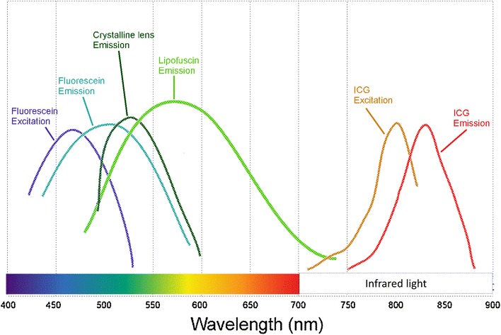
Fundus autofluorescence (FAF) is a non-invasive retinal imaging modality used in clinical practice to provide a density map of lipofuscin, the predominant ocular fluorophore, in the retinal pigment epithelium. Multiple commercially available imaging systems, including the fundus camera, the confocal scanning laser ophthalmoscope, and the ultra-widefield imaging device, are available to the clinician. Each offers unique advantages for evaluating various retinal diseases. The clinical applications of FAF continue to expand. It is now an essential tool for evaluating age related macular degeneration, macular dystrophies, retinitis pigmentosa, white dot syndromes, retinal drug toxicities, and various other retinal disorders. FAF may detect abnormalities beyond those detected on funduscopic exam, fluorescein angiography, or optical coherence tomography, and can be used to elucidate disease pathogenesis, form genotype-phenotype correlations, diagnose and monitor disease, and evaluate novel therapies. Given its ease of use, non-invasive nature, and value in characterizing retinal disease, FAF enjoys increasing clinical relevance. This review summarizes common ocular fluorophores, imaging modalities, and FAF findings for a wide spectrum of retinal disorders.
Overview
The claims and statistics of this paper haven’t been checked yet.
Run the check — free, no account needed. It reads the open-access text and checks the sources it cites.
Integrity signals
Registry
Cites
Statistics
Figures
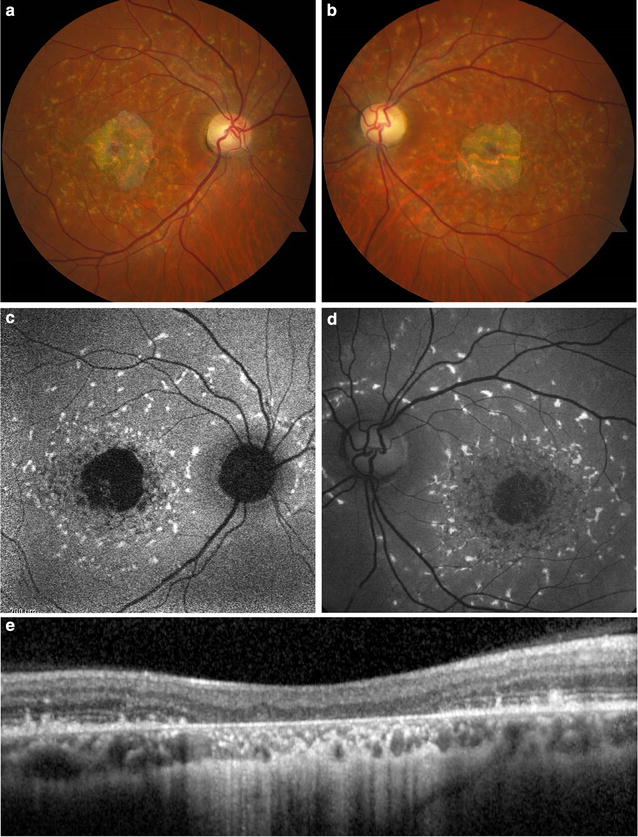
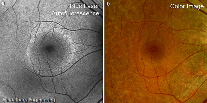
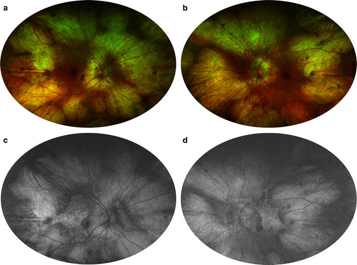
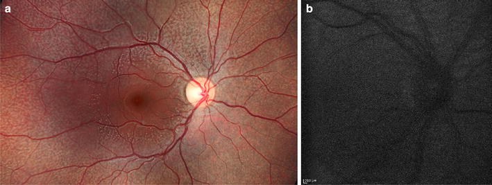
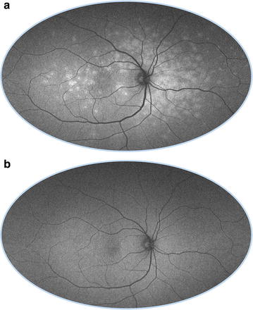
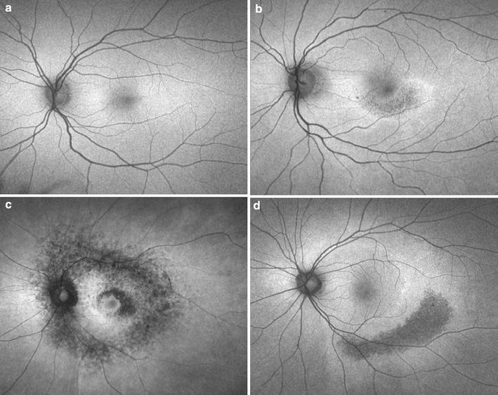
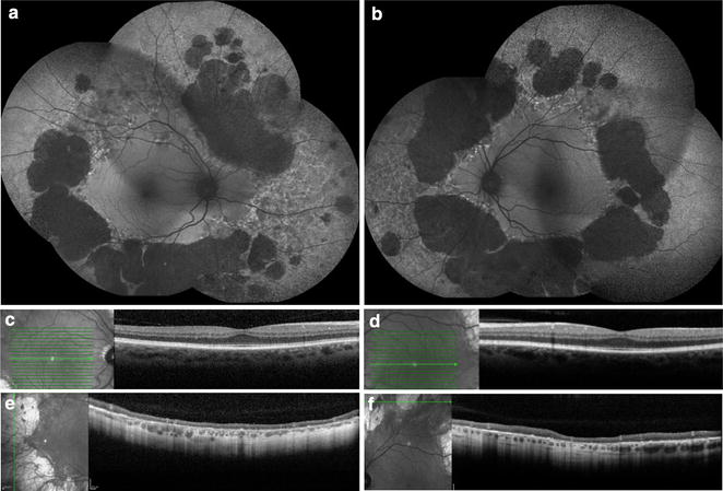
Verification
Claims the paper makes
The claims and statistics of this paper haven’t been checked yet.
Run the check — free, no account needed. It reads the open-access text and checks the sources it cites.
Claims other papers make about this one
Each is checked against this paper’s own text: does it say what the citing paper says it does?
The source backs part of the claim, not all of it — or backs it for a different population, condition or comparison.
In the citing paper
“As A2E can not be further metabolized and as RPE cells are postmitotic, A2E and related bisretinoids accumulate in the RPE in the form of lipofuscin deposits.”
From The Role of Vitamin A in Retinal Diseases (2022) · cites this paper as [75]
Experimental — accuracy not yet measuredThe source backs part of the claim, not all of it — or backs it for a different population, condition or comparison.
In the citing paper
“Although abundant lipofuscin accumulation is involved in the aetiology of certain retinal diseases (e.g., AMD, Best disease and ABCA4-retinopathy), it accumulates at a lower level also with age in the RPE of healthy eyes [9,72,73,74,75].”
From The Role of Vitamin A in Retinal Diseases (2022) · cites this paper as [75]
Experimental — accuracy not yet measuredThe source backs part of the claim, not all of it — or backs it for a different population, condition or comparison.
In the citing paper
“Lipofuscin is an autofluorescent cellular waste product that compromises the function and viability of RPE cells and ultimately results in photoreceptor degeneration and a loss of vision [54,69,73,74,75].”
From The Role of Vitamin A in Retinal Diseases (2022) · cites this paper as [75]
Experimental — accuracy not yet measured
Self-check
Percentages, sums and reported differences re-computed from the paper’s own numbers.
The self-check needs the paper’s full text.
Citations
Cites
Clinical Classification of Age-related Macular Degeneration
Ferris, F. L., Wilkinson, C. P., Bird, A. C., Chakravarthy, U., Chew, E. Y., Csaky, K. G., & Sadda, S. R. (2013) · Ophthalmology · doi:10.1016/j.ophtha.2012.10.036 · Cited by 1,868
Stereoscopic Atlas of Macular Diseases: Diagnosis and Treatment
(1990) · Archives of Ophthalmology · doi:10.1001/archopht.1990.01070060042026 · Cited by 1,014
Delori, F. C., Dorey, C. K., Staurenghi, G., Arend, O., Goger, D. G., & Weiter, J. J. (1995) · PubMed · Cited by 938
Revised Recommendations on Screening for Chloroquine and Hydroxychloroquine Retinopathy
Marmor, M. F., Kellner, U., Lai, T. Y. Y., Lyons, J. S., & Mieler, W. F. (2011) · Ophthalmology · doi:10.1016/j.ophtha.2010.11.017 · Cited by 703
Holz, F. G., Bindewald-Wittich, A., Fleckenstein, M., Dreyhaupt, J., Scholl, H. P. N., & Schmitz-Valckenberg, S. (2007) · American Journal of Ophthalmology · doi:10.1016/j.ajo.2006.11.041 · Cited by 619
Sparrow, J. R., Nakanishi, K., & Parish, C. A. (2000) · PubMed · Cited by 566
Retinal pigment epithelial lipofuscin and melanin and choroidal melanin in human eyes.
Weiter, J. J., Delori, F. C., Wing, G. L., & Fitch, K. A. (1986) · PubMed · Cited by 565
Aging human RPE: morphometric analysis of macular, equatorial, and peripheral cells.
Feeney-Burns, L., Hilderbrand, E. S., & Eldridge, S. (1984) · PubMed · Cited by 561
RPE65 is the isomerohydrolase in the retinoid visual cycle
Moiseyev, G., Chen, Y., Takahashi, Y., Wu, B. X., & Ma, J.-X. (2005) · Proceedings of the National Academy of Sciences · doi:10.1073/pnas.0503460102 · Cited by 536
Prevalence of Age-Related Macular Degeneration in the US Population
Klein, R. (2011) · Archives of Ophthalmology · doi:10.1001/archophthalmol.2010.318 · Cited by 523
Zhang, K., Kniazeva, M., Han, M., Li, W., Yu, Z., Yang, Z., Li, Y., Metzker, M. L., Allikmets, R. L., Zack, D. J., et al. (2001) · Nature Genetics · doi:10.1038/83817 · Cited by 453
Age-related accumulation and spatial distribution of lipofuscin in RPE of normal subjects.
Delori, F. C., Goger, D. G., & Dorey, C. K. (2001) · PubMed · Cited by 424
Near-Infrared Autofluorescence Imaging of the Fundus: Visualization of Ocular Melanin
Keilhauer, C. N., & Delori, F. ̧. C. (2006) · Investigative Ophthalmology & Visual Science · doi:10.1167/iovs.06-0122 · Cited by 398
DRUSEN CHARACTERIZATION WITH MULTIMODAL IMAGING
Spaide, R. F., & Curcio, C. A. (2010) · Retina · doi:10.1097/iae.0b013e3181ee5ce8 · Cited by 397
Sarna, T. (1992) · Journal of Photochemistry and Photobiology B Biology · doi:10.1016/1011-1344(92)85027-r · Cited by 396
The topography and age relationship of lipofuscin concentration in the retinal pigment epithelium.
Wing, G. L., Blanchard, G. C., & Weiter, J. J. (1978) · PubMed · Cited by 394
Feeney, L. (1978) · PubMed · Cited by 389
Zeitz, C., Robson, A. G., & Audo, I. S. (2014) · Progress in Retinal and Eye Research · doi:10.1016/j.preteyeres.2014.09.001 · Cited by 388
Wolfe, F., & Marmor, M. (2010) · Arthritis care & research · doi:10.1002/acr.20133 · Cited by 314
Peripherin. A rim-specific membrane protein of rod outer segment discs.
Molday, R. S., Hicks, D., & Molday, L. L. (1987) · PubMed · Cited by 313
Yamamoto, H., Simon, A., Eriksson, U., Harris, E., Berson, E. L., & Dryja, T. P. (1999) · Nature Genetics · doi:10.1038/9707 · Cited by 298
Delori, F. C., Goger, D. G., Hammond, B. R., Snodderly, D. M., & Burns, S. A. (2001) · Journal of the Optical Society of America A · doi:10.1364/josaa.18.001212 · Cited by 290
A2E-epoxides Damage DNA in Retinal Pigment Epithelial Cells
Sparrow, J. R., Vollmer-Snarr, H. R., Zhou, J., Jang, Y. P., Steffen Jockusch, B., Itagaki, Y., & Nakanishi, K. (2003) · Journal of Biological Chemistry · doi:10.1074/jbc.m300457200 · Cited by 280
Pathology and Pathogenesis of Drusen of the Optic Nervehead
Tso, M. O. M. (1981) · Ophthalmology · doi:10.1016/s0161-6420(81)80038-3 · Cited by 270
Sparrow, J. R., & Cai, B. (2001) · PubMed · Cited by 269
Cideciyan, A. V., Alemán, T. S., Świder, M., Schwartz, S., Steinberg, J. D., Brucker, A. J., Maguire, A. M., Bennett, J., Stone, E. M., & Jacobson, S. G. (2004) · Human Molecular Genetics · doi:10.1093/hmg/ddh048 · Cited by 268
Role of Ocular Melanin in Ophthalmic Physiology and Pathology †
Hu, D., Simon, J. D., & Sarna, T. (2008) · Photochemistry and Photobiology · doi:10.1111/j.1751-1097.2008.00316.x · Cited by 261
Optical density spectra of the macular pigmentin vivo andin vitro
Bone, R. A., Landrum, J. T., & Cains, A. (1992) · Vision Research · doi:10.1016/0042-6989(92)90118-3 · Cited by 247
Stereoscopic Atlas of Macular Diseases: Diagnosis and Treatment
Lane, C. (1988) · British Journal of Ophthalmology · doi:10.1136/bjo.72.9.720-b · Cited by 244
Classification of Fundus Autofluorescence Patterns in Early Age-Related Macular Disease
Bindewald-Wittich, A., Bird, A. C., Dandekar, S. S., Dolar-Szczasny, J., Dreyhaupt, J., Fitzke, F. W., Einbock, W., Holz, F. G., Jorzik, J. J., Keilhauer, C. N., et al. (2005) · Investigative Ophthalmology & Visual Science · doi:10.1167/iovs.04-0430 · Cited by 238
Fundus Autofluorescence and Central Serous Chorioretinopathy
Spaide, R. F., & Klancnik, J. M. (2005) · Ophthalmology · doi:10.1016/j.ophtha.2005.01.003 · Cited by 226
Biosynthetic Studies of A2E, a Major Fluorophore of Retinal Pigment Epithelial Lipofuscin
Ben-Shabat, S., Parish, C. A., Vollmer, H. R., Itagaki, Y., Fishkin, N., Nakanishi, K., & Sparrow, J. R. (2002) · Journal of Biological Chemistry · doi:10.1074/jbc.m108981200 · Cited by 214
AUTOFLUORESCENCE FROM THE OUTER RETINA AND SUBRETINAL SPACE
Spaide, R. F. (2008) · Retina · doi:10.1097/iae.0b013e318158eca4 · Cited by 208
Pang, C. E., Shah, V. P., Sarraf, D., & Freund, K. B. (2014) · American Journal of Ophthalmology · doi:10.1016/j.ajo.2014.04.021 · Cited by 208
Self-citationAutofluorescence distribution associated with drusen in age-related macular degeneration.
Delori, F. C., Fleckner, M. R., Goger, D. G., Weiter, J. J., & Dorey, C. K. (2000) · PubMed · Cited by 207
Absorption of white light in photoreceptors
Warrant, E. J., & Nilsson, D. (1998) · Vision Research · doi:10.1016/s0042-6989(97)00151-x · Cited by 204
Pathogenesis of tears of the retinal pigment epithelium.
Gass, J. D. (1984) · British Journal of Ophthalmology · doi:10.1136/bjo.68.8.513 · Cited by 188
Smith, R. T., Sohrab, M. A., Busuioc, M., & Barile, G. R. (2009) · American Journal of Ophthalmology · doi:10.1016/j.ajo.2009.06.028 · Cited by 187
Quantitative Fundus Autofluorescence in Recessive Stargardt Disease
Burke, T. R., Duncker, T., Woods, R. L., Greenberg, J. P., Zernant, J., Tsang, S. H., Smith, R. T., Allikmets, R. L., Sparrow, J. R., & Delori, F. C. (2014) · Investigative Ophthalmology & Visual Science · doi:10.1167/iovs.13-13624 · Cited by 183
Lois, N., Owens, S. L., Coco, R. M., Hopkins, J. J., Fitzke, F. W., & Bird, A. C. (2002) · American Journal of Ophthalmology · doi:10.1016/s0002-9394(01)01404-0 · Cited by 181
MULTIMODAL IMAGING OF PIGMENT EPITHELIAL DETACHMENT
Mrejen, S., Sarraf, D., Mukkamala, S. K., & Freund, K. B. (2013) · Retina · doi:10.1097/iae.0b013e3182993f66 · Cited by 179
Self-citationFundus autofluorescence in stargardt macular dystrophy–fundus flavimaculatus
Lois, N., Halfyard, A. S., Bird, A. C., Holder, G. E., & Fitzke, F. W. (2004) · American Journal of Ophthalmology · doi:10.1016/j.ajo.2004.02.056 · Cited by 178
Relationship of Senile Macular Degeneration to Ocular Pigmentation
Weiter, J. J., Delori, F. C., Wing, G. L., & Fitch, K. A. (1985) · American Journal of Ophthalmology · doi:10.1016/0002-9394(85)90230-2 · Cited by 178
Pericentral Retinopathy and Racial Differences in Hydroxychloroquine Toxicity
Melles, R. B., & Marmor, M. F. (2014) · Ophthalmology · doi:10.1016/j.ophtha.2014.07.018 · Cited by 175
Histopathologic Findings in Best's Vitelliform Macular Dystrophy
O’Gorman, S. A., Flaherty, W. A., Fishman, G. A., & Berson, E. L. (1988) · Archives of Ophthalmology · doi:10.1001/archopht.1988.01060140421045 · Cited by 170
Chaston, T. B., & Richardson, D. R. (2003) · American Journal of Hematology · doi:10.1002/ajh.10348 · Cited by 168
Freund, K. B., Laud, K., de Lima, L. H. S. G., Spaide, R. F., Zweifel, S. A., & Yannuzzi, L. A. (2010) · Retina · doi:10.1097/iae.0b013e3181ea48ba · Cited by 166
Spaide, R. F., Goldberg, N. R., & Freund, K. B. (2013) · Retina · doi:10.1097/iae.0b013e318286cc77 · Cited by 164
The lipofuscin fluorophore A2E perturbs cholesterol metabolism in retinal pigment epithelial cells
Lakkaraju, A., Finnemann, S. C., & Rodriguez-Boulan, E. (2007) · Proceedings of the National Academy of Sciences · doi:10.1073/pnas.0702504104 · Cited by 162
Weigert, G., Kaya, S., Pemp, B., Sacu, S., Lasta, M., Werkmeister, R. M., Dragostinoff, N., Simader, C., Garhöfer, G., Schmidt-Erfurth, U. M., et al. (2011) · Investigative Ophthalmology & Visual Science · doi:10.1167/iovs.11-7522 · Cited by 160
Kellner, U., Renner, A. B., & Tillack, H. (2006) · Investigative Ophthalmology & Visual Science · doi:10.1167/iovs.05-1290 · Cited by 155
Schmitz-Valckenberg, S., Steinberg, J. S., Fleckenstein, M., Visvalingam, S., Brinkmann, C. K., & Holz, F. G. (2010) · Ophthalmology · doi:10.1016/j.ophtha.2009.10.044 · Cited by 154
Robson, A. G. (2006) · British Journal of Ophthalmology · doi:10.1136/bjo.2005.082487 · Cited by 154
Age-Related Macular Degeneration: Advances in Management and Diagnosis
Yonekawa, Y., Miller, J. W., & Kim, I. K. (2015) · Journal of Clinical Medicine · doi:10.3390/jcm4020343 · Cited by 153
Wu, Y., Yanase, E., Feng, X., Siegel, M. M., & Sparrow, J. R. (2010) · Proceedings of the National Academy of Sciences · doi:10.1073/pnas.0913112107 · Cited by 152
Ablonczy, Z., Higbee, D., Anderson, D. M., Dahrouj, M., Grey, A. C., Gutierrez, D. B., Koutalos, Y., Schey, K. L., Hanneken, A. M., & Crouch, R. K. (2013) · Investigative Ophthalmology & Visual Science · doi:10.1167/iovs.13-12250 · Cited by 149
A Prospective Study of Reticular Macular Disease
Pumariega, N. M., Smith, R. T., Sohrab, M. A., Letien, V., & Souied, E. H. (2011) · Ophthalmology · doi:10.1016/j.ophtha.2011.01.029 · Cited by 145
Rudolf, M., Vogt, S. D., Curcio, C. A., Huisingh, C., McGwin, G., Wägner, A., Grisanti, S., & Read, R. W. (2013) · Ophthalmology · doi:10.1016/j.ophtha.2012.10.007 · Cited by 145
Boon, C. J. F., van Schooneveld, M. J., den Hollander, A. I., van Lith-Verhoeven, J. J. C., Zonneveld-Vrieling, M. N., Theelen, T., Cremers, F. P. M., Hoyng, C. B., & Klevering, B. J. (2007) · British Journal of Ophthalmology · doi:10.1136/bjo.2007.115659 · Cited by 136
Primary amines protect against retinal degeneration in mouse models of retinopathies
Maeda, A., Golczak, M., Chen, Y., Okano, K., Kohno, H., Shiose, S., Ishikawa, K., Harte, W., Palczewska, G., Maeda, T., et al. (2012) · Nature Chemical Biology · doi:10.1038/nchembio.759 · Cited by 135
Nagiel, A., Freund, K. B., Spaide, R. F., Munch, I. C., Larsen, M., & Sarraf, D. (2013) · American Journal of Ophthalmology · doi:10.1016/j.ajo.2013.06.024 · Cited by 133
Self-citationAntioxidant Properties of Melanin in Retinal Pigment Epithelial Cells
Wang, Z., Dillon, J. P., & Gaillard, E. R. (2005) · Photochemistry and Photobiology · doi:10.1562/2005-10-21-ra-725 · Cited by 133
Measuring macular pigment optical density in vivo: a review of techniques
Howells, O., Eperjesi, F., & Bartlett, H. E. (2011) · Graefe s Archive for Clinical and Experimental Ophthalmology · doi:10.1007/s00417-010-1577-5 · Cited by 133
Interpretations of Fundus Autofluorescence from Studies of the Bisretinoids of the Retina
Sparrow, J. R., Yoon, K. D., Wu, Y., & Yamamoto, K. (2010) · Investigative Ophthalmology & Visual Science · doi:10.1167/iovs.10-5852 · Cited by 132
Adult-onset foveomacular vitelliform dystrophy: A fresh perspective
Chowers, I., Tiosano, L., Audo, I. S., Grunin, M. A., & Boon, C. J. F. (2015) · Progress in Retinal and Eye Research · doi:10.1016/j.preteyeres.2015.02.001 · Cited by 129
High-Resolution Adaptive Optics Retinal Imaging of Cellular Structure in Choroideremia
Morgan, J. I. W., Han, G., Klinman, E., Maguire, W., Chung, D. C., Maguire, A. M., & Bennett, J. (2014) · Investigative Ophthalmology & Visual Science · doi:10.1167/iovs.13-13454 · Cited by 123
EXPANDED CLINICAL SPECTRUM OF MULTIPLE EVANESCENT WHITE DOT SYNDROME WITH MULTIMODAL IMAGING
Marsiglia, M., Gallego‐Pinazo, R., de Souza, E. C., Munk, M. R., Yu, S.-Q., Mrejen, S., Cunningham, E. T., Lujan, B. J., Goldberg, N. R., Albini, T. A., et al. (2015) · Retina · doi:10.1097/iae.0000000000000685 · Cited by 123
Murakami, T., Akimoto, M., Ooto, S., Suzuki, T., Ikeda, H. O., Kawagoe, N., Takahashi, M., & Yoshimura, N. (2008) · American Journal of Ophthalmology · doi:10.1016/j.ajo.2007.11.018 · Cited by 121
Popović, P., Jarc-Vidmar, M., & Hawlina, M. (2005) · Graefe s Archive for Clinical and Experimental Ophthalmology · doi:10.1007/s00417-005-1186-x · Cited by 117
Witmer, M. T., Parlitsis, Patel, & Kiss, S. (2013) · Clinical ophthalmology · doi:10.2147/opth.s41731 · Cited by 117
Adult vitelliform macular degeneration: a clinicopathological study
Arnold, J., Sarks, J. P., Killingsworth, M. C., Kettle, E., & Sarks, S. H. (2003) · Eye · doi:10.1038/sj.eye.6700460 · Cited by 115
Sparrow, J. R., Marsiglia, M., Allikmets, R. L., Tsang, S. H., Lee, W., Duncker, T., & Zernant, J. (2015) · Investigative Ophthalmology & Visual Science · doi:10.1167/iovs.15-16763 · Cited by 113
Haimovici, R., D’Amico, D. J., Gragoudas, E. S., & Sokol, S. (2002) · Ophthalmology · doi:10.1016/s0161-6420(01)00947-2 · Cited by 110
Phenotypic Variability in RDH5 Retinopathy (Fundus Albipunctatus)
Sergouniotis, P. I., Sohn, E. H., Li, Z., McBain, V. A., Wright, G. A., Moore, A. T., Robson, A. G., Holder, G. E., & Webster, A. R. (2011) · Ophthalmology · doi:10.1016/j.ophtha.2010.12.031 · Cited by 109
Multimodal Imaging of Optic Disc Drusen
Sato, T., Mrejen, S., & Spaide, R. F. (2013) · American Journal of Ophthalmology · doi:10.1016/j.ajo.2013.03.039 · Cited by 108
Macular dystrophies mimicking age-related macular degeneration
Saksens, N. T. M., Fleckenstein, M., Schmitz-Valckenberg, S., Holz, F. G., den Hollander, A. I., KEUNEN, J. E. E., Boon, C. J. F., & Hoyng, C. B. (2013) · Progress in Retinal and Eye Research · doi:10.1016/j.preteyeres.2013.11.001 · Cited by 104
Race, iris color, and age-related macular degeneration.
Frank, R. N., Puklin, J. E., Stock, C., & La, C. (2000) · Europe PMC (PubMed Central) · Cited by 102
High-Resolution Images of Retinal Structure in Patients with Choroideremia
Syed, R., Sundquist, S. M., Ratnam, K., Zayit‐Soudry, S., Zhang, Y., Crawford, J. B., MacDonald, I. M., Godara, P., Rha, J., Carroll, J., et al. (2013) · Investigative Ophthalmology & Visual Science · doi:10.1167/iovs.12-10707 · Cited by 101
Clinical and Genetic Heterogeneity in Multifocal Vitelliform Dystrophy
Boon, C. J. F. (2007) · Archives of Ophthalmology · doi:10.1001/archopht.125.8.1100 · Cited by 100
Macular pigment: quantitative analysis on autofluorescence images
Trieschmann, M., Spital, G., Lommatzsch, A. P., van Kuijk, E. F., Fitzke, F. W., Bird, A. C., & Pauleikhoff, D. (2003) · Graefe s Archive for Clinical and Experimental Ophthalmology · doi:10.1007/s00417-003-0796-4 · Cited by 100
Duncker, T., Greenberg, J. P., Ramachandran, R., Hood, D. C., Smith, R. T., Hirose, T., Woods, R. L., Tsang, S. H., Delori, F. C., & Sparrow, J. R. (2014) · Investigative Ophthalmology & Visual Science · doi:10.1167/iovs.13-13834 · Cited by 98
Scanning Laser Ophthalmoscopy and Angiography With a Wide-Field Contact Lens System
Staurenghi, G. (2005) · Archives of Ophthalmology · doi:10.1001/archopht.123.2.244 · Cited by 98
Fundus Albipunctatus Associated with Compound Heterozygous Mutations in RPE65
Schatz, P., Preising, M. N., Lorenz, B., Sander, B., Larsen, M., & Rosenberg, T. (2011) · Ophthalmology · doi:10.1016/j.ophtha.2010.09.005 · Cited by 97
Mimoun, G., Soubrane, G., & Coscas, G. (1990) · PubMed · Cited by 96
Robson, A. G., Tufail, A., Fitzke, F. W., Bird, A. C., Moore, A. T., Holder, G. E., & Webster, A. R. (2011) · Retina · doi:10.1097/iae.0b013e318206d155 · Cited by 96
MORPHOLOGY AND FUNCTIONAL CHARACTERISTICS IN ADULT VITELLIFORM MACULAR DYSTROPHY
Renner, A. B., Tillack, H., Kraus, H., Kohl, S., Wissinger, B., MOHR, N., Weber, B. H. F., Kellner, U., & Foerster, M. H. (2004) · Retina · doi:10.1097/00006982-200412000-00014 · Cited by 96
Enhanced accumulation of A2E in individuals homozygous or heterozygous for mutations in BEST1 (VMD2)
Bakall, B., Radu, R. A., Stanton, J. B., Burke, J. M., McKay, B. S., Wadelius, C., Mullins, R. F., Stone, E. M., Travis, G. H., & Marmorstein, A. D. (2007) · Experimental Eye Research · doi:10.1016/j.exer.2007.02.018 · Cited by 95
Butterfly-Shaped Pattern Dystrophy
Zhang, K. (2002) · Archives of Ophthalmology · doi:10.1001/archopht.120.4.485 · Cited by 94
Fundus autofluorescence imaging of retinal dystrophies
Boon, C. J. F., Klevering, B. J., KEUNEN, J. E. E., Hoyng, C. B., & Theelen, T. (2008) · Vision Research · doi:10.1016/j.visres.2008.01.010 · Cited by 93
Tolmachova, T., Tolmachov, O. E., Barnard, A. R., Silva, S. R. D., Lipinski, D. M., Walker, N. J., MacLaren, R. E., & Seabra, M. C. (2013) · Journal of Molecular Medicine · doi:10.1007/s00109-013-1006-4 · Cited by 92
Robson, A. G., Egan, C. A., Luong, V., Bird, A. C., Holder, G. E., & Fitzke, F. W. (2004) · Investigative Ophthalmology & Visual Science · doi:10.1167/iovs.04-0211 · Cited by 87
Carmeli, I., Skákalová, V., Naaman, R., & Vager, Z. (2002) · Angewandte Chemie International Edition · doi:10.1002/1521-3773(20020301)41:5 · Cited by 85
Einbock, W., Moessner, A., Schnurrbusch, U., Holz, F. G., Wolf, S., & for the FAM Study Group (2004) · Graefe s Archive for Clinical and Experimental Ophthalmology · doi:10.1007/s00417-004-1027-3 · Cited by 83
Essex, R. W. (2010) · Archives of Ophthalmology · doi:10.1001/archophthalmol.2010.157 · Cited by 83
The scanning laser ophthalmoscope—a review of its role in bioscience and medicine
Sharp, P. F., Manivannan, A., Xu, H., & Forrester, J. V. (2004) · Physics in Medicine and Biology · doi:10.1088/0031-9155/49/7/001 · Cited by 82
A Review of the Inflammatory Chorioretinopathies: The White Dot Syndromes
Crawford, C. M., & Igboeli, O. C. (2013) · ISRN Inflammation · doi:10.1155/2013/783190 · Cited by 82
Roberts, J. E., Kukielczak, B. M., Hu, D., Miller, D. S., Bilski, P. J., Sik, R. H., Motten, A. G., & Chignell, C. F. (2002) · Photochemistry and Photobiology · doi:10.1562/0031-8655(2002)0750184troaip2.0.co2 · Cited by 79
Virgiliis, S. D., Congia, M., Turco, M., Frau, F., Dessì, C., Argiolu, F., Sorcinelli, R., Sitzia, A., & Cao, A. (1988) · Archives of Disease in Childhood · doi:10.1136/adc.63.3.250 · Cited by 79
Wüstemeyer, H., Moessner, A., Jahn, C., & Wolf, S. (2003) · Graefe s Archive for Clinical and Experimental Ophthalmology · doi:10.1007/s00417-003-0730-9 · Cited by 78
Rozanowski, B., Burke, J. M., Boulton, M. E., Sarna, T., & Rozanowska, M. (2008) · Investigative Ophthalmology & Visual Science · doi:10.1167/iovs.08-1700 · Cited by 76
Subjective and Objective Screening Tests for Hydroxychloroquine Toxicity
Cukras, C. A., Huynh, N., Vitale, S., Wong, W. T., Ferris, F. L., & Sieving, P. A. (2014) · Ophthalmology · doi:10.1016/j.ophtha.2014.07.056 · Cited by 72
Sarraf, D., Joseph, A., & Rahimy, E. (2014) · Europe PMC (PubMed Central) · Cited by 72
Self-citationWarburton, S., Davis, W. E., Southwick, K., Xin, H., Woolley, A. T., Burton, G. F., & Thulin, C. D. (2007) · PubMed Central · Cited by 70
A2-Rhodopsin: a new fluorophore isolated from photoreceptor outer segments
Fishkin, N. E., Jang, Y. P., Itagaki, Y., Sparrow, J. R., & Nakanishi, K. (2003) · Organic & Biomolecular Chemistry · doi:10.1039/b212213h · Cited by 69
Jorzik, J. J., Bindewald-Wittich, A., Dithmar, S., & Holz, F. G. (2005) · Retina · doi:10.1097/00006982-200506000-00003 · Cited by 69
Nonmydriatic fluorescence-based quantitative imaging of human macular pigment distributions
Sharifzadeh, M., Bernstein, P. S., & Gellermann, W. (2006) · Journal of the Optical Society of America A · doi:10.1364/josaa.23.002373 · Cited by 68
MultiColor imaging in the evaluation of geographic atrophy due to age-related macular degeneration
Moussa, N. B., Georges, A., Capuano, V., Merle, B., Souied, E. H., & Querques, G. (2015) · British Journal of Ophthalmology · doi:10.1136/bjophthalmol-2014-305643 · Cited by 68
Ogura, S., Yasukawa, T., Kato, A., Usui, H., Hirano, Y., Yoshida, M., & Ogura, Y. (2014) · American Journal of Ophthalmology · doi:10.1016/j.ajo.2014.07.021 · Cited by 67
Diagnostic imaging in patients with retinitis pigmentosa
Mitamura, Y., Mitamura-Aizawa, S., Nagasawa, T., Katome, T., Eguchi, H., & Naito, T. (2012) · The Journal of Medical Investigation · doi:10.2152/jmi.59.1 · Cited by 66
Greenstein, V. C., Duncker, T., Holopigian, K., Carr, R. E., Greenberg, J. P., Tsang, S. H., & Hood, D. C. (2011) · Retina · doi:10.1097/iae.0b013e31821dfc17 · Cited by 63
Sundelin, S. P., NILSSON, S. E. G., & Brunk, U. T. (2001) · Free Radical Biology and Medicine · doi:10.1016/s0891-5849(00)00444-5 · Cited by 62
Gene Therapy for Choroideremia Using an Adeno-Associated Viral (AAV) VectorPreprint
Barnard, A. R., Groppe, M., & MacLaren, R. E. (2014) · Cold Spring Harbor Perspectives in Medicine · doi:10.1101/cshperspect.a017293 · Cited by 62
Vaclavik, V., Vujosevic, S., Dandekar, S. S., Bunce, C., Pető, T., & Bird, A. C. (2007) · Ophthalmology · doi:10.1016/j.ophtha.2007.04.023 · Cited by 62
Acute exudative polymorphous vitelliform maculopathy.
Gass, J. D. M., Chuang, E. L., & Granek, H. (1988) · PubMed · Cited by 61
Microperimetry, fundus autofluorescence, and retinal layer changes in progressing geographic atrophy
Pilotto, E., Benetti, E., Convento, E., Guidolin, F., Longhin, E., Parrozzani, R., & Midena, E. (2013) · Canadian Journal of Ophthalmology · doi:10.1016/j.jcjo.2013.03.022 · Cited by 61
Sandberg, M. A., Gaudio, A. R., Miller, S., & Weiner, A. (1994) · PubMed · Cited by 60
Fundus autofluorescence in exudative age-related macular degeneration
McBain, V. A., Townend, J., & Lois, N. (2006) · British Journal of Ophthalmology · doi:10.1136/bjo.2006.095109 · Cited by 59
Feng, J., Chen, X., Sun, X., Wang, F., & Sun, X. (2014) · Ophthalmic Research · doi:10.1159/000363387 · Cited by 57
Crouch, R. K., Koutalos, Y., Kono, M., Schey, K. L., & Ablonczy, Z. (2015) · Progress in molecular biology and translational science · doi:10.1016/bs.pmbts.2015.06.005 · Cited by 56
Preising, M. N., Wegscheider, E., Friedburg, C., Poloschek, C. M., Wabbels, B., & Lorenz, B. (2009) · Ophthalmology · doi:10.1016/j.ophtha.2009.01.016 · Cited by 56
Analysis of visual pigment by fundus autofluorescence
Theelen, T., Berendschot, T. T. J. M., Boon, C. J. F., Hoyng, C. B., & Klevering, B. J. (2007) · Experimental Eye Research · doi:10.1016/j.exer.2007.10.022 · Cited by 54
Iacono, P., Parodi, M. B., Papayannis, A., Spina, C. L., Varano, M., & Bandello, F. M. (2015) · Graefe s Archive for Clinical and Experimental Ophthalmology · doi:10.1007/s00417-014-2899-5 · Cited by 54
Fleckenstein, M., Issa, P. C., Fuchs, H., Finger, R. P., Helb, H. –., Scholl, H. P. N., & Holz, F. G. (2008) · Eye · doi:10.1038/eye.2008.59 · Cited by 54
FUNDUS AUTOFLUORESCENCE AND MULTIPLE EVANESCENT WHITE DOT SYNDROME
Furino, C., Boscia, F., Cardascia, N., Alessio, G., & Sborgia, C. (2009) · Retina · doi:10.1097/iae.0b013e31818c5e04 · Cited by 53
Fundus Autofluorescence and Photoreceptor Bleaching in Multiple Evanescent White Dot Syndrome
Joseph, A., Rahimy, E., Freund, K. B., Sorenson, J. A., & Sarraf, D. (2013) · Ophthalmic surgery, lasers & imaging retina · doi:10.3928/23258160-20131105-08 · Cited by 53
Self-citationFundus autofluorescence imaging compared with different confocal scanning laser ophthalmoscopes
Bellmann, C. (2003) · British Journal of Ophthalmology · doi:10.1136/bjo.87.11.1381 · Cited by 52
Early Paracentral Visual Field Loss in Patients Taking Hydroxychloroquine
Elder, M. (2006) · Archives of Ophthalmology · doi:10.1001/archopht.124.12.1729 · Cited by 50
Drusen detection in retro-mode imaging by a scanning laser ophthalmoscope
Acton, J. H., Cubbidge, R. P., King, H., Galsworthy, P., & Gibson, J. M. (2011) · Acta Ophthalmologica · doi:10.1111/j.1755-3768.2011.02123.x · Cited by 48
Fundus autofluorescence in acute and chronic central serous chorioretinopathy
Dinç, U. A., Tatlıpınar, S., Yenerel, N. M., Görgün, E., & Çiftçi, F. (2011) · Clinical and Experimental Optometry · doi:10.1111/j.1444-0938.2011.00598.x · Cited by 47
A Clinicopathologic Report of the Retinal Lesions Associated With Didanosine
Whitcup, S. M. (1994) · Archives of Ophthalmology · doi:10.1001/archopht.1994.01090240100033 · Cited by 47
Genead, M. A., Fishman, G. A., & Lindeman, M. (2010) · Ophthalmic Genetics · doi:10.3109/13816810903584971 · Cited by 46
Acute Exudative Paraneoplastic Polymorphous Vitelliform Maculopathy in Five Cases
Al-Dahmash, S. A., Shields, C. L., Bianciotto, C. G., Witkin, A. J., Witkin, S. R., & Shields, J. A. (2012) · Ophthalmic surgery, lasers & imaging retina · doi:10.3928/15428877-20120712-01 · Cited by 46
Autofluorescence of the crystalline lens in early and late onset diabetes.
Sparrow, J. M., Bron, A. J., Brown, N., & Neil, H. A. W. (1992) · British Journal of Ophthalmology · doi:10.1136/bjo.76.1.25 · Cited by 46
Fundus Autofluorescence Patterns in Best Vitelliform Macular Dystrophy
Parodi, M. B., Iacono, P., Campa, C., Turco, C. D., & Bandello, F. M. (2014) · American Journal of Ophthalmology · doi:10.1016/j.ajo.2014.07.026 · Cited by 46
Multimodal assessment of choroideremia patients defines pre-treatment characteristics
Seitz, I. P., Zhour, A., Kohl, S., Llavona, P., Peter, T., Wilhelm, B., Zrenner, E., Ueffing, M., Bartz‐Schmidt, K. U., & Fischer, M. D. (2015) · Graefe s Archive for Clinical and Experimental Ophthalmology · doi:10.1007/s00417-015-2976-4 · Cited by 45
Teke, M. Y., Elgin, U., Nalcacioglu-Yüksekkaya, P., Şen, E. M., Ozdal, P. C., & Öztürk, F. (2014) · Europe PMC (PubMed Central) · doi:10.3980/j.issn.2222-3959.2014.02.29 · Cited by 45
Abnormal Fundus Autofluorescence Results of Patients in Long-term Treatment with Deferoxamine
Viola, F., Barteselli, G., Dell’Arti, L., Vezzola, D., Villani, E., Mapelli, C., Zanaboni, L., Cappellini, M. D., & Ratiglia, R. (2012) · Ophthalmology · doi:10.1016/j.ophtha.2012.01.039 · Cited by 45
Paraneoplastic Cloudy Vitelliform Submaculopathy in Primary Vitreoretinal Lymphoma
Pang, C. E., Shields, C. L., Jumper, J. M., & Yannuzzi, L. A. (2014) · American Journal of Ophthalmology · doi:10.1016/j.ajo.2014.08.031 · Cited by 44
Dynamic analyses reveal cytoprotection by RPE melanosomes against non-photic stress.
Burke, J. M., Kaczara, P., Skumatz, C. M. B., Zaręba, M., Raciti, M. W., & Sarna, T. (2011) · PubMed Central · Cited by 44
LACK OF AUTOFLUORESCENCE IN FUNDUS ALBIPUNCTATUS ASSOCIATED WITH MUTATIONS IN RDH5
Schatz, P., Preising, M., Lorenz, B., Sander, B., Larsen, M., Eckstein, C., & Rosenberg, T. (2010) · Retina · doi:10.1097/iae.0b013e3181dc050a · Cited by 44
Retinal toxicity associated with didanosine in HIV-infected adults
Cobo, J., Ruiz, M. F., Figueroa, M. S., Antela, A., Quereda, C., Pérez-Elı́as, M. J., Corral, Í., & Guerrero, A. (1996) · AIDS · doi:10.1097/00002030-199609000-00022 · Cited by 43
Autofluorescence Findings in Acute Exudative Polymorphous Vitelliform Maculopathy
Vaclavik, V. (2007) · Archives of Ophthalmology · doi:10.1001/archopht.125.2.274 · Cited by 42
Iris colour, ethnic origin and progression of age‐related macular degeneration
Nicolas, C. M., Robman, L. D., Tikellis, G., Dimitrov, P. N., Dowrick, A. S., Guymer, R. H., & McCarty, C. A. (2003) · Clinical and Experimental Ophthalmology · doi:10.1046/j.1442-9071.2003.00711.x · Cited by 40
A2E, A Fluorophore of RPE Lipofuscin, Can Destabilize Membrane
Sparrow, J. R., Cai, B., Jang, Y. P., Zhou, J., & Nakanishi, K. (2007) · Springer eBooks · doi:10.1007/0-387-32442-9_10 · Cited by 39
Relationship between Autofluorescence and Advanced Glycation End Products in Diabetic Lenses
Abiko, T., Abiko, A., Ishiko, S., Takeda, M., Horiuchi, S., & Yoshida, A. (1999) · Experimental Eye Research · doi:10.1006/exer.1998.0615 · Cited by 39
Wang, N.-K., Chuang, L.-H., Lai, C.-C., Chou, C. L., Chu, H.-Y., Yeung, L., Chen, Y.-P., Chen, K.-J., Wu, W.-C., Chen, T.-L., et al. (2012) · Documenta Ophthalmologica · doi:10.1007/s10633-012-9336-z · Cited by 39
Multimodal analysis of the progression of Best vitelliform macular dystrophy.
Querques, G., Zerbib, J., Georges, A., Massamba, N., Forte, R., Querques, L., Rozet, J., Kaplan, J., & Souied, E. H. (2014) · Europe PMC (PubMed Central) · Cited by 38
Effect of Cataract in Evaluation of Macular Pigment Optical Density by Autofluorescence Spectrometry
Sasamoto, Y., Gomi, F., Sawa, M., Sakaguchi, H., Tsujikawa, M., & Nishida, K. (2010) · Investigative Ophthalmology & Visual Science · doi:10.1167/iovs.10-5664 · Cited by 37
Multimodal Imaging in Hereditary Retinal Diseases
Pichi, F., Morara, M., Veronese, C., Nucci, P., & Ciardella, A. P. (2013) · Journal of Ophthalmology · doi:10.1155/2013/634351 · Cited by 36
AUTOFLUORESCENCE IN ADULT-ONSET FOVEOMACULAR VITELLIFORM DYSTROPHY
Parodi, M. B., Iacono, P., PEDIO, M., PECE, A., ISOLA, V., FACHIN, A., PINTO, M., & RAVALICO, G. (2008) · Retina · doi:10.1097/iae.0b013e31816f859d · Cited by 35
Didanosine-Associated Retinal Toxicity in Adults Infected With Human Immunodeficiency Virus
Gabrielian, A., MacCumber, M. W., Kukuyev, A., Mitsuyasu, R. T., Holland, G. N., & Sarraf, D. (2013) · JAMA Ophthalmology · doi:10.1001/jamaophthalmol.2013.579 · Cited by 35
Self-citationKlettner, A., Koinzer, S., Waetzig, V., Herdegen, T., & Roider, J. B. (2010) · Cutaneous and Ocular Toxicology · doi:10.3109/15569521003745685 · Cited by 34
Morgan, J. I. W., & Pugh, E. N. (2013) · Investigative Ophthalmology & Visual Science · doi:10.1167/iovs.12-11089 · Cited by 33
Holz, F. G., Steinberg, J. S., Göbel, A. P., Fleckenstein, M., & Schmitz-Valckenberg, S. (2014) · Graefe s Archive for Clinical and Experimental Ophthalmology · doi:10.1007/s00417-014-2858-1 · Cited by 33
Fleckenstein, M. (2008) · Archives of Ophthalmology · doi:10.1001/archopht.126.10.1461 · Cited by 33
Excited state properties and in vitro phototoxicity studies of three phenothiazine derivatives
F., E., L., L., G., A., et al. (2002) · Pure Amsterdam UMC · doi:10.1562/0031-8655(2002)075 · Cited by 33
True color scanning laser ophthalmoscopy and optical coherence tomography handheld probe
LaRocca, F., Nankivil, D., Farsiu, S., & Izatt, J. A. (2014) · Biomedical Optics Express · doi:10.1364/boe.5.003204 · Cited by 31
Wang, H., Lemire, B. D., Cass, C. E., Weiner, J. H., Michalak, M., Penn, A. M. W., & Fliegel, L. (1996) · Biochimica et Biophysica Acta (BBA) - Molecular Basis of Disease · doi:10.1016/0925-4439(96)00015-4 · Cited by 30
Landa, G., Rosen, R. B., Pilavas, J., & Garcia, P. M. T. (2011) · Ophthalmic Research · doi:10.1159/000324988 · Cited by 29
You, Q. S., Bartsch, D., Espina, M., Alam, M. M. R., Camacho, N., Mendoza, N., & Freeman, W. R. (2015) · Retina · doi:10.1097/iae.0000000000000893 · Cited by 27
The Fundus Autofluorescence Spectrum of Punctate Inner Choroidopathy
Li, M., Zhang, X., & Wen, F. (2015) · Journal of Ophthalmology · doi:10.1155/2015/202097 · Cited by 27
Macular vitelliform lesion in desferrioxamine-related retinopathy
Genead, M. A., Fishman, G. A., Anastasakis, A., & Lindeman, M. (2010) · Documenta Ophthalmologica · doi:10.1007/s10633-010-9236-z · Cited by 27
Dépistage du décollement de rétine par l’utilisation d’une imagerie panrétinienne
Bonnay, G., Nguyen, F., Meunier, I., Ducasse, A., Hamel, C., & Arndt, C. (2011) · Journal Français d Ophtalmologie · doi:10.1016/j.jfo.2011.02.012 · Cited by 26
Bull's eye maculopathy with deferoxamine
Bansal, V., Elgarbly, I., Ghanchi, F. D., & Atkinson, P. L. (2003) · European Journal Of Haematology · doi:10.1034/j.1600-0609.2003.00073.x · Cited by 26
Furino, C., Boscia, F., Cardascia, N., Sborgia, L., & Sborgia, C. (2008) · Ophthalmologica · doi:10.1159/000130427 · Cited by 26
Drusen detection by confocal aperture-modulated infrared scanning laser ophthalmoscopy
Diniz, B. C., Ribeiro, R., Rodger, D. C., Maia, M., & Sadda, S. R. (2012) · British Journal of Ophthalmology · doi:10.1136/bjophthalmol-2012-302575 · Cited by 26
Autofluorescence imaging of macular pigment: influence and correction of ocular media opacities
Sharifzadeh, M., Obana, A., & Gellermann, W. (2014) · Journal of Biomedical Optics · doi:10.1117/1.jbo.19.9.096010 · Cited by 25
Grajewski, R. S., Schuler‐Thurner, B., Mauch, C., Kreuzberg, N., Koch, K. R., Bergua, A., Cursiefen, C., & Heindl, L. M. (2014) · Graefe s Archive for Clinical and Experimental Ophthalmology · doi:10.1007/s00417-013-2563-5 · Cited by 25
Atypical Presentation of Multiple Evanescent White Dot Syndrome (MEWDS)
Yenerel, N. M., Küçümen, R. B., Görgün, E., & Dinç, U. A. (2008) · Ocular Immunology and Inflammation · doi:10.1080/09273940802026060 · Cited by 25
Autofluorescence Patterns as a Predictive Factor for Neovascularization
Batoğlu, F., Demirel, S., Özmert, E., Oğuz, Y. G., & Özyol, P. (2014) · Optometry and Vision Science · doi:10.1097/opx.0000000000000321 · Cited by 24
INDOCYANINE-GREEN ANGIOGRAPHY IN ACUTE IDIOPATHIC EXUDATIVE POLYMORPHOUS VITELLIFORM MACULOPATHY
Vianna, R. N. G., Muralha, A., & Muralha, L. (2003) · Retina · doi:10.1097/00006982-200308000-00016 · Cited by 22
Prieto, P. M., McLellan, J. S., & Burns, S. A. (2005) · Vision Research · doi:10.1016/j.visres.2005.01.023 · Cited by 22
Mendis, R., & Lois, N. (2014) · Graefe s Archive for Clinical and Experimental Ophthalmology · doi:10.1007/s00417-013-2549-3 · Cited by 22
Diabetic retinopathy assessed by dynamic light scattering and corneal autofluorescence
Rovati, L. L., Fankhauser, F., Docchio, F., & van Best, J. A. (1998) · Journal of Biomedical Optics · doi:10.1117/1.429882 · Cited by 21
Autofluorescence of the Diabetic and Healthy Human Corneain vivoat Different Excitation Wavelengths
SCHAIK, H. J. V., Alkemade, C. T. J., Swart, W., & van Best, J. A. (1999) · Experimental Eye Research · doi:10.1006/exer.1998.0575 · Cited by 20
Zhang, P., Wang, H., Zhang, Z.-F., Sun, D., Zhu, J., Li, J., & Wang, Y. (2015) · Europe PMC (PubMed Central) · doi:10.3980/j.issn.2222-3959.2015.05.27 · Cited by 18
Smith, R. T., Bernstein, P. S., & Curcio, C. A. (2013) · Investigative Ophthalmology & Visual Science · doi:10.1167/iovs.13-12798 · Cited by 17
RESOLUTION OF A GIANT PIGMENT EPITHELIAL DETACHMENT WITH HALF-DOSE AFLIBERCEPT
Nagiel, A., Sadda, S. R., Schwartz, S. D., & Sarraf, D. (2015) · Retinal Cases & Brief Reports · doi:10.1097/icb.0000000000000184 · Cited by 17
Self-citationFundus Autofluorescence Imaging Findings in Retinal Pigment Epithelial Tear
Karadimas, P., Paleokastritis, G. P., & Bouzas, E. A. (2006) · European Journal of Ophthalmology · doi:10.1177/112067210601600520 · Cited by 17
Park, S. P., Siringo, F. S., Pensec, N., Hong, I. H., Sparrow, J. R., Barile, G. R., Tsang, S. H., & Chang, S. (2013) · Ophthalmic surgery, lasers & imaging retina · doi:10.3928/23258160-20131105-04 · Cited by 16
Fundus Autofluorescence Imaging in Punctate Inner Choroidopathy with Blind Spot Enlargement
Riaz, K. M., Jampol, L. M., & Mirza, R. G. (2012) · Ocular Immunology and Inflammation · doi:10.3109/09273948.2012.723108 · Cited by 15
Stabilized cone imaging with adaptive optics optical coherence tomography
Kocaoglu, O. P., Ferguson, R. D., Liu, Z., Jonnal, R. S., Wang, Q., Hammer, D. X., & Miller, D. T. (2013) · Investigative Ophthalmology & Visual Science · Cited by 14
Klufas, M. A., O’Hearn, T. M., & Sarraf, D. (2015) · Retinal Cases & Brief Reports · doi:10.1097/icb.0000000000000203 · Cited by 13
Self-citationCamacho, N., Barteselli, G., Nezgoda, J. T., El-Emam, S., Cheng, L., Bartsch, D., & Freeman, W. R. (2015) · British Journal of Ophthalmology · doi:10.1136/bjophthalmol-2014-306226 · Cited by 12
Deli, A., Moetteli, L., Ambresin, A., & Mantel, I. (2013) · International Ophthalmology · doi:10.1007/s10792-013-9749-z · Cited by 11
Şermet, F. (2017) · Güncel Retina Dergisi (Current Retina Journal) · doi:10.37783/crj-0009 · Cited by 11
Yamamoto, M., Kohno, T., & Shiraki, K. (2009) · PubMed · Cited by 10
Shin, J. Y., Choi, H. J., Lee, J., Choi, M., Chung, B., & Byeon, S. H. (2015) · Graefe s Archive for Clinical and Experimental Ophthalmology · doi:10.1007/s00417-015-3244-3 · Cited by 10
Lois, N., & Forrester, J. V. (2009) · Cited by 9
Noemi Lois and John V. Forrester: Fundus Autofluorescence
Krebs, I. (2010) · Graefe s Archive for Clinical and Experimental Ophthalmology · doi:10.1007/s00417-010-1330-0 · Cited by 8
A woman with didanosine retinopathy and non-cirrhotic portal hypertension
Pinto, R., Lino, S., Nogueira, V., Fonseca, A., & Ornelas, C. (2013) · International Journal of STD & AIDS · doi:10.1177/0956462412472817 · Cited by 8
Salvatore, S., Vingolo, E. M., Salducci, Librando, A., & Huang, Y.-H. (2011) · Clinical ophthalmology · doi:10.2147/opth.s23080 · Cited by 6
Nagiel, A., Freund, K. B., Jung, J. J., Bhavsar, K. V., Spaide, R. F., & Sarraf, D. (2014) · Investigative Ophthalmology & Visual Science · Cited by 4
Self-citationACUTE EXUDATIVE POLYMORPHOUS VITELLIFORM MACULOPATHY IN A YOUNG WOMAN
Wiklund, A. L., & Peebo, B. B. (2012) · Retinal Cases & Brief Reports · doi:10.1097/icb.0b013e31825956dc · Cited by 4
Spectral and time-resolved studies on ocular structures
Schweitzer, D., Jentsch, S., Schenke, S., Hammer, M., Biskup, C. U., & Gaillard, E. R. (2007) · Diagnostic Optical Spectroscopy in Biomedicine · doi:10.1364/ecbo.2007.6628_6 · Cited by 4
Fundus Autofluorescence Is Not the Best Early Screen for Hydroxychloroquine Toxicity—Reply
Gorovoy, I. R., & Gorovoy, M. S. (2013) · JAMA Ophthalmology · doi:10.1001/jamaophthalmol.2013.5077 · Cited by 2
Research works printed but not linked: 2
- Park SP, Siringo FS, Pensec N, et al. Comparison of fundus autofluorescence between fundus camera and confocal scanning laser ophthalmoscope-based systems. Ophthalmic Surgery Lasers Imaging Retina. 2013;44(6):536–43. · DOI
- Espina M, Barteselli G, Ma F, et al. Noncontact ultra-wide field lens system by Heidelberg spectralis. Invest Ophthalmol Vis Sci. 2014;55(13):1615.
Cited by
Sorted by citations
Fluorescence lifetime imaging ophthalmoscopy
Dysli, C., Wolf, S., Berezin, M. Y., Sauer, L., Hammer, M., & Zinkernagel, M. S. (2017) · Progress in Retinal and Eye Research · doi:10.1016/j.preteyeres.2017.06.005 · Cited by 203
Retinal imaging in Alzheimer's and neurodegenerative diseases
Snyder, P. J., Alber, J., Alt, C., Bain, L. J., Bouma, B. E., Bouwman, F. H., DeBuc, D. C., Campbell, M. C. W., Carrillo, M. C., Chew, E. Y., et al. (2020) · Alzheimer s & Dementia · doi:10.1002/alz.12179 · Cited by 197
Silva, S. R. D., Arno, G., Robson, A. G., Fakin, A., Pontikos, N., Mohamed, M. D., Bird, A. C., Moore, A. T., Michaelides, M., Webster, A. R., et al. (2020) · Progress in Retinal and Eye Research · doi:10.1016/j.preteyeres.2020.100898 · Cited by 154
Smart Contact Lenses as Wearable Ophthalmic Devices for Disease Monitoring and Health Management
Seo, H., Chung, W. G., Kwon, Y. W., Kim, S., Hong, Y., Park, W., Kim, E., Lee, J., Lee, S., Kim, M., et al. (2023) · Chemical Reviews · doi:10.1021/acs.chemrev.3c00290 · Cited by 157
The Role of Vitamin A in Retinal Diseases
Sajovic, J., Meglič, A., Glavač, D., Markelj, Š., Hawlina, M., & Fakin, A. (2022) · International Journal of Molecular Sciences · doi:10.3390/ijms23031014 · Cited by 109
Metadata
- Authors
- Yung, M., Klufas, M. A., & Sarraf, D.
- Affiliations
- University of California, Los Angeles (US)
- Corresponding author
- Sarraf, David
- Venue
- International Journal of Retina and Vitreous
- Published
- 2016-04-08
- Volume
- 2
- Issue
- 1
- Pages
- 12-12
- ISSN
- 2056-9920 (ISSN-L)
- PMC
- PMC5088473
- OpenAlex
- W2329521208
- Cited by
- 236
- References
- 198
- Citations by year
- 2026: 15 · 2025: 23 · 2024: 22 · 2023: 20 · 2022: 29 · 2021: 40 · 2020: 31 · 2019: 24 · 2018: 22 · 2017: 8 · 2016: 1
- FWCI
- 3.58
- Type
- journal article
- Language
- en
- Open access
- gold
- doi.org
- Licence
- cc-by
- Locations
- International Journal of Retina and Vitreous (publishedVersion, cc-by)
- PubMed Central (publishedVersion, cc-by)
- PubMed (publishedVersion)
- Funders
- Regeneron Pharmaceuticals
- Genentech
- Topics
- Health Sciences › Medicine › Ophthalmology › Retinal Diseases and Treatments
- Health Sciences › Medicine › Radiology, Nuclear Medicine and Imaging › Retinal Imaging and Analysis
- Health Sciences › Medicine › Ophthalmology › Retinal Development and Disorders
- Keywords
- fundus autofluorescence, lipofuscin, retinal pigment epithelium, age-related macular degeneration, macular dystrophy, retinitis pigmentosa, white dot syndromes, ultra-widefield imaging, confocal scanning laser ophthalmoscopy, genotype-phenotype correlation
About these checks
Registry, retraction, Cites and statistics results are computed, not judged, with one exception: a printed reference that no index lists is read once by a model before it counts as a citation error. Claims are extracted by a model and kept only when their quote appears verbatim in the text; each cited source is then checked, with a model’s help, for whether it says what the claim relies on. Those verdicts can be wrong — read the claim’s quote and the cited source before relying on one.
Statistics and arithmetic are recomputed, not judged.
Confidence bands are shown only for fields calibrated on our annotated test set. None is calibrated yet, so no band is shown.
What counts as a claim ›How we measure confidence ›How we check this ›
Are you an author, or do you see a problem with this page? Request a review
