

The DOI matches its Crossref record; 195 of the 198 references it cites were found in a registry or an index, and 1 are not checked yet.
Clinical applications of fundus autofluorescence in retinal disease
Open accessAbstract
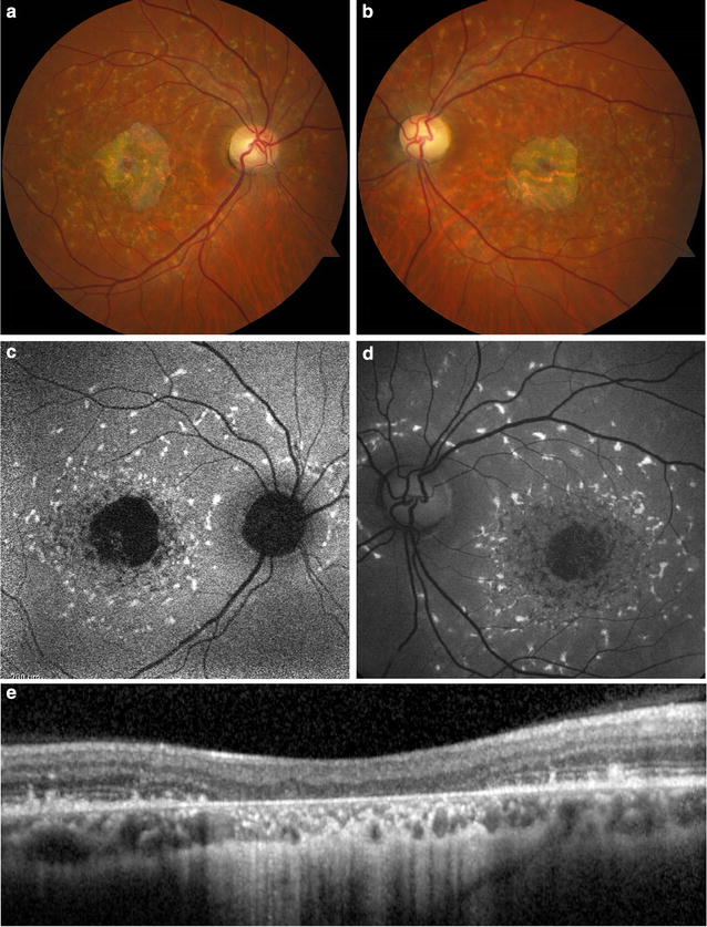
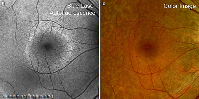
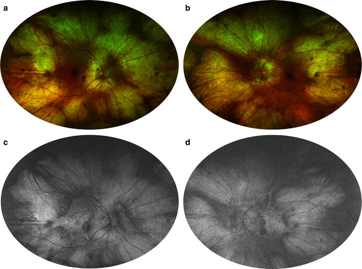
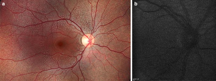
Fundus autofluorescence (FAF) is a non-invasive retinal imaging modality used in clinical practice to provide a density map of lipofuscin, the predominant ocular fluorophore, in the retinal pigment epithelium. Multiple commercially available imaging systems, including the fundus camera, the confocal scanning laser ophthalmoscope, and the ultra-widefield imaging device, are available to the clinician. Each offers unique advantages for evaluating various retinal diseases. The clinical applications of FAF continue to expand. It is now an essential tool for evaluating age related macular degeneration, macular dystrophies, retinitis pigmentosa, white dot syndromes, retinal drug toxicities, and various other retinal disorders. FAF may detect abnormalities beyond those detected on funduscopic exam, fluorescein angiography, or optical coherence tomography, and can be used to elucidate disease pathogenesis, form genotype-phenotype correlations, diagnose and monitor disease, and evaluate novel therapies. Given its ease of use, non-invasive nature, and value in characterizing retinal disease, FAF enjoys increasing clinical relevance. This review summarizes common ocular fluorophores, imaging modalities, and FAF findings for a wide spectrum of retinal disorders.
Overview
The claims and statistics of this paper haven’t been checked yet.
Run the check — free, no account needed. It reads the open-access text and checks the sources it cites.
Integrity signals
Registry
Cites
Statistics
Figures
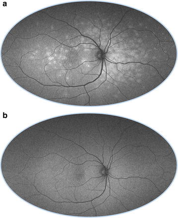
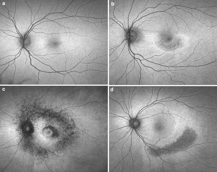
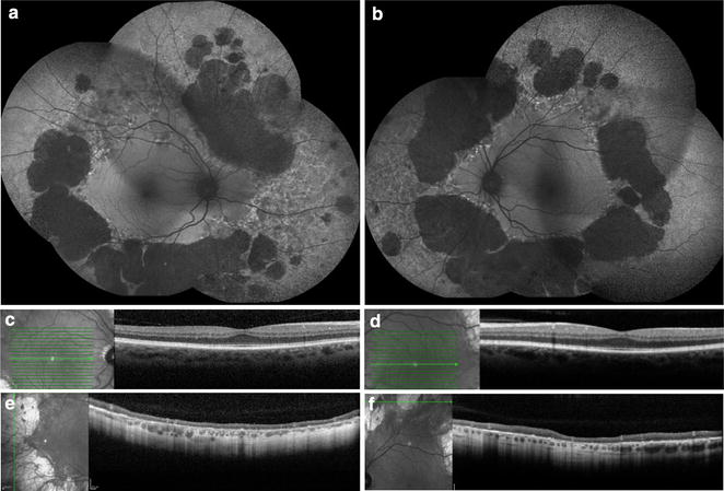
Verification
Claims the paper makes
The claims and statistics of this paper haven’t been checked yet.
Run the check — free, no account needed. It reads the open-access text and checks the sources it cites.
Claims other papers make about this one
Each is checked against this paper’s own text: does it say what the citing paper says it does?
The source backs part of the claim, not all of it — or backs it for a different population, condition or comparison.
In the citing paper
“As A2E can not be further metabolized and as RPE cells are postmitotic, A2E and related bisretinoids accumulate in the RPE in the form of lipofuscin deposits.”
From The Role of Vitamin A in Retinal Diseases (2022) · cites this paper as [75]
Experimental — accuracy not yet measuredThe source backs part of the claim, not all of it — or backs it for a different population, condition or comparison.
In the citing paper
“Although abundant lipofuscin accumulation is involved in the aetiology of certain retinal diseases (e.g., AMD, Best disease and ABCA4-retinopathy), it accumulates at a lower level also with age in the RPE of healthy eyes [9,72,73,74,75].”
From The Role of Vitamin A in Retinal Diseases (2022) · cites this paper as [75]
Experimental — accuracy not yet measuredThe source backs part of the claim, not all of it — or backs it for a different population, condition or comparison.
In the citing paper
“Lipofuscin is an autofluorescent cellular waste product that compromises the function and viability of RPE cells and ultimately results in photoreceptor degeneration and a loss of vision [54,69,73,74,75].”
From The Role of Vitamin A in Retinal Diseases (2022) · cites this paper as [75]
Experimental — accuracy not yet measured
Self-check
Percentages, sums and reported differences re-computed from the paper’s own numbers.
The self-check needs the paper’s full text.
Citations
Cites
Clinical Classification of Age-related Macular Degeneration
Ferris, F. L., Wilkinson, C. P., Bird, A. C., Chakravarthy, U., Chew, E. Y., Csaky, K. G., & Sadda, S. R. (2013) · Ophthalmology · doi:10.1016/j.ophtha.2012.10.036 · Cited by 1,868
Stereoscopic Atlas of Macular Diseases: Diagnosis and Treatment
(1990) · Archives of Ophthalmology · doi:10.1001/archopht.1990.01070060042026 · Cited by 1,014
Delori, F. C., Dorey, C. K., Staurenghi, G., Arend, O., Goger, D. G., & Weiter, J. J. (1995) · PubMed · Cited by 938
Revised Recommendations on Screening for Chloroquine and Hydroxychloroquine Retinopathy
Marmor, M. F., Kellner, U., Lai, T. Y. Y., Lyons, J. S., & Mieler, W. F. (2011) · Ophthalmology · doi:10.1016/j.ophtha.2010.11.017 · Cited by 703
Holz, F. G., Bindewald-Wittich, A., Fleckenstein, M., Dreyhaupt, J., Scholl, H. P. N., & Schmitz-Valckenberg, S. (2007) · American Journal of Ophthalmology · doi:10.1016/j.ajo.2006.11.041 · Cited by 619
Sparrow, J. R., Nakanishi, K., & Parish, C. A. (2000) · PubMed · Cited by 566
Retinal pigment epithelial lipofuscin and melanin and choroidal melanin in human eyes.
Weiter, J. J., Delori, F. C., Wing, G. L., & Fitch, K. A. (1986) · PubMed · Cited by 565
Aging human RPE: morphometric analysis of macular, equatorial, and peripheral cells.
Feeney-Burns, L., Hilderbrand, E. S., & Eldridge, S. (1984) · PubMed · Cited by 561
RPE65 is the isomerohydrolase in the retinoid visual cycle
Moiseyev, G., Chen, Y., Takahashi, Y., Wu, B. X., & Ma, J.-X. (2005) · Proceedings of the National Academy of Sciences · doi:10.1073/pnas.0503460102 · Cited by 536
Prevalence of Age-Related Macular Degeneration in the US Population
Klein, R. (2011) · Archives of Ophthalmology · doi:10.1001/archophthalmol.2010.318 · Cited by 523
Research works printed but not linked: 2
- Park SP, Siringo FS, Pensec N, et al. Comparison of fundus autofluorescence between fundus camera and confocal scanning laser ophthalmoscope-based systems. Ophthalmic Surgery Lasers Imaging Retina. 2013;44(6):536–43. · DOI
- Espina M, Barteselli G, Ma F, et al. Noncontact ultra-wide field lens system by Heidelberg spectralis. Invest Ophthalmol Vis Sci. 2014;55(13):1615.
Cited by
Sorted by citations
Fluorescence lifetime imaging ophthalmoscopy
Dysli, C., Wolf, S., Berezin, M. Y., Sauer, L., Hammer, M., & Zinkernagel, M. S. (2017) · Progress in Retinal and Eye Research · doi:10.1016/j.preteyeres.2017.06.005 · Cited by 203
Retinal imaging in Alzheimer's and neurodegenerative diseases
Snyder, P. J., Alber, J., Alt, C., Bain, L. J., Bouma, B. E., Bouwman, F. H., DeBuc, D. C., Campbell, M. C. W., Carrillo, M. C., Chew, E. Y., et al. (2020) · Alzheimer s & Dementia · doi:10.1002/alz.12179 · Cited by 197
Silva, S. R. D., Arno, G., Robson, A. G., Fakin, A., Pontikos, N., Mohamed, M. D., Bird, A. C., Moore, A. T., Michaelides, M., Webster, A. R., et al. (2020) · Progress in Retinal and Eye Research · doi:10.1016/j.preteyeres.2020.100898 · Cited by 154
Smart Contact Lenses as Wearable Ophthalmic Devices for Disease Monitoring and Health Management
Seo, H., Chung, W. G., Kwon, Y. W., Kim, S., Hong, Y., Park, W., Kim, E., Lee, J., Lee, S., Kim, M., et al. (2023) · Chemical Reviews · doi:10.1021/acs.chemrev.3c00290 · Cited by 157
The Role of Vitamin A in Retinal Diseases
Sajovic, J., Meglič, A., Glavač, D., Markelj, Š., Hawlina, M., & Fakin, A. (2022) · International Journal of Molecular Sciences · doi:10.3390/ijms23031014 · Cited by 109
Serous business: Delineating the broad spectrum of diseases with subretinal fluid in the macula
van Dijk, E. H. C., & Boon, C. J. F. (2021) · Progress in Retinal and Eye Research · doi:10.1016/j.preteyeres.2021.100955 · Cited by 88
CLINICOPATHOLOGIC CORRELATION OF GEOGRAPHIC ATROPHY SECONDARY TO AGE-RELATED MACULAR DEGENERATION
Li, M., Dolz‐Marco, R., Huisingh, C., Messinger, J. D., Feist, R. M., Ferrara, D., Freund, K. B., & Curcio, C. A. (2019) · Retina · doi:10.1097/iae.0000000000002461 · Cited by 84
A Systematic Review of Carotenoids in the Management of Age-Related Macular Degeneration
Lem, D. W., Davey, P. G., Gierhart, D. L., & Rosen, R. B. (2021) · Antioxidants · doi:10.3390/antiox10081255 · Cited by 85
Challenges and opportunities in clinical translation of biomedical optical spectroscopy and imaging
Wilson, B. C., Jermyn, M., & Leblond, F. (2018) · Journal of Biomedical Optics · doi:10.1117/1.jbo.23.3.030901 · Cited by 84
Multimodal Imaging of Nonneovascular Age-Related Macular Degeneration
Garrity, S. T., Sarraf, D., Freund, K. B., & Sadda, S. R. (2018) · Investigative Ophthalmology & Visual Science · doi:10.1167/iovs.18-24158 · Cited by 78
Self-citationFundus autofluorescence imaging in hereditary retinal diseases
Pichi, F., Abboud, E. B., Ghazi, N. G., & Khan, A. O. (2017) · Acta Ophthalmologica · doi:10.1111/aos.13602 · Cited by 76
Gambril, J. A., Sloan, K. R., Swain, T. A., Huisingh, C., Zarubina, A. V., Messinger, J. D., Ach, T., & Curcio, C. A. (2019) · Investigative Ophthalmology & Visual Science · doi:10.1167/iovs.19-26949 · Cited by 79
Lessons learned from quantitative fundus autofluorescence
Sparrow, J. R., Duncker, T., Schuerch, K., Paavo, M., & de Carvalho, J. R. L. (2019) · Progress in Retinal and Eye Research · doi:10.1016/j.preteyeres.2019.100774 · Cited by 76
Quantitative approaches in multimodal fundus imaging: State of the art and future perspectives
Arrigo, A., Aragona, E., Parodi, M. B., & Bandello, F. M. (2022) · Progress in Retinal and Eye Research · doi:10.1016/j.preteyeres.2022.101111 · Cited by 68
Lochocki, B., Boon, B. D. C., Verheul, S. R., Zada, L., Hoozemans, J. J. M., Ariese, F., & de Boer, J. F. (2021) · Communications Biology · doi:10.1038/s42003-021-01981-x · Cited by 61
Review of clinical approaches in fluorescence lifetime imaging ophthalmoscopy
Sauer, L., Andersen, K. M., Dysli, C., Zinkernagel, M. S., Bernstein, P. S., & Hammer, M. (2018) · Journal of Biomedical Optics · doi:10.1117/1.jbo.23.9.091415 · Cited by 61
Autofluorescence‐based optical biopsy: An effective diagnostic tool in hepatology
Croce, A. C., Ferrigno, A., Bottiroli, G. F., & Vairetti, M. (2018) · Liver International · doi:10.1111/liv.13753 · Cited by 60
Monitoring progression of retinitis pigmentosa: current recommendations and recent advances
Menghini, M., Cehajic‐Kapetanovic, J., & MacLaren, R. E. (2020) · Expert Opinion on Orphan Drugs · doi:10.1080/21678707.2020.1735352 · Cited by 59
Treder, M., Lauermann, J. L., & Eter, N. (2018) · Graefe s Archive for Clinical and Experimental Ophthalmology · doi:10.1007/s00417-018-4098-2 · Cited by 58
Hydroxychloroquine-induced Retinal Toxicity
Yusuf, I. H., Issa, P. C., & Ahn, S. J. (2023) · Frontiers in Pharmacology · doi:10.3389/fphar.2023.1196783 · Cited by 61
Habibalahi, A., Bala, C., Allende, A., Anwer, A. G., & Goldys, E. M. (2019) · The Ocular Surface · doi:10.1016/j.jtos.2019.03.003 · Cited by 56
Saleh, G. A., Batouty, N. M., Haggag, S., Elnakib, A. A., Khalifa, F., Taher, F., Mohamed, M. A., Farag, R. K., Sandhu, H. S., Sewelam, A., et al. (2022) · Bioengineering · doi:10.3390/bioengineering9080366 · Cited by 54
Biswas, S., Gaviria-Bravo, M. L., Malheiro, L., Marques, J. P., Giordano, V., & Liang, H. (2018) · Ophthalmology and Therapy · doi:10.1007/s40123-018-0146-6 · Cited by 52
Age, lipofuscin and melanin oxidation affect fundus near-infrared autofluorescence
Taubitz, T., Fang, Y., Biesemeier, A., Julien, S., & Schraermeyer, U. (2019) · EBioMedicine · doi:10.1016/j.ebiom.2019.09.048 · Cited by 51
Ophthalmic sensing technologies for ocular disease diagnostics
Shi, Y., Jiang, N., Bikkannavar, P., Cordeiro, M. F., & Yetisen, A. K. (2021) · The Analyst · doi:10.1039/d1an01244d · Cited by 50
Nanodiagnostics and Nanotherapeutics for age-related macular degeneration
Sarkar, A., & Dyawanapelly, S. (2020) · Journal of Controlled Release · doi:10.1016/j.jconrel.2020.10.054 · Cited by 48
Advances in Retinal Optical Imaging
Li, Y., Xia, X., & Paulus, Y. M. (2018) · Photonics · doi:10.3390/photonics5020009 · Cited by 46
Advanced retinal imaging and applications for clinical practice: A consensus review
Fogel-Levin, M., Sadda, S. R., Rosenfeld, P. J., Waheed, N. K., Querques, G., Freund, K. B., & Sarraf, D. (2022) · Survey of Ophthalmology · doi:10.1016/j.survophthal.2022.02.004 · Cited by 42
Self-citationA Systematic Review of Carotenoids in the Management of Diabetic Retinopathy
Lem, D. W., Gierhart, D. L., & Davey, P. G. (2021) · Nutrients · doi:10.3390/nu13072441 · Cited by 45
Camino, A., Wang, Z., Wang, J., Pennesi, M. E., Yang, P. G., Huang, D., Li, D., & Jia, Y. (2018) · Biomedical Optics Express · doi:10.1364/boe.9.003092 · Cited by 41
Chloroquine and Hydroxychloroquine Toxicity
Song, W., Muste, J. C., Greenlee, T. E., & Singh, R. P. (2020) · American Journal of Ophthalmic Clinical Trials · doi:10.25259/ajoct_2_2020 · Cited by 42
Chen, L., Messinger, J. D., Ferrara, D., Freund, K. B., & Curcio, C. A. (2020) · Ophthalmology Retina · doi:10.1016/j.oret.2020.11.006 · Cited by 41
Borrelli, E., Lei, J., Balasubramanian, S., Uji, A., Cozzi, M., Sarao, V., Lanzetta, P., Staurenghi, G., & Sadda, S. R. (2017) · British Journal of Ophthalmology · doi:10.1136/bjophthalmol-2017-310881 · Cited by 40
Zhong, Y., Mohan, K., Liu, J., Al‐Attar, A., Lin, P., Flight, R. M., Sun, Q., Warmoes, M. O., Deshpande, R. R., Liu, H., et al. (2020) · Biochimica et Biophysica Acta (BBA) - Molecular Basis of Disease · doi:10.1016/j.bbadis.2020.165883 · Cited by 36
Updates on ophthalmic imaging features of optic disc drusen, papilledema, and optic disc edema
Yan, Y., & Liao, Y. J. (2020) · Current Opinion in Neurology · doi:10.1097/wco.0000000000000881 · Cited by 36
Romano, F., Airaldi, M., Cozzi, M., Oldani, M., Riva, E., Bertoni, A. I., Dautaj, A., Bertelli, M., Staurenghi, G., & Salvetti, A. P. (2021) · Ophthalmology Science · doi:10.1016/j.xops.2021.100016 · Cited by 36
Next-generation phenotyping of inherited retinal diseases from multimodal imaging with Eye2Gene
Pontikos, N., Woof, W. A., Lin, S., Ghoshal, B., Mendes, B. S., Veturi, Y. A., Nguyen, Q., Javanmardi, B., Georgiou, M., Hustinx, A., et al. (2025) · Nature Machine Intelligence · doi:10.1038/s42256-025-01040-8 · Cited by 38
AbdelMaksoud, E., Barakat, S., & Elmogy, M. M. (2020) · Computers in Biology and Medicine · doi:10.1016/j.compbiomed.2020.104039 · Cited by 35
Vienola, K. V., Zhang, M., Snyder, V. C., Sahel, J., Dansingani, K. K., & Rossi, E. A. (2020) · Scientific Reports · doi:10.1038/s41598-020-66581-x · Cited by 32
Characterization of Retinitis Pigmentosa Using Fluorescence Lifetime Imaging Ophthalmoscopy (FLIO)
Andersen, K. M., Sauer, L., Gensure, R. H., Hammer, M., & Bernstein, P. S. (2018) · Translational Vision Science & Technology · doi:10.1167/tvst.7.3.20 · Cited by 31
Carotenoids in the Management of Glaucoma: A Systematic Review of the Evidence
Lem, D. W., Gierhart, D. L., & Davey, P. G. (2021) · Nutrients · doi:10.3390/nu13061949 · Cited by 35
Viggiano, P., Boscia, G., Sadeghi, E., Cheung, G., Borrelli, E., Alessio, G., Chhablani, J. K., & Boscia, F. (2025) · Progress in Retinal and Eye Research · doi:10.1016/j.preteyeres.2025.101372 · Cited by 30
Vessey, K. A., Ho, T., Jobling, A. I., Mills, S. A., Tran, M. X., Brandli, A. A., Lam, J., Guymer, R. H., & Fletcher, E. L. (2018) · Investigative Ophthalmology & Visual Science · doi:10.1167/iovs.17-23098 · Cited by 30
Ultra-Widefield Fundus Autofluorescence Imaging of Patients with Retinitis Pigmentosa
Hariri, A. H., Gui, W., O’Keefe, G. A. D., Ip, M. S., Sadda, S. R., & Gorin, M. B. (2017) · Ophthalmology Retina · doi:10.1016/j.oret.2017.10.018 · Cited by 29
Jidigam, V. K., Singh, R., Batoki, J. C., Milliner, C., Sawant, O. B., Bonilha, V. L., & Rao, S. (2021) · Graefe s Archive for Clinical and Experimental Ophthalmology · doi:10.1007/s00417-021-05460-1 · Cited by 29
Heiferman, M. J., & Fawzi, A. A. (2016) · Retina · doi:10.1097/iae.0000000000001254 · Cited by 29
Multimodal Imaging Features of Optic Disc Drusen
Yan, Y., Ludwig, C. A., & Liao, Y. J. (2021) · American Journal of Ophthalmology · doi:10.1016/j.ajo.2020.12.023 · Cited by 28
Agarwal, K., Vinekar, A. S., Chandra, P., Padhi, T. R., Nayak, S., Jayanna, S., Panchal, B. U., Jalali, S., & Das, T. (2021) · Indian Journal of Ophthalmology · doi:10.4103/ijo.ijo_1917_20 · Cited by 28
Chavan, R., & Pete, D. J. (2024) · Journal of Engineering and Applied Science · doi:10.1186/s44147-023-00335-0 · Cited by 27
Oishi, A., Miyata, M., Numa, S., Otsuka, Y., Oishi, M., & Tsujikawa, A. (2019) · International Journal of Retina and Vitreous · doi:10.1186/s40942-019-0173-z · Cited by 27
Ryu, J., Statz, J. P., Chan, W. K., Burch, F. C., Brigande, J. V., Kempton, B., Porsov, E. V., Renner, L. M., McGill, T., Burwitz, B. J., et al. (2022) · Scientific Reports · doi:10.1038/s41598-022-13689-x · Cited by 26
Gocuk, S. A., Jolly, J. K., Edwards, T. L., & Ayton, L. N. (2023) · Progress in Retinal and Eye Research · doi:10.1016/j.preteyeres.2023.101190 · Cited by 27
Campbell, J. M., Gosnell, M. E., Agha, A., Handley, S., Knab, A., Anwer, A. G., Bhargava, A., & Goldys, E. M. (2024) · Advanced Materials · doi:10.1002/adma.202403761 · Cited by 29
Imaging the eye as a window to brain health: frontier approaches and future directions
Banna, H. U., Slayo, M., Armitage, J. A., del Rosal, B., Giummarra, L., & Spencer, S. J. (2024) · Journal of Neuroinflammation · doi:10.1186/s12974-024-03304-3 · Cited by 30
Ahn, S. J., Joung, J., & Lee, B. R. (2019) · American Journal of Ophthalmology · doi:10.1016/j.ajo.2019.09.008 · Cited by 26
Wizenty, J., Schumann, T., Theil, D., Stockmann, M., Pratschke, J., Tacke, F., Aigner, F., & Wuensch, T. (2020) · Molecules · doi:10.3390/molecules25092095 · Cited by 26
Oximetry using multispectral imaging: theory and application
MacKenzie, L. E., & Harvey, A. R. (2018) · Journal of Optics · doi:10.1088/2040-8986/aab74c · Cited by 25
Zhang, X., Henneman, N. F., Girardot, P. E., Sellers, J. T., Chrenek, M. A., Li, Y., Wang, J., Brenner, C., Nickerson, J. M., & Boatright, J. H. (2020) · Investigative Ophthalmology & Visual Science · doi:10.1167/iovs.61.10.47 · Cited by 26
Borrelli, E., Bandello, F. M., Boon, C. J. F., Carelli, V., Lenaers, G., Reibaldi, M., Sadda, S. R., Sadun, A. A., Sarraf, D., Yu‐Wai‐Man, P., et al. (2024) · Progress in Retinal and Eye Research · doi:10.1016/j.preteyeres.2024.101264 · Cited by 25
Self-citationHabibalahi, A., Allende, A., Bala, C., Anwer, A. G., Mukhopadhyay, S. C., & Goldys, E. M. (2019) · IEEE Access · doi:10.1109/access.2019.2942959 · Cited by 24
Arslan, J., Samarasinghe, G., Sowmya, A., Benke, K. K., Hodgson, L., Guymer, R. H., & Baird, P. N. (2021) · Translational Vision Science & Technology · doi:10.1167/tvst.10.8.2 · Cited by 22
Fumi, D., Ruggeri, F., Fasciolo, D., Antonello, E., Burtini, G., & Abdolrahimzadeh, S. (2025) · Vision · doi:10.3390/vision9010019 · Cited by 20
Li, D. Q., & Choudhry, N. (2020) · Current Opinion in Ophthalmology · doi:10.1097/icu.0000000000000653 · Cited by 20
Tisi, A., Passacantando, M., Lozzi, L., & Maccarone, R. (2020) · Experimental Eye Research · doi:10.1016/j.exer.2020.108169 · Cited by 20
Novel imaging techniques for hydroxychloroquine retinopathy
Yusuf, I. H., Issa, P. C., & Ahn, S. J. (2022) · Frontiers in Medicine · doi:10.3389/fmed.2022.1026934 · Cited by 19
Fukushima, M., Inoue, T., Miyai, T., & Obata, R. (2019) · European Journal of Ophthalmology · doi:10.1177/1120672119832183 · Cited by 19
Ultra-Wide-Field Imaging in Uveitis
Cunningham, E. T., Munk, M. R., Kiss, S., & Zierhut, M. (2019) · Ocular Immunology and Inflammation · doi:10.1080/09273948.2019.1605264 · Cited by 19
Oh, J. K., Lima de Carvalho, J. R., Ryu, J., Tsang, S. H., & Sparrow, J. R. (2020) · Scientific Reports · doi:10.1038/s41598-020-65763-x · Cited by 18
Valiente‐Soriano, F. J., Salinas‐Navarro, M., Pierdomenico, J. D., García‐Ayuso, D., Lucas‐Ruiz, F., Pinilla, I., Cuenca, N., Vidal‐Sanz, M., Villegas‐Pérez, M. P., & Agudo‐Barriuso, M. (2020) · Scientific Reports · doi:10.1038/s41598-020-64131-z · Cited by 18
Multimodal imaging in infectious and noninfectious intermediate, posterior and panuveitis
Thomas, A. S., & Lin, P. (2021) · Current Opinion in Ophthalmology · doi:10.1097/icu.0000000000000762 · Cited by 17
From mouse to human: Accessing the biochemistry of vision in vivo by two-photon excitation
Palczewska, G., Wojtkowski, M., & Palczewski, K. (2023) · Progress in Retinal and Eye Research · doi:10.1016/j.preteyeres.2023.101170 · Cited by 17
Hydroxychloroquine Retinal Toxicity
Proano, C. E., & KIMBALL, G. P. (2019) · New England Journal of Medicine · doi:10.1056/nejmicm1304542 · Cited by 16
Review: Emerging Eye-Based Diagnostic Technologies for Traumatic Brain Injury
Harris, G., Rickard, J. J. S., Butt, G. F., Kelleher, L., Blanch, R. J., Cooper, J. M., & Oppenheimer, P. G. (2022) · IEEE Reviews in Biomedical Engineering · doi:10.1109/rbme.2022.3161352 · Cited by 19
Multicolor imaging in macular telangiectasia—a comparison with fundus autofluorescence
Govindahari, V., Fraser‐Bell, S., Ayachit, A., Invernizzi, A., Nair, U., Nair, D. V., Lupidi, M., Singh, S. R., Rajendran, A., Zur, D., et al. (2020) · Graefe s Archive for Clinical and Experimental Ophthalmology · doi:10.1007/s00417-020-04878-3 · Cited by 15
Applications of fundus autofluorescence and widefield angiography in clinical practice
Banda, H. K., Shah, G. K., & Blinder, K. J. (2018) · Canadian Journal of Ophthalmology · doi:10.1016/j.jcjo.2018.10.003 · Cited by 15
Wong, D. T., Berger, A. R., Bourgault, S., Chen, J. L., Colleaux, K. M., Cruess, A. F., Dookeran, R. I., Gauthier, D., Hurley, B. R., Kapusta, M. A., et al. (2021) · Ophthalmologica · doi:10.1159/000516108 · Cited by 14
Cheng, Y., Fleckenstein, M., Schmitz-Valckenberg, S., Lu, J., Liu, Z., Herrera, G., Gregori, G., Wang, R. K., Rosenfeld, P. J., & Trivizki, O. (2024) · American Journal of Ophthalmology · doi:10.1016/j.ajo.2024.10.002 · Cited by 13
Klufas, M. A., O’Hearn, T. M., & Sarraf, D. (2015) · Retinal Cases & Brief Reports · doi:10.1097/icb.0000000000000203 · Cited by 13
Self-citationFluorescence Lifetime Imaging Ophthalmoscopy (FLIO)
Bernstein, P. S., Dysli, C., Fischer, J., Hammer, M., Katayama, Y., Sauer, L., & Zinkernagel, M. S. (2019) · Springer eBooks · doi:10.1007/978-3-030-16638-0_10 · Cited by 14
Berlin, A., Clark, M. E., Swain, T. A., Fischer, N. A., McGwin, G., Sloan, K. R., Owsley, C. J., & Curcio, C. A. (2022) · Translational Vision Science & Technology · doi:10.1167/tvst.11.10.23 · Cited by 13
Russell, M. W., Muste, J. C., Seth, K., Kumar, M., Rich, C., Singh, R. P., & Traboulsi, E. I. (2022) · Ophthalmic Genetics · doi:10.1080/13816810.2022.2144903 · Cited by 12
Bader, I., Groot, C., Tan, H. S., Milongo, J.-M. A., den Haan, J., Verberk, I. M. W., Yong, K. X. X., Orellina, J. A., Campbell, S. R., Wilson, D., et al. (2024) · Alzheimer s Research & Therapy · doi:10.1186/s13195-024-01545-1 · Cited by 12
Horng, H., O’Brien, K., Lamont, A., Sochol, R. D., Pfefer, T. J., & Chen, Y. (2021) · Optics Letters · doi:10.1364/ol.412849 · Cited by 12
Mitchell, M. E., Cook, L. C., Shiers, S. I., Tavares‐Ferreira, D., Akopian, A. N., Dussor, G. O., & Price, T. J. (2023) · The Journal of Comparative Neurology · doi:10.1002/cne.25463 · Cited by 12
Mota, S. E. H.-D., Melo-Granados, E. A. R., Fromow-Guerra, J., Béjar-Cornejo, F., Gallego‐Pinazo, R., & Rodríguez-Ayala, E. (2019) · European Journal of Ophthalmology · doi:10.1177/1120672119833267 · Cited by 12
Borrelli, E., Battista, M., Zuccaro, B., Sacconi, R., Brambati, M., Querques, L., Prascina, F., Sadda, S. R., Bandello, F. M., & Querques, G. (2020) · Journal of Clinical Medicine · doi:10.3390/jcm9082388 · Cited by 12
Ultra-Widefield Autofluorescence Imaging of Retinal Detachment Compared to Retinoschisis
Nadelmann, J. B., Patel, M. M., Kiss, S., Askin, G., Chan, R. V. P., Papakostas, T., D'Amico, D. J., & Orlin, A. (2019) · Ophthalmic surgery, lasers & imaging retina · doi:10.3928/23258160-20190905-03 · Cited by 11
Alex, D., Anantharaman, G., Gopalakrishnan, M., Manayath, G. J., Amar, S. P., Raman, R., Sreenivasan, R., Ayachit, A., & Sivaprasad, S. (2020) · Retina · doi:10.1097/iae.0000000000002954 · Cited by 11
Nafar, Z., Wen, R., & Jiao, S. (2018) · Biomedical Optics Express · doi:10.1364/boe.9.003768 · Cited by 10
Long term follow-up of a family with GUCY2D dominant cone dystrophy
Tsokolas, G., Almuhtaseb, H., Griffiths, H., Shawkat, F. S., Pengelly, R. J., Ennis, S., & Lotery, A. J. (2018) · International Journal of Ophthalmology · doi:10.18240/ijo.2018.12.12 · Cited by 10
Yu, W., Kang, L., Tsang, V. T. C., Zhang, Y., Wong, I. H. M., & Wong, T. T. W. (2022) · eLife · doi:10.7554/elife.81015 · Cited by 10
Froines, C. P., Saunders, T. F., Heathcote, J., Pak, J. W., Chew, E. Y., Blodi, B. A., & Domalpally, A. (2024) · Translational Vision Science & Technology · doi:10.1167/tvst.13.11.1 · Cited by 9
Bubis, E., Sher, I., Skaat, A., Sharvit‐Ginon, I., Szalapak, A. M., Moroz, I., Kalter‐Leibovici, O., & Rotenstreich, Y. (2019) · Translational Vision Science & Technology · doi:10.1167/tvst.8.1.26 · Cited by 8
Ometto, G., Montesano, G., Afgeh, S. S., Lazaridis, G., Liu, X., Keane, P. A., Crabb, D. P., & Denniston, A. K. (2020) · Translational Vision Science & Technology · doi:10.1167/tvst.9.9.38 · Cited by 9
Exploring retinal conditions through blue light reflectance imaging
Guerra, R. L. L., Guerra, C. L. L., Meirelles, M. G. B., Barbosa, G. C. S., Novais, E. A., Badaró, E., Lucatto, L. F. A., & Roisman, L. (2025) · Progress in Retinal and Eye Research · doi:10.1016/j.preteyeres.2024.101326 · Cited by 8
Lee, J., Han, M., Wang, K., Butler, L. R., & Sinclair, D. A. (2026) · Progress in Retinal and Eye Research · doi:10.1016/j.preteyeres.2026.101442 · Cited by 7
Nafar, Z., Wen, R., & Jiao, S. (2018) · Experimental Biology and Medicine · doi:10.1177/1535370218813529 · Cited by 8
Abdelbaky, M. I. M., Maamoun, T. A. E., Mabrouk, F. I., & Hassan, R. M. (2021) · The Egyptian Journal of Internal Medicine · doi:10.1186/s43162-021-00047-y · Cited by 8
Thomson, K., Karouta, C., Sabeti, F., Anstice, N. S., Leung, M., Jong, T., Maddess, T., Morgan, I. G., Game, J., & Ashby, R. (2022) · Clinical and Translational Science · doi:10.1111/cts.13392 · Cited by 8
Rošar, A. P., Casalino, G., Cozzi, M., Pellegrini, M., Bottoni, F., Dell'Arti, L., Lavrič, A., Umek, L., Petrovic, M. G., Pavesio, C. E., et al. (2021) · Retina · doi:10.1097/iae.0000000000003247 · Cited by 8
Metadata
- Authors
- Yung, M., Klufas, M. A., & Sarraf, D.
- Affiliations
- University of California, Los Angeles (US)
- Corresponding author
- Sarraf, David
- Venue
- International Journal of Retina and Vitreous
- Published
- 2016-04-08
- Volume
- 2
- Issue
- 1
- Pages
- 12-12
- ISSN
- 2056-9920 (ISSN-L)
- PMC
- PMC5088473
- OpenAlex
- W2329521208
- Cited by
- 236
- References
- 198
- Citations by year
- 2026: 15 · 2025: 23 · 2024: 22 · 2023: 20 · 2022: 29 · 2021: 40 · 2020: 31 · 2019: 24 · 2018: 22 · 2017: 8 · 2016: 1
- FWCI
- 3.58
- Type
- journal article
- Language
- en
- Open access
- gold
- doi.org
- Licence
- cc-by
- Locations
- International Journal of Retina and Vitreous (publishedVersion, cc-by)
- PubMed Central (publishedVersion, cc-by)
- PubMed (publishedVersion)
- Funders
- Regeneron Pharmaceuticals
- Genentech
- Topics
- Health Sciences › Medicine › Ophthalmology › Retinal Diseases and Treatments
- Health Sciences › Medicine › Radiology, Nuclear Medicine and Imaging › Retinal Imaging and Analysis
- Health Sciences › Medicine › Ophthalmology › Retinal Development and Disorders
- Keywords
- fundus autofluorescence, lipofuscin, retinal pigment epithelium, age-related macular degeneration, macular dystrophy, retinitis pigmentosa, white dot syndromes, ultra-widefield imaging, confocal scanning laser ophthalmoscopy, genotype-phenotype correlation
About these checks
Registry, retraction, Cites and statistics results are computed, not judged, with one exception: a printed reference that no index lists is read once by a model before it counts as a citation error. Claims are extracted by a model and kept only when their quote appears verbatim in the text; each cited source is then checked, with a model’s help, for whether it says what the claim relies on. Those verdicts can be wrong — read the claim’s quote and the cited source before relying on one.
Statistics and arithmetic are recomputed, not judged.
Confidence bands are shown only for fields calibrated on our annotated test set. None is calibrated yet, so no band is shown.
What counts as a claim ›How we measure confidence ›How we check this ›
Are you an author, or do you see a problem with this page? Request a review
