

The DOI matches its Crossref record; 38 of the 38 references it cites were found in a registry or an index.
Autoantibodies to MOG in a distinct subgroup of adult multiple sclerosis
Open accessAbstract
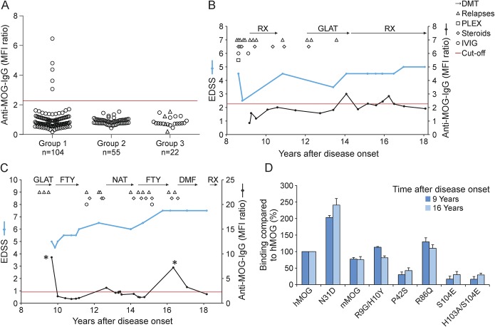
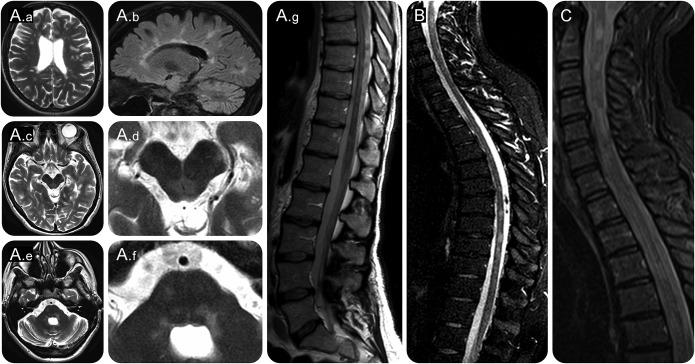
OBJECTIVES: To evaluate the presence of antibodies to conformation-intact myelin oligodendrocyte glycoprotein (MOG) in a subgroup of adult patients with clinically definite multiple sclerosis (MS) preselected for a specific clinical phenotype including severe spinal cord, optic nerve, and brainstem involvement. METHODS: Antibodies to MOG were investigated using a cell-based assay in 3 groups of patients: 104 preselected patients with MS (group 1), 55 age- and sex-matched, otherwise unselected patients with MS (group 2), and in 22 brain-biopsied patients with demyelinating diseases of the CNS (n = 19 with MS), 4 of whom classified as MS type II (group 3). Recognized epitopes were identified with mutated variants of MOG. RESULTS: Antibodies to MOG were found in about 5% (5/104) of preselected adult patients with MS. In contrast, in groups 2 and 3, none of the patients tested positive for MOG antibodies. Patients with MS with antibodies to MOG predominantly manifested with concomitant severe brainstem and spinal cord involvement and had a severe disease course with high relapse rates and failure to several disease-modifying therapies. Three of them had been treated with plasma exchange with a favorable response. All anti-MOG-positive patients with MS showed typical MS lesions on brain MRI. Longitudinal analysis up to 9 years revealed fluctuations and reappearance of anti-MOG reactivity. Epitope mapping indicated interindividual heterogeneity, yet intraindividual stability of the antibody response. CONCLUSIONS: Antibodies to MOG can be found in a distinct subgroup of adult MS with a specific clinical phenotype and may indicate disease heterogeneity.
Overview
The claims and statistics of this paper haven’t been checked yet.
Run the check — free, no account needed. It reads the open-access text and checks the sources it cites.
Integrity signals
Registry
Cites
Statistics
Figures
Verification
Claims the paper makes
The claims and statistics of this paper haven’t been checked yet.
Run the check — free, no account needed. It reads the open-access text and checks the sources it cites.
Self-check
Percentages, sums and reported differences re-computed from the paper’s own numbers.
The self-check needs the paper’s full text.
Citations
Cites
Diagnostic criteria for multiple sclerosis: 2010 revisions to the McDonald criteria
Polman, C. H., Reingold, S. C., Banwell, B., Clanet, M., Cohen, J. A., Filippi, M., Fujihara, K., Havrdova, E., Hutchinson, M., Kappos, L., et al. (2011) · Annals of Neurology · doi:10.1002/ana.22366 · Cited by 9,802
International consensus diagnostic criteria for neuromyelitis optica spectrum disorders
Wingerchuk, D. M., Banwell, B., Bennett, J. L., Cabre, P., Carroll, W., Chitnis, T., de Seze, J., Fujihara, K., Greenberg, B., Jacob, A., et al. (2015) · Neurology · doi:10.1212/wnl.0000000000001729 · Cited by 4,649
New Multiple Sclerosis Phenotypic Classification
LUBLIN, F. D. (2014) · European Neurology · doi:10.1159/000367614 · Cited by 4,277
Ruiter, A. M., Meilof, J. F., Somanje-Bolweg, R. R. J., van Gorsel, E., & Kalkers, N. F. (2017) · Case Reports in Neurology · doi:10.1159/000455942 · Cited by 1,762
von Büdingen, H. ‐., Palanichamy, A., Lehmann‐Horn, K., Michel, B., & Zamvil, S. S. (2015) · European Neurology · doi:10.1159/000377675 · Cited by 1,475
Multiple Sclerosis Severity Score: Using disability and disease duration to rate disease severity
Roxburgh, R. H. S. R., Seaman, S. R., Masterman, T., Hensiek, A. E., Sawcer, S. J., Vukusic, S., Achiti, I., Confavreux, C., Coustans, M., le Page, E., et al. (2005) · Neurology · doi:10.1212/01.wnl.0000156155.19270.f8 · Cited by 963
Overlapping demyelinating syndromes and anti–N‐methyl‐D‐aspartate receptor encephalitis
Titulaer, M. J., Höftberger, R., Iizuka, T., Leypoldt, F., McCracken, L., Cellucci, T., Benson, L. A., Shu, H., Irioka, T., Hirano, M., et al. (2014) · Annals of Neurology · doi:10.1002/ana.24117 · Cited by 518
Mader, S., Gredler, V., Schanda, K., Rostasy, K. M., Dujmović, I., Pfaller, K., Lutterotti, A., Jarius, S., Pauli, F. D., Kuenz, B., et al. (2011) · Journal of Neuroinflammation · doi:10.1186/1742-2094-8-184 · Cited by 449
Myelin-oligodendrocyte glycoprotein antibodies in adults with a neuromyelitis optica phenotype
Kitley, J., Woodhall, M., Waters, P., Leite, M. I., Devenney, E., Craig, J., Palace, J., & Vincent, A. (2012) · Neurology · doi:10.1212/wnl.0b013e31826aac4e · Cited by 445
Self-antigen tetramers discriminate between myelin autoantibodies to native or denatured protein
O’Connor, K. C., McLaughlin, K. A., Jager, P. L. D., Chitnis, T., Bettelli, E., Xu, C., Robinson, W. H., Cherry, S. V., Bar‐Or, A., Banwell, B., et al. (2007) · Nature Medicine · doi:10.1038/nm1488 · Cited by 419
Cited by
Sorted by citations
Items 101–200 of 206
Challenges associated with using extracellular vesicles as biomarkers in neurodegenerative disease
Couch, Y. (2023) · Expert Review of Molecular Diagnostics · doi:10.1080/14737159.2023.2277373 · Cited by 23
Emerging drugs for the treatment of neuromyelitis optica
Duchow, A. S., Chien, C., Paul, F., & Bellmann–Strobl, J. (2020) · Expert Opinion on Emerging Drugs · doi:10.1080/14728214.2020.1803828 · Cited by 20
De Masi, R., & Orlando, S. (2022) · International Journal of Molecular Sciences · doi:10.3390/ijms23137373 · Cited by 20
Salama, S., Pardo, S., & Levy, M. (2019) · Multiple Sclerosis and Related Disorders · doi:10.1016/j.msard.2019.02.023 · Cited by 19
Fernández, I. M., Macrini, C., Krumbholz, M., Hensbergen, P. J., Ederveen, A. L. H., Winklmeier, S., Vural, A., Kurne, A., Jenne, D. E., Kamp, F., et al. (2019) · Frontiers in Immunology · doi:10.3389/fimmu.2019.01189 · Cited by 19
Self-citationCross, H., Sabiq, F., Ackermans, N., Mattar, A. A. G., Au, S., Woodhall, M. R., Sun, B., Devonshire, V. A., Carruthers, R. L., Sayao, A., et al. (2021) · Frontiers in Neurology · doi:10.3389/fneur.2020.525933 · Cited by 20
Tzartos, J. S., Karagiorgou, K., Tzanetakos, D., Breza, M., Evangelopoulos, M., Pelidou, S., Bakirtzis, C., Nikolaidis, I., Koutsis, G., Notas, K., et al. (2020) · Journal of the Neurological Sciences · doi:10.1016/j.jns.2020.116673 · Cited by 22
Myelin oligodendrocyte glycoprotein immunoglobulin G‐associated disease: An overview
Fujihara, K., Sato, D. K., Nakashima, I., Takahashi, T., Kaneko, K., Ogawa, R., Akaishi, T., Matsumoto, Y., Takai, Y., Nishiyama, S., et al. (2018) · Clinical and Experimental Neuroimmunology · doi:10.1111/cen3.12434 · Cited by 16
Lin, T.-Y., Chien, C., Lu, A., Paul, F., & Zimmermann, H. G. (2021) · Expert Review of Neurotherapeutics · doi:10.1080/14737175.2021.1982697 · Cited by 16
Gontika, M. P., & Anagnostouli, M. C. (2018) · Multiple Sclerosis International · doi:10.1155/2018/8487471 · Cited by 17
Antibody signatures in patients with histopathologically defined multiple sclerosis patterns
Stork, L., Ellenberger, D., Ruprecht, K., Reindl, M., Beißbarth, T., Friede, T., Kümpfel, T., Gerdes, L. A., Gloth, M., Liman, T., et al. (2020) · Acta Neuropathologica · doi:10.1007/s00401-019-02120-x · Cited by 15
Self-citationCase Series: Myelin Oligodendrocyte Glycoprotein-Immunoglobulin G-Related Disease Spectrum
Alshamrani, F., Alnajashi, H. A., Shosha, E., Casserly, C. S., & Morrow, S. A. (2020) · Frontiers in Neurology · doi:10.3389/fneur.2020.00089 · Cited by 14
Lotan, I., Hellmann, M. A., Benninger, F., Stiebel‐Kalish, H., & Steiner, I. (2018) · Journal of Neuroimmunology · doi:10.1016/j.jneuroim.2018.09.010 · Cited by 14
Fujihara, K. (2019) · Multiple Sclerosis Journal · doi:10.1177/1352458519895236 · Cited by 13
Server, A., Sakinis, T., Pfeiffer, H. C. V., Sandvig, I., Barlinn, J., & Marthinsen, P. B. (2020) · Radiographics · doi:10.1148/rg.2020200032 · Cited by 13
Fragoso, D. C., Salles, L., Apóstolos‐Pereira, S. L., Callegaro, D., Sato, D. K., & Rimkus, C. D. M. (2023) · Arquivos de Neuro-Psiquiatria · doi:10.1055/s-0043-1768669 · Cited by 12
Marchionatti, A., Hansel, G., Avila, G. U., & Sato, D. K. (2021) · Frontiers in Immunology · doi:10.3389/fimmu.2021.642272 · Cited by 12
Yanagida, A., Iizuka, T., Nagai, T., Usui, R., Kaneko, J., Akutsu, T., Hanajima, R., Kanazawa, N., & Nishiyama, K. (2017) · Journal of the Neurological Sciences · doi:10.1016/j.jns.2017.09.020 · Cited by 12
Clinical utility of anti-MOG antibody testing in a Danish cohort
Papp, V., Langkilde, A. R., Blinkenberg, M., Schreiber, K. I., Jensen, P. E., & Sellebjerg, F. T. (2018) · Multiple Sclerosis and Related Disorders · doi:10.1016/j.msard.2018.09.010 · Cited by 11
Biomarkers of Multiple Sclerosis
Pastare, D., Bennour, M. R., Polunosika, E., & Karelis, G. (2019) · The Open Immunology Journal · doi:10.2174/1874226201909010001 · Cited by 13
Anti-MOG-associated demyelinating disorders: two sides of the same coin
Maniscalco, G. T., Allegorico, L., Alfieri, G., Napolitano, M., Ranieri, A., Renna, R., Servillo, G., Pezzella, M., Capone, E., Altomare, L., et al. (2020) · Neurological Sciences · doi:10.1007/s10072-020-04892-7 · Cited by 12
Pediatric demyelinating disease and anti‐MOG antibody
Tenembaum, S. N. (2021) · Clinical and Experimental Neuroimmunology · doi:10.1111/cen3.12627 · Cited by 12
Manzano, G. S., Levy, M., Salky, R., Mateen, F. J., Klawiter, E. C., Chitnis, T., Vasileiou, E. S., Sotirchos, E. S., Gibbons, E. L., Huda, S., et al. (2024) · Journal of Neurology · doi:10.1007/s00415-024-12585-w · Cited by 11
Do Myelin Oligodendrocyte Glycoprotein Antibodies Represent a Distinct Syndrome?
Chen, J. J.-W., & Fraser, C. L. (2019) · Journal of Neuro-Ophthalmology · doi:10.1097/wno.0000000000000779 · Cited by 11
Zheng, Y., Cai, M., Li, E., Fang, W. J., Shen, C., & Zhang, Y. (2021) · Frontiers in Immunology · doi:10.3389/fimmu.2021.671425 · Cited by 11
MR imaging in children with transverse myelitis and acquired demyelinating syndromes
Naggar, I. E., Cleaveland, R., Wendel, E., Bertolini, A., Schanda, K., Karenfort, M., Thiels, C., Marina, A. D., Schimmel, M., Leiz, S., et al. (2022) · Multiple Sclerosis and Related Disorders · doi:10.1016/j.msard.2022.104068 · Cited by 11
Self-citationMarcucci, S., Elkasaby, M., Walch, R., Zare-Shahabadi, A., Mahammedi, A., Abboud, H., & Zabeti, A. (2021) · Multiple Sclerosis and Related Disorders · doi:10.1016/j.msard.2021.102964 · Cited by 12
Eichinger, A., Neumaier, I., & Skerra, A. (2021) · Biological Chemistry · doi:10.1515/hsz-2021-0122 · Cited by 11
Lee, Y., Nam, S. O., Ko, A., Kong, J., & Byun, S. Y. (2020) · Korean Journal of Pediatrics · doi:10.3345/cep.2019.01305 · Cited by 10
Cellina, M. I., Vincenza, F., Ciocca, M., Pirovano, M., & Oliva, G. (2017) · The Neuroradiology Journal · doi:10.1177/1971400917698856 · Cited by 10
Veselaj, K., Kamber, N., Briner, M., Friedli, C. D., Diem, L., Guse, K., Miclea, A., Wiest, R., Wagner, F., Grabe, H., et al. (2020) · CNS Neuroscience & Therapeutics · doi:10.1111/cns.13461 · Cited by 10
Zheng, S., Wang, Y., Geng, J., Liu, X., & Huo, L. (2024) · Frontiers in Immunology · doi:10.3389/fimmu.2024.1278867 · Cited by 9
Abdullah, S., Wong, W. F., & Tan, C. T. (2017) · Multiple Sclerosis International · doi:10.1155/2017/1359761 · Cited by 9
B-cells as therapeutic targets in neuro-inflammatory diseases
Hohlfeld, R. (2017) · Clinical Immunology · doi:10.1016/j.clim.2017.07.013 · Cited by 10
Self-citationAtypical MOG antibody disease presenting with typical multiple sclerosis lesions
Dolbec, K. S., Chalkley, J. J., & Sudhakar, P. (2020) · Multiple Sclerosis and Related Disorders · doi:10.1016/j.msard.2020.102342 · Cited by 8
Biomarkers in Rare Demyelinating Disease of the Central Nervous System
Boziki, M. K., Sintila, S.-A., Ioannidis, P., & Grigoriadis, N. (2020) · International Journal of Molecular Sciences · doi:10.3390/ijms21218409 · Cited by 8
Brain Atrophy in Relapsing Optic Neuritis Is Associated With Crion Phenotype
Navarro-Cantó, L., Carratalà‐Boscà, S., Vicente, C. A., Gil‐Perotín, S., Miralles, F. C. P., Castillo-Villalba, J., Cubas-Núñez, L., & Estruch, B. C. (2019) · Frontiers in Neurology · doi:10.3389/fneur.2019.01157 · Cited by 8
Yılmaz, Ü., Gücüyener, K., Yavuz, M., Öncel, I. H., Canpolat, M., Saltık, S., Ünver, O., Kurt, A. N. Ç., Tosun, A. F., Yılmaz, S., et al. (2022) · European Journal of Paediatric Neurology · doi:10.1016/j.ejpn.2022.08.006 · Cited by 8
Ierich, J. C. M., Brum, D. G., Moraes, A. D. S., Higa, A. M., Garcia, P. S., Miyazaki, C. M., Ferreira, M., Peroni, L. A., Oliveira, G. S., Franca, E. D. F., et al. (2019) · Scientific Reports · doi:10.1038/s41598-018-36578-8 · Cited by 7
Olbert, E., Brunner, C., Alhani, N., Našel, C., & Struhal, W. (2022) · eNeurologicalSci · doi:10.1016/j.ensci.2022.100432 · Cited by 7
Myelin Oligodendrocyte Glycoprotein (MOG) Associated Diseases: Updates in Pediatric Practice
Khoshnood, M. M., & Santoro, J. D. (2023) · Seminars in Pediatric Neurology · doi:10.1016/j.spen.2023.101056 · Cited by 6
Moritz, C. P., Stoevesandt, O., Tholance, Y., Camdessanché, J., & Antoine, J.-C. G. (2020) · New Biotechnology · doi:10.1016/j.nbt.2020.08.007 · Cited by 6
Remlinger, J., Bagnoud, M., Meli, I., Massy, M., Linington, C., Chan, A., Bennett, J. L., Hoepner, R., Enzmann, V., & Salmen, A. (2023) · Multiple Sclerosis and Related Disorders · doi:10.1016/j.msard.2023.104892 · Cited by 6
Trivedi, R. R., Archambault, A. S., Pavlak, C., Gastaldi, M., Cantoni, C., Ghezzi, L., Cross, A. H., Miller, T. M., & Wu, G. F. (2024) · Journal of the Neurological Sciences · doi:10.1016/j.jns.2024.123041 · Cited by 7
Liu, J., Xu, L., Chen, Z., Li, M., Yi, H., & Peng, F. (2017) · Bosnian Journal of Basic Medical Sciences · doi:10.17305/bjbms.2017.2255 · Cited by 6
Krankheitsverlauf und Prognose
Flachenecker, P., & Zettl, U. K. (2015) · Elsevier eBooks · doi:10.1016/b978-3-437-22083-8.00007-9 · Cited by 5
Posporis, C., Beltrán, E., Dunning, M., Espadas, I., Gillespie, S., Barry, A. T., & Wessmann, A. (2019) · Frontiers in Veterinary Science · doi:10.3389/fvets.2019.00415 · Cited by 8
Transient MOG antibody seroconversion associated with immunomodulating therapy
Pawlitzki, M., Campe, C., Rolfes, L., Heinze, H., Leypoldt, F., Wandinger, K., Reindl, M., Wildemann, B., Jarius, S., & Körtvélyessy, P. (2019) · Multiple Sclerosis and Related Disorders · doi:10.1016/j.msard.2019.101420 · Cited by 6
Uveitis, optic neuritis and MOG
Gupta, P., Goyal, V., Srivastava, A. K., Pandit, A. K., & Prasad, K. (2020) · Multiple Sclerosis Journal - Experimental Translational and Clinical · doi:10.1177/2055217320925107 · Cited by 5
Lotan, I., Oertel, F. C., Chien, C., Asseyer, S., Paul, F., & Stiebel‐Kalish, H. (2020) · Clinical and Experimental Neuroimmunology · doi:10.1111/cen3.12623 · Cited by 5
Adivi, A., Lucero, J., Simpson, N., McDonald, J. D., & Lund, A. K. (2022) · Neurotoxicology and Teratology · doi:10.1016/j.ntt.2022.107071 · Cited by 5
Krankheitsverlauf und Prognose
Flachenecker, P., & Zettl, U. K. (2018) · Elsevier eBooks · doi:10.1016/b978-3-437-22084-5.00007-x · Cited by 5
Optische Kohärenztomografie in der Neurologie – Methodik und Anwendung in Forschung und Klinik
Zimmermann, H. G., Brandt, A. U., & Paul, F. (2017) · Klinische Neurophysiologie · doi:10.1055/s-0043-118781 · Cited by 4
Neuromyelitis-optica-Spektrum-Erkrankung und Schwangerschaft
Borisow, N., Hellwig, K., & Paul, F. (2018) · Der Nervenarzt · doi:10.1007/s00115-018-0486-4 · Cited by 4
Magnetic Resonance Imaging and Analysis in Multiple Sclerosis
Jakimovski, D., Ramasamy, D. P., & Zivadinov, R. (2019) · Current clinical neurology · doi:10.1007/978-3-030-24436-1_6 · Cited by 5
Sawada, J., Katayama, T., Toyoshima, S., Nitamizu, S., Yamamoto, K., Fukuura, A., Kikuchi, S., Kano, K., Asanome, A., Takahashi, K., et al. (2018) · Clinical and Experimental Neuroimmunology · doi:10.1111/cen3.12459 · Cited by 4
Optische Kohärenztomographie bei Neuromyelitis optica-Spektrum-Erkrankungen
Oertel, F. C., Zimmermann, H. G., Brandt, A. U., & Paul, F. (2017) · Der Nervenarzt · doi:10.1007/s00115-017-0444-6 · Cited by 4
Potential Pathological Biomarkers in Multiple SclerosisPreprint
Mathur, D., Rout, S., Mishra, B., Rodas, G., Vallamkondu, J., Kandimalla, R., & Casanova, B. (2020) · Preprints.org · doi:10.20944/preprints202009.0293.v1 · Cited by 1
Dai, Y., Yin, Q. S., Han, B., Yuan, T., Su, K., Dai, W. H., Wang, D., & Yang, L. (2025) · Journal of Neurology · doi:10.1007/s00415-025-13278-8 · Cited by 3
Ciampi, E. (2025) · Biomedicines · doi:10.3390/biomedicines13102344 · Cited by 3
Vilaseca, A., Filippatou, A. G., Said, Y., Sechi, E., Syc‐Mazurek, S. B., Cacciaguerra, L., Guo, K., Vorasoot, N., Niederschweiberer, M. A., Thakolwiboon, S., et al. (2026) · Neurology Neuroimmunology & Neuroinflammation · doi:10.1212/nxi.0000000000200563 · Cited by 3
Miao, J., Aboagye, D. E., Chulpayev, B., Liu, L., Ishkanian, G., Kolanuvada, B., Alaie, D., & Petrillo, R. L. (2018) · American Journal of Case Reports · doi:10.12659/ajcr.906150 · Cited by 3
Frequency of myelin oligodendrocyte glycoprotein antibodies in pediatric onset multiple sclerosis
Solmaz, İ., Doran, T., Yousefi, M., Konuşkan, B., Öncel, I. H., Vural, A., & Anlar, B. (2022) · Multiple Sclerosis and Related Disorders · doi:10.1016/j.msard.2022.104097 · Cited by 3
Nicot, A. B., Harb, J., Garcia, A., Guillot, F., Mai, H., Mathé, C., Morille, J., Vallino, A., Dugast, E., Shah, S. P., et al. (2023) · Brain Communications · doi:10.1093/braincomms/fcad044 · Cited by 3
Differenzialdiagnostik autoimmun-entzündlicher Rückenmarkserkrankungen
Oertel, F. C., Scheel, M., Chien, C., Bischof, A., Finke, C., & Paul, F. (2021) · Der Nervenarzt · doi:10.1007/s00115-021-01092-2 · Cited by 3
Asano, T., Saito, Y., Matsuoka, N., Temmoku, J., Fujita, Y., Hattori, K., Kobayashi, S., Ojima, A., Takahashi, T., Matsumoto, H., et al. (2021) · Medicine · doi:10.1097/md.0000000000024889 · Cited by 3
Multiple Sklerose und andere autoimmune ZNS-Erkrankungen
Stangel, M., Kleinschnitz, C., Mäurer, M., Raab, P., Sühs, K., & Trebst, C. (2018) · Springer eBooks · doi:10.1007/978-3-662-55530-9_1 · Cited by 3
Myelin oligodendrocyte glycoprotein immunoglobulin G-associated encephalomyelitis
Eliseeva, D. D., Vasiliev, A. V., Шабалина, А. А., Simaniv, T. O., & Zakharova, M. N. (2020) · S S Korsakov Journal of Neurology and Psychiatry · doi:10.17116/jnevro202012007213 · Cited by 3
The observations on the influence of a small dose of x-rays on the conjugating paramecia.
Komala, Z. (1968) · PubMed · Cited by 3
Diagnosis of multiple sclerosis in times of MOG and AQP4 autoantibody testing – A monocentric study
Weber, J., Bernsdorff, N., Robinson, T., Endres, D., Rauer, S., & Berger, B. (2020) · Journal of the Neurological Sciences · doi:10.1016/j.jns.2020.117289 · Cited by 2
Molecular biomarkers in multiple sclerosis
Michaličková, D., Kübra, Ö., Das, D., Osama, B., & Slanař, O. (2022) · Arhiv za farmaciju · doi:10.5937/arhfarm72-36165 · Cited by 23
Myelin Oligodendrocyte Glycoprotein Antibody Associated Disease
Palace, J. A., & Fadda, G. (2023) · Elsevier eBooks · doi:10.1016/b978-0-323-95702-1.00057-9 · Cited by 2
Pediatric inflammatory leukoencephalopathies
Abdel‐Mannan, O., & Hacohen, Y. (2024) · Handbook of clinical neurology · doi:10.1016/b978-0-323-99209-1.00001-6 · Cited by 2
Uveitis: A snapshot in the MOG antibody spectrum
Fabiani, C., Tosi, G. M., Damato, V., Ulivelli, M., Rufa, A., Cerase, A., Stefano, N. D., & Cortese, R. (2024) · Multiple Sclerosis Journal · doi:10.1177/13524585241285173 · Cited by 2
Çakan, M., Çimen, B., Acar, N. P., Sara, Y., Karabudak, R., & Tuncer, A. (2025) · Multiple Sclerosis and Related Disorders · doi:10.1016/j.msard.2025.106312 · Cited by 2
Seok, H. Y. (2025) · Neurological Sciences · doi:10.1007/s10072-025-08194-8 · Cited by 3
CNS Demyelination Diseases Following Exposure to Urban Air Pollution
Ehsanifar, M. (2022) · Journal of Biomedical Research & Environmental Sciences · doi:10.37871/jbres1423 · Cited by 2
Clinical Course and Treatment in MOG Antibody Seropositive Children: A Case Series
Kim, G., Saxena, S., & Chitnis, T. (2017) · Journal of Pediatric Neurology · doi:10.1055/s-0037-1608795 · Cited by 2
Brinkmeyer‐Langford, C., Li, J., Welsh, C. J., & Tiffany‐Castiglioni, E. (2017) · Elsevier eBooks · doi:10.1016/b978-0-12-801238-3.65384-9 · Cited by 2
Testing for Myelin Oligodendrocyte Glycoprotein Antibody (MOG-IgG) in typical MS
Lopez-Chiriboga, A. S., Pittock, S. J., Weinshenker, B. G., Chen, J. J.-W., & Flanagan, E. P. (2019) · Multiple Sclerosis and Related Disorders · doi:10.1016/j.msard.2019.06.037 · Cited by 2
Kitagawa, S., Osada, T., Kaneko, K., Takahashi, T., Suzuki, N., & Nakahara, J. (2018) · Rinsho Shinkeigaku · doi:10.5692/clinicalneurol.cn-001184 · Cited by 1
Analysis of probable predictors of aggressive multiple sclerosis
Спирин, Н. Н., Kiseleva, E., & Спирина, Н. Н. (2020) · Bulletin of Siberian Medicine · doi:10.20538/1682-0363-2019-4-127-135 · Cited by 1
Pache, F. C., Wildemann, B., Paul, F., & Jarius, S. (2018) · Neuroradiologie Scan · doi:10.1055/a-0578-3693
Ji, H., Kim, H., Lee, S., Lee, H., Cho, S., Ko, I., Kim, J., Yun, J., Jeong, H., Kim, S., et al. (2023) · Journal of Korean Medicine Rehabilitation · doi:10.18325/jkmr.2023.33.4.215 · Cited by 1
Myelin-Oligodendrocyte Glycoprotein Antibodies Spectrum Disorders
Etemadifar, M., & Fazli, A. (2017) · Caspian Journal of Neurological Sciences · doi:10.29252/nirp.cjns.3.11.231 · Cited by 1
Filippi, M., & Rocca, M. A. (2020) · Springer eBooks · doi:10.1007/978-3-030-38621-4_4 · Cited by 1
Otto, F., Moser, T., Feige, J., Seiberl, M., Bieler, L., & Sellner, J. (2020) · Multiple Sclerosis and Related Disorders · doi:10.1016/j.msard.2020.102108 · Cited by 1
Clinical Reasoning: A man with rapidly ascending paralysis
Rosenberg, J. B., Aradi, S. D., & Pruitt, A. A. (2017) · Neurology · doi:10.1212/wnl.0000000000004156
Garcia, P. S. (2019) · Digital Library of Theses and Dissertations (Universidade de São Paulo) · doi:10.11606/t.99.2019.tde-11022019-090657
Amarante, A. M. (2019) · Digital Library of Theses and Dissertations (Universidade de São Paulo) · doi:10.11606/t.76.2019.tde-20052019-141004
Anatomie pathologique de la sclérose en plaques et des maladies apparentées
Seilhean, D., & Merkler, D. (2019) · Elsevier eBooks · doi:10.1016/b978-2-294-71420-7.00004-5
Inflammatory Conditions of the Hypothalamus
Lovera, J. F., Feria, O. V., & Losada, V. (2020) · Contemporary Endocrinology · doi:10.1007/978-3-030-62187-2_20
Clinical Approach to Pediatric Demyelinating Disease
Santoro, J. D., & Chitnis, T. (2021) · Springer eBooks · doi:10.1007/978-3-030-61883-4_29
Kulmala, K., Vaitiniemi, R., Blad, H., & Karrenbauer, V. D. (2021) · Research Square · doi:10.21203/rs.3.rs-471793/v1
Veauthier, C., & Paul, F. (2019) · Elsevier eBooks · doi:10.1016/b978-0-12-813014-8.00016-0
Pathophysiology and Imaging Diagnosis of Demyelinating Disorders
Bernitsas, E. (2018) · MDPI eBooks · doi:10.3390/books978-3-03842-944-9
Greenwood, R. (2019) · Faculty Opinions – Post-Publication Peer Review of the Biomedical Literature · doi:10.3410/f.727344630.793555826
Selin, K., Vaitiniemi, R., Blad, H., & Karrenbauer, V. D. (2021) · Research Square · doi:10.21203/rs.3.rs-471793/v2
Pediatric Multiple Sclerosis: Clinical Presentation, Diagnosis and Management
Saucier, L. E., & Chitnis, T. (2024) · Elsevier eBooks · doi:10.1016/b978-0-323-95702-1.00160-3
Metadata
- Authors
- Spadaro, M., Gerdes, L. A., Krumbholz, M., Ertl-Wagner, B., Thaler, F. S., Schuh, E., Metz, I., Blaschek, A., Dick, A., Brück, W., Hohlfeld, R., Meinl, E., & Kümpfel, T.
- Affiliations
- Universitätsklinikum Tübingen (DE)
- Corresponding author
- Kümpfel, Tania
- ORCID
- Venue
- Neurology Neuroimmunology & Neuroinflammation
- Published
- 2016-10
- Volume
- 3
- Issue
- 5
- Pages
- e257-e257
- ISSN
- 2332-7812 (ISSN-L)
- PMC
- PMC4949775
- OpenAlex
- W2462819126
- Cited by
- 206
- References
- 38
- Citations by year
- 2026: 2 · 2025: 7 · 2024: 9 · 2023: 8 · 2022: 11 · 2021: 18 · 2020: 34 · 2019: 41 · 2018: 35 · 2017: 37 · 2016: 2
- FWCI
- 34.84
- Type
- journal article
- Language
- en
- Open access
- gold
- doi.org
- Licence
- cc-by-nc-nd
- Locations
- Neurology Neuroimmunology & Neuroinflammation (publishedVersion, cc-by-nc-nd)
- PubMed Central (publishedVersion, cc-by-nc-nd)
- PubMed (publishedVersion)
- Open access LMU (Ludwid Maxmilian's Universitat Munchen) (submittedVersion)
- Funders
- Deutsche Forschungsgemeinschaft (TR‐128)
- Gemeinnützige Hertie-Stiftung
- Werner Reichenberger Stiftung
- Topics
- Health Sciences › Medicine › Clinical Neurology › Multiple Sclerosis Research Studies
- Health Sciences › Medicine › Clinical Neurology › Autoimmune Neurological Disorders and Treatments
- Health Sciences › Medicine › Clinical Neurology › Peripheral Neuropathies and Disorders
- Keywords
- myelin oligodendrocyte glycoprotein, anti-MOG antibodies, multiple sclerosis, disease heterogeneity, brainstem involvement, spinal cord involvement, optic nerve involvement, plasma exchange, epitope mapping, relapse
About these checks
Registry, retraction, Cites and statistics results are computed, not judged, with one exception: a printed reference that no index lists is read once by a model before it counts as a citation error. Claims are extracted by a model and kept only when their quote appears verbatim in the text; each cited source is then checked, with a model’s help, for whether it says what the claim relies on. Those verdicts can be wrong — read the claim’s quote and the cited source before relying on one.
Statistics and arithmetic are recomputed, not judged.
Confidence bands are shown only for fields calibrated on our annotated test set. None is calibrated yet, so no band is shown.
What counts as a claim ›How we measure confidence ›How we check this ›
Are you an author, or do you see a problem with this page? Request a review
