

The DOI matches its Crossref record; 39 of the 39 references it cites were found in a registry or an index.
Genomic and serologic characterization of enterovirus A71 brainstem encephalitis
Open accessAbstract
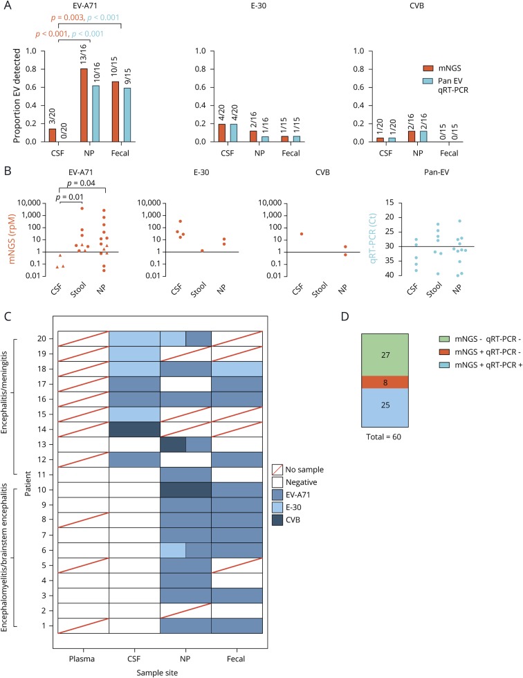
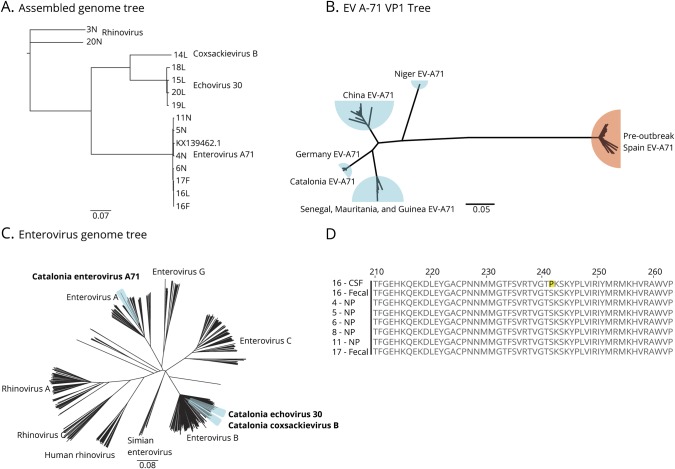
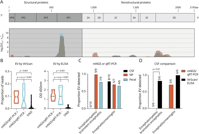
Objective In 2016, Catalonia experienced a pediatric brainstem encephalitis outbreak caused by enterovirus A71 (EV-A71). Conventional testing identified EV in the periphery but rarely in CSF. Metagenomic next-generation sequencing (mNGS) and CSF pan-viral serology (VirScan) were deployed to enhance viral detection and characterization. Methods RNA was extracted from the CSF (n = 20), plasma (n = 9), stool (n = 15), and nasopharyngeal samples (n = 16) from 10 children with brainstem encephalitis and 10 children with meningitis or encephalitis. Pathogens were identified using mNGS. Available CSF from cases (n = 12) and pediatric other neurologic disease controls (n = 54) were analyzed with VirScan with a subset (n = 9 and n = 50) validated by ELISA. Results mNGS detected EV in all samples positive by quantitative reverse transcription polymerase chain reaction (qRT-PCR) (n = 25). In qRT-PCR-negative samples (n = 35), mNGS found virus in 23% (n = 8, 3 CSF samples). Overall, mNGS enhanced EV detection from 42% (25/60) to 57% (33/60) (p-value = 0.013). VirScan and ELISA increased detection to 92% (11/12) compared with 46% (4/12) for CSF mNGS and qRT-PCR (p-value = 0.023). Phylogenetic analysis confirmed the EV-A71 strain clustered with a neurovirulent German EV-A71. A single amino acid substitution (S241P) in the EVA71 VP1 protein was exclusive to the CNS in one subject. Conclusion mNGS with VirScan significantly increased the CNS detection of EVs relative to qRT-PCR, and the latter generated an antigenic profile of the acute EV-A71 immune response. Genomic analysis confirmed the close relation of the outbreak EV-A71 and neuroinvasive German EV-A71. A S241P substitution in VP1 was found exclusively in the CSF.
Overview
The claims and statistics of this paper haven’t been checked yet.
Run the check — free, no account needed. It reads the open-access text and checks the sources it cites.
Integrity signals
Registry
Cites
Statistics
Figures
Verification
Claims the paper makes
The claims and statistics of this paper haven’t been checked yet.
Run the check — free, no account needed. It reads the open-access text and checks the sources it cites.
Claims other papers make about this one
Each is checked against this paper’s own text: does it say what the citing paper says it does?
The cited source says what the claim attributes to it.
In the citing paper
“Another study used mNGS, VirScan, and serology and found a high rate of enterovirus detection (up to 92%) relative to routine qRT-PCR and mNGS (36).”
From Viruses Causing Aseptic Meningitis: A Tertiary Medical Center Experience With a Multiplex PCR Assay (2020) · cites this paper as [36]
Self-check
Percentages, sums and reported differences re-computed from the paper’s own numbers.
The self-check needs the paper’s full text.
Citations
Cites
SPAdes: A New Genome Assembly Algorithm and Its Applications to Single-Cell Sequencing
Bankevich, A., Nurk, S., Antipov, D., Gurevich, A. A., Dvorkin, M. V., Kulikov, A. S., Lesin, V. M., Nikolenko, S., Pham, S., Prjibelski, A. D., et al. (2012) · Journal of Computational Biology · doi:10.1089/cmb.2012.0021 · Cited by 26,312
Clinical Metagenomic Sequencing for Diagnosis of Meningitis and Encephalitis
Wilson, M. R., Sample, H. A., Zorn, K. C., Arevalo, S., Yu, G., Neuhaus, J., Federman, S., Stryke, D., Briggs, B., Langelier, C., et al. (2019) · New England Journal of Medicine · doi:10.1056/nejmoa1803396 · Cited by 1,067
Self-citationActionable Diagnosis of Neuroleptospirosis by Next-Generation Sequencing
Wilson, M. R., Naccache, S. N., Samayoa, E., Biagtan, M., Bashir, H., Yu, G., Salamat, S. M., Somasekar, S., Federman, S., Miller, S., et al. (2014) · New England Journal of Medicine · doi:10.1056/nejmoa1401268 · Cited by 977
Self-citationLeber, A. L., Everhart, K., Balada-Llasat, J.-M., Cullison, J., Daly, J., Holt, S., Lephart, P., Salimnia, H., Schreckenberger, P. C., DesJarlais, S., et al. (2016) · Journal of Clinical Microbiology · doi:10.1128/jcm.00730-16 · Cited by 609 · cited 2×
“The subjects 1–10 were negative by the BioFire FilmArray Meningitis/Encephalitis panel.(2) All EVpositive subjects (by clinical PCR) were genotyped at the Enterovirus Unit of the Spanish National Centre for Microbiology according to a previously described procedure.”
BackgroundMethodMiller, S. A., Naccache, S. N., Samayoa, E., Messacar, K., Arevalo, S., Federman, S., Stryke, D., Pham, E., Fung, B., Bolosky, W. J., et al. (2019) · Genome Research · doi:10.1101/gr.238170.118 · Cited by 593
Comprehensive serological profiling of human populations using a synthetic human virome
Xu, G. J., Kula, T., Xu, Q., Li, M. Z., Vernon, S. D., Ndung’u, T., Ruxrungtham, K., Sánchez, J., Brander, C., Chung, R. T., et al. (2015) · Science · doi:10.1126/science.aaa0698 · Cited by 487
Human P-selectin glycoprotein ligand-1 is a functional receptor for enterovirus 71
Nishimura, Y., Shimojima, M., Tano, Y., Miyamura, T., Wakita, T., & Shimizu, H. (2009) · Nature Medicine · doi:10.1038/nm.1961 · Cited by 415
Virus genomes reveal factors that spread and sustained the Ebola epidemic
Dudas, G., Carvalho, L. M., Bedford, T., Tatem, A. J., Baele, G., Faria, N. R., Park, D. J., Ladner, J. T., Arias, A., Asogun, D., et al. (2017) · Nature · doi:10.1038/nature22040 · Cited by 400
Greninger, A. L., Naccache, S. N., Messacar, K., Clayton, A., Yu, G., Somasekar, S., Federman, S., Stryke, D., Anderson, C., Yagi, S., et al. (2015) · The Lancet Infectious Diseases · doi:10.1016/s1473-3099(15)70093-9 · Cited by 387
Gu, W., Crawford, E. D., O’Donovan, B. D., Wilson, M. R., Chow, E. D., Retallack, H., & DeRisi, J. L. (2016) · Genome biology · doi:10.1186/s13059-016-0904-5 · Cited by 361
Self-citationChronic Meningitis Investigated via Metagenomic Next-Generation Sequencing
Wilson, M. R., O’Donovan, B. D., Gelfand, J. M., Sample, H. A., Chow, F. C., Betjemann, J. P., Shah, M. P., Richie, M. B., Gorman, M., Hajj‐Ali, R. A., et al. (2018) · JAMA Neurology · doi:10.1001/jamaneurol.2018.0463 · Cited by 305
Self-citationNaccache, S. N., Peggs, K. S., Mattes, F. M., Phadke, R., Garson, J. A., Grant, P., Samayoa, E., Federman, S., Miller, S., Lunn, M. P., et al. (2015) · Clinical Infectious Diseases · doi:10.1093/cid/ciu912 · Cited by 294
Frequency and Dynamics of Recombination within Different Species of Human Enteroviruses
Peter Simmonds, K., & Welch, J. (2005) · Journal of Virology · doi:10.1128/jvi.80.1.483-493.2006 · Cited by 269
Understanding the Direction of Bias in Studies of Diagnostic Test Accuracy
Kohn, M. A., Carpenter, C. R., & Newman, T. B. (2013) · Academic Emergency Medicine · doi:10.1111/acem.12255 · Cited by 247
Outbreak of Neurologic Enterovirus Type 71 Disease: A Diagnostic Challenge
Pérez‐Vélez, C. M., Anderson, M., Robinson, C. C., McFarland, E. J., Nix, W. A., Pallansch, M. A., Oberste, M. S., & Glodé, M. P. (2007) · Clinical Infectious Diseases · doi:10.1086/521895 · Cited by 204
Verstrepen, W. A., Kuhn, S., Kockx, M. M., Van De Vyvere, M. E., & Mertens, A. H. (2001) · Journal of Clinical Microbiology · doi:10.1128/jcm.39.11.4093-4096.2001 · Cited by 177
Leitch, E. C. M., Cabrerizo, M., Cardosa, J., Harvala, H., Ivanova, O. E., Koike, S., Kroes, A. C. M., Lukashev, A. N., Perera, D., Roivainen, M., et al. (2011) · Journal of Virology · doi:10.1128/jvi.06065-11 · Cited by 137
Self-citationA novel cause of chronic viral meningoencephalitis: C ache Valley virus
Wilson, M. R., Suan, D., Duggins, A., Schubert, R. D., Khan, L. M., Sample, H. A., Zorn, K. C., Hoffmann, A. R., Blick, A. K., Shingde, M. V., et al. (2017) · Annals of Neurology · doi:10.1002/ana.24982 · Cited by 133
Self-citationCabrerizo, M., Tarragó, D., Muñoz‐Almagro, C., del Amo, E., Domínguez‐Gil, M., Eiros, J. M., López-Miragaya, I., Pérez, C., Reina, J., Otero, A., et al. (2013) · Clinical Microbiology and Infection · doi:10.1111/1469-0691.12361 · Cited by 132
Self-citationPan-viral serology implicates enteroviruses in acute flaccid myelitis
Schubert, R. D., Hawes, I. A., Ramachandran, P., Ramesh, A., Crawford, E. D., Pak, J. E., Wu, W. C. H., Cheung, C. K., O’Donovan, B. D., Tato, C. M., et al. (2019) · Nature Medicine · doi:10.1038/s41591-019-0613-1 · Cited by 130
Self-citationNishimura, Y., Lee, H., Hafenstein, S. L., Kataoka, C., Wakita, T., Bergelson, J. M., & Shimizu, H. (2013) · PLoS Pathogens · doi:10.1371/journal.ppat.1003511 · Cited by 116
Messacar, K., Davizon, E. S., Osborne, C. M., Press, C. A., Schreiner, T., Martin, J. A., Messer, R., Maloney, J. A., Burakoff, A. W., Barnes, M., et al. (2019) · The Lancet Infectious Diseases · doi:10.1016/s1473-3099(19)30632-2 · Cited by 112
Severe paediatric conditions linked with EV-A71 and EV-D68, France, May to October 2016
Antona, D., Kossorotoff, M., Schuffenecker, I., Mirand, A., Leruez‐Ville, M., Bassi, C., Aubart, M., Moulin, F., Lévy-Bruhl, D., Henquell, C., et al. (2016) · Eurosurveillance · doi:10.2807/1560-7917.es.2016.21.46.30402 · Cited by 100
Cabrerizo, M., Echevarría, J. E., González, I., de Miguel, T., & Trallero, G. (2008) · Journal of Medical Virology · doi:10.1002/jmv.21197 · Cited by 95
Self-citationAntibodies to Enteroviruses in Cerebrospinal Fluid of Patients with Acute Flaccid Myelitis
Mishra, N., Ng, T. F. F., Marine, R. L., Jain, K., Ng, J., Thakkar, R., Caciula, A., Price, A., Garcia, J. A., Burns, J. C., et al. (2019) · mBio · doi:10.1128/mbio.01903-19 · Cited by 86
Casas‐Alba, D., de Sevilla, M. F., Valero, A., Fortuny, C., García‐García, J. J., Ortez, C., Muchart, J., Armangué, T., Jordán, I., Luaces, C., et al. (2017) · Clinical Microbiology and Infection · doi:10.1016/j.cmi.2017.03.016 · Cited by 80
Self-citationCell and tissue tropism of enterovirus 71 and other enteroviruses infections
Lin, J., & Shih, S. (2014) · Journal of Biomedical Science · doi:10.1186/1423-0127-21-18 · Cited by 75
Sun, L., Huanying, Z., Zheng, H.-Z., Xue, G., He, J., Da-wei, G., Min, K., Liu, Z., Chang-wen, K., Li, J., et al. (2011) · Japanese Journal of Infectious Diseases · doi:10.7883/yoken.64.13 · Cited by 57
González-Sanz, R., Casas‐Alba, D., Launes, C., Muñoz‐Almagro, C., Ruiz-García, M. M., Alonso, M., González-Abad, M. J., Megías, G., Rabella, N., del Cuerpo, M., et al. (2019) · Eurosurveillance · doi:10.2807/1560-7917.es.2019.24.7.1800089 · Cited by 54
Self-citationJohnson, T. P., Larman, H. B., Lee, M., Whitehead, S. S., Kowalak, J. A., Toro, C., Lau, C. C., Kim, J., Johnson, K. R., Reoma, L. B., et al. (2019) · Annals of Neurology · doi:10.1002/ana.25588 · Cited by 51
A severe pediatric infection with a novel enterovirus A71 strain, Thuringia, Germany
Karrasch, M., Fischer, E., Scholten, M., Sauerbrei, A., Henke, A., Renz, D. M., Mentzel, H., Böer, K., Böttcher, S., Diedrich, S., et al. (2016) · Journal of Clinical Virology · doi:10.1016/j.jcv.2016.09.007 · Cited by 44
Recombinant Enterovirus A71 Subgenogroup C1 Strains, Germany, 2015
Böttcher, S., Obermeier, P., Neubauer, K., & Diedrich, S. (2016) · Emerging infectious diseases · doi:10.3201/eid2210.160357 · Cited by 40
Midgley, S., Nielsen, A. G., Trebbien, R., Poulsen, M. W., Andersen, P. H., & Fischer, T. K. (2017) · Eurosurveillance · doi:10.2807/1560-7917.es.2017.22.26.30565 · Cited by 31
Launes, C., Casas-Alba, D., Fortuny, C., Valero-Rello, A., Cabrerizo, M., & Muñoz-Almagro, C. (2017) · Journal of Clinical Microbiology · doi:10.1128/jcm.01931-16 · Cited by 26
Self-citationGenetic Characterization of Enterovirus A71 Circulating in Africa
Fernández-García, M. D., Volle, R., Joffret, M., Sadeuh‐Mba, S. A., Gouandjika‐Vasilache, I., Kébé, O., Wiley, M. R., Majumdar, M., Simon‐Lorière, E., Sakuntabhai, A., et al. (2018) · Emerging infectious diseases · doi:10.3201/eid2404.171783 · Cited by 24
Mutations in VP1 and 5′-UTR affect enterovirus 71 virulence
Chang, C., Wu, S.-R., Chen, Y.-C., Lee, K.-J., Chung, N., Lu, Y.-J., Yu, S., Liu, C., & Chow, Y. (2018) · Scientific Reports · doi:10.1038/s41598-018-25091-7 · Cited by 24
Wilson, M. R., Fedewa, G., Stenglein, M. D., Olejnik, J., Rennick, L. J., Nambulli, S., Feldmann, F., Duprex, W. P., Connor, J. H., Mühlberger, E., et al. (2016) · mSystems · doi:10.1128/msystems.00058-16 · Cited by 17
Self-citationSelva, L., Martínez-Planas, A., García‐García, J. J., Casadevall, R., Cubells, C. L., & Muñoz‐Almagro, C. (2011) · European Journal of Clinical Microbiology & Infectious Diseases · doi:10.1007/s10096-011-1364-1 · Cited by 11
Self-citationAuthor Correction: Mutations in VP1 and 5′-UTR affect enterovirus 71 virulence
Chang, C., Wu, S.-R., Chen, Y.-C., Lee, K.-J., Chung, N., Lu, Y.-J., Yu, S., Liu, C., & Chow, Y. (2018) · Scientific Reports · doi:10.1038/s41598-018-26833-3 · Cited by 1
Cited by
Sorted by citations
Metagenomics for neurological infections — expanding our imagination
Ramachandran, P., & Wilson, M. R. (2020) · Nature Reviews Neurology · doi:10.1038/s41582-020-0374-y · Cited by 182
Self-citationGraff, K. E., Dominguez, S. R., & Messacar, K. (2021) · Journal of the Pediatric Infectious Diseases Society · doi:10.1093/jpids/piab067 · Cited by 44
Steffen, T. L., Stone, E. T., Hassert, M., Geerling, E., Grimberg, B. T., Espino, A. M., Pantoja, P., Climent, C., Hoft, D. F., George, S. L., et al. (2020) · doi:10.1101/2020.08.21.261727 · Cited by 25
Qu, C., Chen, Y., Ouyang, Y., Huang, W., Liu, F., Yan, L., Lu, R., Zeng, Y., & Liu, Z. (2022) · Frontiers in Neurology · doi:10.3389/fneur.2022.989280 · Cited by 34
Viruses Causing Aseptic Meningitis: A Tertiary Medical Center Experience With a Multiplex PCR Assay
Aldriweesh, M. A., Shafaay, E. A., Alwatban, S. M., Alkethami, O. M., Aljuraisi, F. N., Bosaeed, M., & Alharbi, N. K. (2020) · Frontiers in Neurology · doi:10.3389/fneur.2020.602267 · Cited by 30
“Another study used mNGS, VirScan, and serology and found a high rate of enterovirus detection (up to 92%) relative to routine qRT-PCR and mNGS (36).”
Result
Metadata
- Authors
- Leon, K. E., Schubert, R. D., Casas-Alba, D., Hawes, I. A., Ramachandran, P. S., Ramesh, A., Pak, J. E., Wu, W., Cheung, C. K., Crawford, E. D., Khan, L. M., Launes, C., Sample, H. A., Zorn, K. C., Cabrerizo, M., Valero-Rello, A., Langelier, C., Muñoz-Almagro, C., DeRisi, J. L., & Wilson, M. R.
- Affiliations
- University of California, San Francisco (US)
- From the Medical Scientist Training Program (K.E.L.), University of California, San Francisco; Biomedical Sciences Graduate Program (K.E.L., I.A.H.), University of California, San Francisco; Weill Institute for Neurosciences (R.D.S., I.A.H., P.S.R., A.R., M.R.W.), University of California, San Francisco; Department of Neurology (R.D.S., I.A.H., P.S.R., A.R., M.R.W.), University of California, San Francisco; Institut de Recerca Pediàtrica Hospital Sant Joan de Déu (D.C.-A., C.L., A.V.-R., C.M.-A.),...
- Corresponding author
- Wilson, Michael R.
- ORCID
- Venue
- Neurology Neuroimmunology & Neuroinflammation
- Published
- 2020-05
- Volume
- 7
- Issue
- 3
- ISSN
- 2332-7812 (ISSN-L)
- PMC
- PMC7136061
- OpenAlex
- W3010465504
- Cited by
- 32
- References
- 39
- Citations by year
- 2026: 4 · 2025: 3 · 2024: 5 · 2023: 6 · 2022: 4 · 2021: 6 · 2020: 4
- FWCI
- 0.66
- Type
- journal article
- Language
- en
- Update notices
- correction (2025-10-06)
- Open access
- gold
- doi.org
- Licence
- cc-by-nc-nd
- Locations
- Neurology Neuroimmunology & Neuroinflammation (publishedVersion, cc-by-nc-nd)
- PubMed Central (publishedVersion, cc-by-nc-nd)
- PubMed (publishedVersion)
- Hispana (publishedVersion, cc-by-nc-nd)
- Repository of Digital Objects for Teaching Research and Culture (University of Valencia) (submittedVersion, cc-by-nc)
- LA Referencia (Red Federada de Repositorios Institucionales de Publicaciones Científicas) (submittedVersion, cc-by-nc-nd)
- eScholarship (California Digital Library) (submittedVersion)
- eScholarship (California Digital Library) (submittedVersion, cc-by-nc-nd)
- Funders
- Multiple Sclerosis Society
- National Institutes of Health
- University of California, San Francisco
- European Regional Development Fund
- National Institute of General Medical Sciences (T32GM007618)
- National Institute of Neurological Disorders and Stroke (F31NS113432, K08NS096117)
- Topics
- Health Sciences › Medicine › Infectious Diseases › Infectious Encephalopathies and Encephalitis
- Health Sciences › Medicine › Infectious Diseases › Viral Infections and Immunology Research
- Health Sciences › Medicine › Public Health, Environmental and Occupational Health › Mosquito-borne diseases and control
- Keywords
- enterovirus 71, brainstem encephalitis, cerebrospinal fluid viral detection, metagenomic next-generation sequencing
- MeSH terms
- Brain Stem, Child, Preschool, Enterovirus Infections, Enzyme-Linked Immunosorbent Assay, Female, Humans, Infant, Male, Meningitis, Viral, Phylogeny, RNA, Viral, Cohort Studies, Sequence Analysis, RNA, Encephalitis, Viral, Reverse Transcriptase Polymerase Chain Reaction, Enterovirus A, Human, High-Throughput Nucleotide Sequencing
About these checks
Registry, retraction, Cites and statistics results are computed, not judged, with one exception: a printed reference that no index lists is read once by a model before it counts as a citation error. Claims are extracted by a model and kept only when their quote appears verbatim in the text; each cited source is then checked, with a model’s help, for whether it says what the claim relies on. Those verdicts can be wrong — read the claim’s quote and the cited source before relying on one.
Statistics and arithmetic are recomputed, not judged.
Confidence bands are shown only for fields calibrated on our annotated test set. None is calibrated yet, so no band is shown.
What counts as a claim ›How we measure confidence ›How we check this ›
Are you an author, or do you see a problem with this page? Request a review
