

The DOI matches its Crossref record; 5 of the 5 references it cites were found in a registry or an index.
International consensus diagnostic criteria for neuromyelitis optica spectrum disorders
Open accessAbstract
Neuromyelitis optica (NMO) is an inflammatory CNS syndrome distinct from multiple sclerosis (MS) that is associated with serum aquaporin-4 immunoglobulin G antibodies (AQP4-IgG). Prior NMO diagnostic criteria required optic nerve and spinal cord involvement but more restricted or more extensive CNS involvement may occur. The International Panel for NMO Diagnosis (IPND) was convened to develop revised diagnostic criteria using systematic literature reviews and electronic surveys to facilitate consensus. The new nomenclature defines the unifying term NMO spectrum disorders (NMOSD), which is stratified further by serologic testing (NMOSD with or without AQP4-IgG). The core clinical characteristics required for patients with NMOSD with AQP4-IgG include clinical syndromes or MRI findings related to optic nerve, spinal cord, area postrema, other brainstem, diencephalic, or cerebral presentations. More stringent clinical criteria, with additional neuroimaging findings, are required for diagnosis of NMOSD without AQP4-IgG or when serologic testing is unavailable. The IPND also proposed validation strategies and achieved consensus on pediatric NMOSD diagnosis and the concepts of monophasic NMOSD and opticospinal MS.
Overview
The claims and statistics of this paper haven’t been checked yet.
Run the check — free, no account needed. It reads the open-access text and checks the sources it cites.
Integrity signals
Registry
Cites
Statistics
Figures
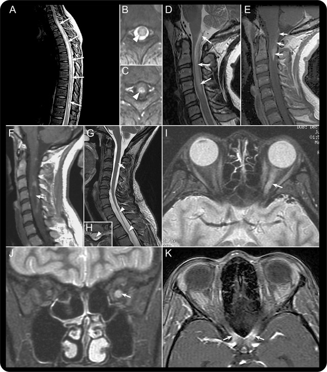
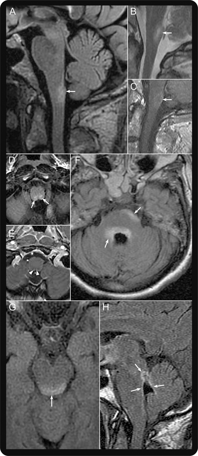
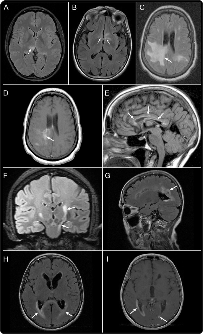
Verification
Claims the paper makes
The claims and statistics of this paper haven’t been checked yet.
Run the check — free, no account needed. It reads the open-access text and checks the sources it cites.
Claims other papers make about this one
Each is checked against this paper’s own text: does it say what the citing paper says it does?
The source backs part of the claim, not all of it — or backs it for a different population, condition or comparison.
In the citing paper
“Although CSF examination has been de-emphasized in successive iterations of the McDonald Criteria, it remains a valuable diagnostic test. 57”
From Diagnosis of multiple sclerosis: 2017 revisions of the McDonald criteria (2018) · cites this paper as [57]
Experimental — accuracy not yet measuredThe cited source says what the claim attributes to it.
In the citing paper
“A major discovery was the association between neuromyelitis optica spectrum disorder (NMOSD) and serum aquaporin-4 IgG (AQP4-IgG), confirming that NMOSD is a different disease from MS requiring distinct treatments.4”
From Diagnosis of multiple sclerosis: progress and challenges (2017) · cites this paper as [4]
The cited source says what the claim attributes to it.
In the citing paper
“NMOSD is an inflammatory astrocytopathy of the CNS with clinical and radiological features that overlap with MS. 4”
From Diagnosis of multiple sclerosis: progress and challenges (2017) · cites this paper as [4]
Self-check
Percentages, sums and reported differences re-computed from the paper’s own numbers.
The self-check needs the paper’s full text.
Citations
Cites
Krupp, L. B., Tardieu, M., Amato, M. P., Banwell, B., Chitnis, T., Dale, R. C., Ghezzi, A., Hintzen, R., Kornberg, A., Pohl, D., et al. (2013) · Multiple Sclerosis Journal · doi:10.1177/1352458513484547 · Cited by 1,130
Self-citationDistinction between MOG antibody-positive and AQP4 antibody-positive NMO spectrum disorders
Sato, D. K., Callegaro, D., Lana-Peixoto, M. A., Waters, P. J., Jorge, F. M. D. H., Takahashi, T., Nakashima, I., Apostolos-Pereira, S. L., Talim, N., Simm, R. F., et al. (2014) · Neurology · doi:10.1212/wnl.0000000000000101 · Cited by 850
Serologic diagnosis of NMO: A multicenter comparison of aquaporin-4-IgG assays
Waters, P. J., McKeon, A., Leite, M. I., Rajasekharan, S., Lennon, V. A., Villalobos, A., Palace, J., Mandrekar, J. N., Vincent, A., Bar-Or, A., et al. (2012) · Neurology · doi:10.1212/wnl.0b013e318248dec1 · Cited by 526
Self-citationNeuromyelitis optica unique area postrema lesions: Nausea, vomiting, and pathogenic implications
Popescu, B. F. G., Lennon, V. A., Parisi, J. E., Howe, C. L., Weigand, S. D., Cabrera-Gómez, J. A., Newell, K., Mandler, R. N., Pittock, S. J., Weinshenker, B. G., et al. (2011) · Neurology · doi:10.1212/wnl.0b013e318214332c · Cited by 296
Self-citationDistinction of seropositive NMO spectrum disorder and MS brain lesion distribution
Matthews, L., Marasco, R., Jenkinson, M., Küker, W., Luppe, S., Leite, M. I., Giorgio, A., De Stefano, N., Robertson, N., Johansen-Berg, H., et al. (2013) · Neurology · doi:10.1212/wnl.0b013e3182887957 · Cited by 225
Research works printed but not linked: 55
- 10.1016/S1474-4422(07)70216-8 · DOI
- 10.1212/01.wnl.0000216139.44259.74 · DOI
- 10.1186/1742-2094-10-8 · DOI
- 10.1212/WNL.53.5.1107 · DOI
- 10.1016/S0140-6736(04)17551-X · DOI · Paper page
- 10.1084/jem.20050304 · DOI · Paper page
- 10.1016/S1474-4422(12)70133-3 · DOI
- 10.1212/WNL.0b013e3182190f74 · DOI
- 10.1016/j.jns.2007.06.044 · DOI
- 10.1007/s00415-009-0009-0 · DOI
- 10.1212/WNL.26.6_Part_2.8 · DOI
- 10.1111/bpa.12084 · DOI
- 10.1212/WNL.0b013e318296ea08 · DOI
- 10.1016/j.msard.2012.06.002 · DOI · Paper page
- 10.1001/archneurol.2011.216 · DOI
- 10.1177/1352458511431973 · DOI
- 10.1177/1352458506070732 · DOI
- 10.1212/01.wnl.0000303817.82134.da · DOI
- 10.1002/ana.20770 · DOI
- 10.1136/jnnp-2011-300403 · DOI
- 10.1212/01.wnl.0000183151.19351.82 · DOI
- 10.1002/ana.22121 · DOI
- 10.1177/1352458513507822 · DOI
- 10.1191/1352458505ms1200cr · DOI
- 10.1212/WNL.48.1.58 · DOI
- 10.1177/1352458511404917 · DOI
- 10.1212/01.wnl.0000343001.36493.ae · DOI
- 10.1001/archneurol.2012.112 · DOI
- 10.1212/01.wnl.0000254502.87233.9a · DOI
- 10.1001/jamaneurol.2014.2137 · DOI · Paper page
- 10.1177/1352458510376640 · DOI
- 10.1177/1352458513506953 · DOI
- 10.1001/archneurol.2011.949 · DOI
- 10.1001/archneur.63.10.1398 · DOI
- 10.1186/1742-2094-9-14 · DOI
- 10.1001/archneur.63.3.390 · DOI
- 10.1001/archneur.63.7.964 · DOI
- 10.1177/1352458509103301 · DOI
- 10.1001/archneurol.2009.152 · DOI
- 10.1016/j.jns.2010.12.001 · DOI
- 10.1111/cen3.12107 · DOI
- 10.1001/jamaneurol.2014.1581 · DOI · Paper page
- 10.1212/WNL.0b013e318296e917 · DOI
- 10.1212/WNL.0b013e31826aac4e · DOI · Paper page
- 10.1001/jamaneurol.2013.5857 · DOI · Paper page
- 10.1093/brain/awn240 · DOI
- 10.1212/WNL.0b013e3182a6cb5c · DOI
- 10.1212/WNL.0b013e318211c379 · DOI
- 10.1016/j.jns.2011.03.038 · DOI · Paper page
- 10.1212/WNL.0b013e3181f39a66 · DOI
- 10.1212/01.wnl.0000284600.80782.d5 · DOI
- 10.1212/01.wnl.0000314832.24682.c6 · DOI
- 10.1177/1352458511431077 · DOI
- 10.1111/bpa.12099 · DOI
- 10.1212/WNL.0000000000001126 · DOI
Cited by
Sorted by citations
Diagnosis of multiple sclerosis: 2017 revisions of the McDonald criteria
Thompson, A. J., Banwell, B. L., Barkhof, F., Carroll, W. M., Coetzee, T., Comi, G., Correale, J., Fazekas, F., Filippi, M., Freedman, M. S., et al. (2018) · The Lancet Neurology · doi:10.1016/s1474-4422(17)30470-2 · Cited by 8,167
Self-citationDiagnosis of multiple sclerosis: progress and challenges
Brownlee, W. J., Hardy, T. A., Fazekas, F., & Miller, D. H. (2017) · The Lancet · doi:10.1016/s0140-6736(16)30959-x · Cited by 701
Traboulsee, A., Greenberg, B. M., Bennett, J. L., Szczechowski, L., Fox, E., Shkrobot, S., Yamamura, T., Terada, Y., Kawata, Y., Wright, P., et al. (2020) · The Lancet Neurology · doi:10.1016/s1474-4422(20)30078-8 · Cited by 513
Self-citationZhang, C., Zhang, M., Qiu, W., Ma, H., Zhang, X., Zhu, Z., Yang, C.-S., Jia, D., Zhang, T.-X., Yuan, M., et al. (2020) · The Lancet Neurology · doi:10.1016/s1474-4422(20)30070-3 · Cited by 287
Self-citationAutoantibodies to MOG in a distinct subgroup of adult multiple sclerosis
Spadaro, M., Gerdes, L. A., Krumbholz, M., Ertl-Wagner, B., Thaler, F. S., Schuh, E., Metz, I., Blaschek, A., Dick, A., Brück, W., et al. (2016) · Neurology Neuroimmunology & Neuroinflammation · doi:10.1212/nxi.0000000000000257 · Cited by 206
Metadata
- Authors
- Wingerchuk, D. M., Banwell, B., Bennett, J. L., Cabre, P., Carroll, W., Chitnis, T., de Seze, J., Fujihara, K., Greenberg, B., Jacob, A., Jarius, S., Lana-Peixoto, M., Levy, M., Simon, J. H., Tenembaum, S., Traboulsee, A. L., Waters, P., Wellik, K. E., & Weinshenker, B. G.
- Affiliations
- Mayo Clinic (US)
- From the Departments of Neurology (D.M.W.) and Library Services (K.E.W.), Mayo Clinic, Scottsdale, AZ; the Children's Hospital of Philadelphia (B.B.), PA; the Departments of Neurology and Ophthalmology (J.L.B.), University of Colorado Denver, Aurora; the Service de Neurologie (P.C.), Centre Hospitalier Universitaire de Fort de France, Fort-de-France, Martinique; Department of Neurology (W.C.), Sir Charles Gairdner Hospital, Perth, Australia; the Department of Neurology (T.C.), Massachusetts General...
- Corresponding author
- Wingerchuk, Dean M.
- Weinshenker, Brian G.
- ORCID
- Venue
- Neurology
- Published
- 2015-07-14
- Volume
- 85
- Issue
- 2
- Pages
- 177-189
- ISSN
- 0028-3878 (ISSN-L)
- 1526-632X
- PMC
- PMC4515040
- OpenAlex
- W2129229339
- Cited by
- 4,649
- References
- 5
- Citations by year
- 2026: 434 · 2025: 457 · 2024: 496 · 2023: 432 · 2022: 476 · 2021: 524 · 2020: 477 · 2019: 388 · 2018: 353 · 2017: 308 · 2016: 241 · 2015: 56
- FWCI
- 253.64
- Type
- journal article
- Language
- en
- Open access
- hybrid
- doi.org
- Licence
- cc-by-nc-nd
- Locations
- Neurology (publishedVersion, cc-by-nc-nd)
- Europe PMC (PubMed Central) (publishedVersion, cc-by-nc-nd)
- PubMed Central (publishedVersion, cc-by-nc-nd)
- PubMed (publishedVersion)
- eScholarship (California Digital Library) (submittedVersion, cc-by-nc-nd)
- HAL (submittedVersion, cc-by-nc-nd)
- eScholarship (California Digital Library) (submittedVersion)
- Funders
- GlaxoSmithKline
- Sanofi
- Biogen
- Viropharma
- Teva Pharmaceutical Industries
- Alexion Pharmaceuticals
- Guthy-Jackson Charitable Foundation
- European Committee for Treatment and Research in Multiple Sclerosis
- Acorda Therapeutics
- Multiple Sclerosis Society of Canada
- Eisai
- Ministry of Education, Science and Technology
- Astellas Pharma
- Teijin Pharma
- Mitsubishi Tanabe Pharma Corporation
- Ono Pharmaceutical
- National Institutes of Health
- Novartis Pharma
- Chugai Pharmaceutical
- National Eye Institute (R01EY022936)
- National Institute of Neurological Disorders and Stroke (K08NS078555)
- Daiichi Sankyo Europe
- Topics
- Health Sciences › Medicine › Clinical Neurology › Multiple Sclerosis Research Studies
- Health Sciences › Medicine › Clinical Neurology › Peripheral Neuropathies and Disorders
- Health Sciences › Medicine › Ophthalmology › Ocular Diseases and Behçet’s Syndrome
- Keywords
- neuromyelitis optica spectrum disorder, neuromyelitis optica, diagnostic criteria, aquaporin-4 antibodies, opticospinal multiple sclerosis, neuroimaging findings, area postrema, brainstem involvement, serologic testing
- MeSH terms
- Humans, Neuromyelitis Optica
About these checks
Registry, retraction, Cites and statistics results are computed, not judged, with one exception: a printed reference that no index lists is read once by a model before it counts as a citation error. Claims are extracted by a model and kept only when their quote appears verbatim in the text; each cited source is then checked, with a model’s help, for whether it says what the claim relies on. Those verdicts can be wrong — read the claim’s quote and the cited source before relying on one.
Statistics and arithmetic are recomputed, not judged.
Confidence bands are shown only for fields calibrated on our annotated test set. None is calibrated yet, so no band is shown.
What counts as a claim ›How we measure confidence ›How we check this ›
Are you an author, or do you see a problem with this page? Request a review
