

The DOI matches its Crossref record; 40 of the 41 references it cites were found in a registry or an index.
Dysregulated mitophagy and mitochondrial organization in optic atrophy due to OPA1 mutations
Open accessAbstract
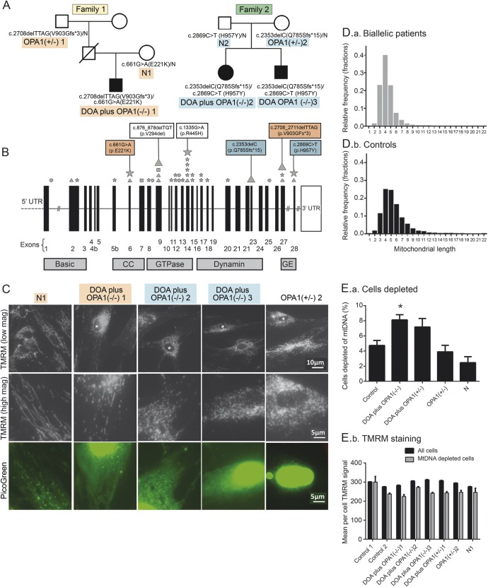
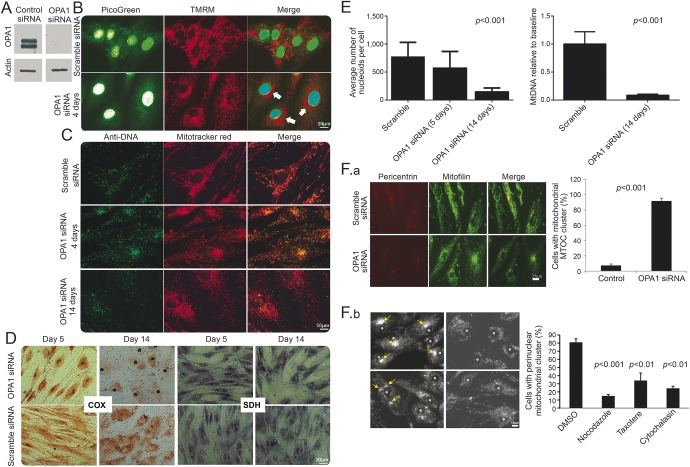
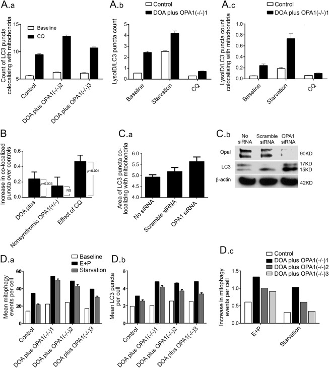
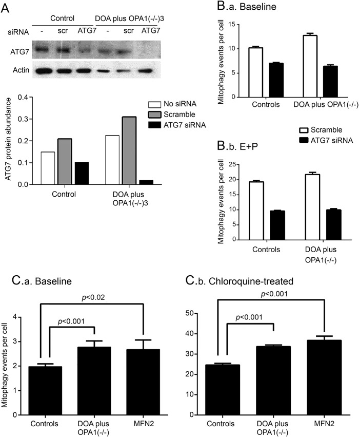
OBJECTIVE: To investigate mitophagy in 5 patients with severe dominantly inherited optic atrophy (DOA), caused by depletion of OPA1 (a protein that is essential for mitochondrial fusion), compared with healthy controls. METHODS: Patients with severe DOA (DOA plus) had peripheral neuropathy, cognitive regression, and epilepsy in addition to loss of vision. We quantified mitophagy in dermal fibroblasts, using 2 high throughput imaging systems, by visualizing colocalization of mitochondrial fragments with engulfing autophagosomes. RESULTS: Fibroblasts from 3 biallelic OPA1(-/-) patients with severe DOA had increased mitochondrial fragmentation and mitochondrial DNA (mtDNA)-depleted cells due to decreased levels of OPA1 protein. Similarly, in siRNA-treated control fibroblasts, profound OPA1 knockdown caused mitochondrial fragmentation, loss of mtDNA, impaired mitochondrial function, and mitochondrial mislocalization. Compared to controls, basal mitophagy (abundance of autophagosomes colocalizing with mitochondria) was increased in (1) biallelic patients, (2) monoallelic patients with DOA plus, and (3) OPA1 siRNA-treated control cultures. Mitophagic flux was also increased. Genetic knockdown of the mitophagy protein ATG7 confirmed this by eliminating differences between patient and control fibroblasts. CONCLUSIONS: We demonstrated increased mitophagy and excessive mitochondrial fragmentation in primary human cultures associated with DOA plus due to biallelic OPA1 mutations. We previously found that increased mitophagy (mitochondrial recycling) was associated with visual loss in another mitochondrial optic neuropathy, Leber hereditary optic neuropathy (LHON). Combined with our LHON findings, this implicates excessive mitochondrial fragmentation, dysregulated mitophagy, and impaired response to energetic stress in the pathogenesis of mitochondrial optic neuropathies, potentially linked with mitochondrial mislocalization and mtDNA depletion.
Overview
The claims and statistics of this paper haven’t been checked yet.
Run the check — free, no account needed. It reads the open-access text and checks the sources it cites.
Integrity signals
Registry
Cites
Statistics
Figures
Verification
Claims the paper makes
The claims and statistics of this paper haven’t been checked yet.
Run the check — free, no account needed. It reads the open-access text and checks the sources it cites.
Claims other papers make about this one
Each is checked against this paper’s own text: does it say what the citing paper says it does?
In the citing paper
“Opa1 deficiency has been reported to induce low mtDNA copy number and nucleoids abundance in human fibroblasts, which suggests that those alterations are not just restricted to muscle cells (Liao et al, 2017).”
From Mitochondrial DNA and TLR9 drive muscle inflammation upon Opa1 deficiency (2018) · cites this paper as Liao et al, 2017
Self-check
Percentages, sums and reported differences re-computed from the paper’s own numbers.
The self-check needs the paper’s full text.
Citations
Cites
Guidelines for the use and interpretation of assays for monitoring autophagy (3rd edition).
Klionsky, D. J., Abdelmohsen, K., Abe, A., Abedin, M. J., Abeliovich, H., Acevedo Arozena, A., Adachi, H., Adams, C. M., Adams, P. D., Adeli, K., et al. (2016) · Autophagy · doi:10.1080/15548627.2015.1100356 · Cited by 5,894
Parkin is recruited selectively to impaired mitochondria and promotes their autophagy
Narendra, D., Tanaka, A., Suen, D.-F., & Youle, R. J. (2008) · The Journal of Cell Biology · doi:10.1083/jcb.200809125 · Cited by 4,026
Fission and selective fusion govern mitochondrial segregation and elimination by autophagy
Twig, G., Elorza, A., Molina, A. J. A., Mohamed, H., Wikstrom, J. D., Walzer, G., Stiles, L., Haigh, S. E., Katz, S., Las, G., et al. (2008) · The EMBO Journal · doi:10.1038/sj.emboj.7601963 · Cited by 3,082
PINK1 Is Selectively Stabilized on Impaired Mitochondria to Activate Parkin
Narendra, D. P., Jin, S. M., Tanaka, A., Suen, D.-F., Gautier, C. A., Shen, J., Cookson, M. R., & Youle, R. J. (2010) · PLoS Biology · doi:10.1371/journal.pbio.1000298 · Cited by 2,944
During autophagy mitochondria elongate, are spared from degradation and sustain cell viability
Gomes, L. C., Benedetto, G. D., & Scorrano, L. (2011) · Nature Cell Biology · doi:10.1038/ncb2220 · Cited by 1,735
Gegg, M. E., Cooper, J. M., Chau, K.-Y., Rojo, M., Schapira, A. H. V., & Taanman, J.-W. (2010) · Human Molecular Genetics · doi:10.1093/hmg/ddq419 · Cited by 910
Lysosomal positioning coordinates cellular nutrient responses
Korolchuk, V. I., Saiki, S., Lichtenberg, M., Siddiqi, F. H., Roberts, E. A., Imarisio, S., Jahreiss, L., Sarkar, S., Futter, M., Menzies, F. M., et al. (2011) · Nature Cell Biology · doi:10.1038/ncb2204 · Cited by 891
Ebneth, A., Godemann, R., Stamer, K., Illenberger, S., Trinczek, B., Mandelkow, E.-M., & Mandelkow, E. (1998) · The Journal of Cell Biology · doi:10.1083/jcb.143.3.777 · Cited by 820
SLP‐2 is required for stress‐induced mitochondrial hyperfusion
Tondera, D., Grandemange, S., Jourdain, A. A., Karbowski, M., Mattenberger, Y., Herzig, S., Cruz, S. D., Clerc, P., Lauria, I., Merkwirth, C., et al. (2009) · The EMBO Journal · doi:10.1038/emboj.2009.89 · Cited by 796
OPA1 mutations induce mitochondrial DNA instability and optic atrophy 'plus' phenotypes
Amati-Bonneau, P., Valentino, M. L., Reynier, P., Gallardo, M. E., Bornstein, B., Boissiere, A., Campos, Y., Rivera, H., de la Aleja, J. G., Carroccia, R., et al. (2008) · Brain · doi:10.1093/brain/awm298 · Cited by 520
Cited by
Sorted by citations
Mitochondrial dynamics in health and disease
Yapa, N. M. B., Lisnyak, V., Reljić, B., & Ryan, M. T. (2021) · FEBS Letters · doi:10.1002/1873-3468.14077 · Cited by 323
Mitochondrial diseases: from molecular mechanisms to therapeutic advances
Wen, H., Deng, H., Li, B., Chen, J., Zhu, J., Zhang, X., Yoshida, S., & Zhou, Y. (2025) · Signal Transduction and Targeted Therapy · doi:10.1038/s41392-024-02044-3 · Cited by 284
Mitochondrial DNA and TLR9 drive muscle inflammation upon Opa1 deficiency
Rodríguez‐Nuevo, A., Díaz‐Ramos, A., Noguera, E., Díaz‐Sáez, F., Duran, X., Muñoz, J. P., Romero, M., Plana, N., Sebastián, D., Tezze, C., et al. (2018) · The EMBO Journal · doi:10.15252/embj.201796553 · Cited by 207
Regulation of Mammalian Mitochondrial Dynamics: Opportunities and Challenges
Yu, R., Lendahl, U., Nistér, M., & Zhao, J. (2020) · Frontiers in Endocrinology · doi:10.3389/fendo.2020.00374 · Cited by 183
Eight human OPA1 isoforms, long and short: What are they for?
Dotto, V. D., Fogazza, M., Carelli, V., Rugolo, M., & Zanna, C. (2018) · Biochimica et Biophysica Acta (BBA) - Bioenergetics · doi:10.1016/j.bbabio.2018.01.005 · Cited by 174
Xian, H., & Liou, Y. (2020) · Cell Death and Differentiation · doi:10.1038/s41418-020-00657-z · Cited by 177
MitophAging: Mitophagy in Aging and Disease
Bakula, D., & Scheibye‐Knudsen, M. (2020) · Frontiers in Cell and Developmental Biology · doi:10.3389/fcell.2020.00239 · Cited by 152
The Balance of MFN2 and OPA1 in Mitochondrial Dynamics, Cellular Homeostasis, and Disease
Zanfardino, P., Amati, A., Perrone, M., & Petruzzella, V. (2025) · Biomolecules · doi:10.3390/biom15030433 · Cited by 129
Exercise effects on physiological function during aging
Radak, Z. O., Torma, F., Berkes, I., Goto, S., Mimura, T., Pósa, A., Balogh, L., Boldogh, I. R., Suzuki, K., Higuchi, M., et al. (2018) · Free Radical Biology and Medicine · doi:10.1016/j.freeradbiomed.2018.10.444 · Cited by 118
Optic neuropathies: the tip of the neurodegeneration iceberg
Carelli, V., Morgia, C. L., Ross‐Cisneros, F. N., & Sadun, A. A. (2017) · Human Molecular Genetics · doi:10.1093/hmg/ddx273 · Cited by 116
Arribat, Y., Broskey, N. T., Greggio, C., Boutant, M., Conde Alonso, S., Kulkarni, S. S., Lagarrigue, S., Carnero, E. A., Besson, C., Cantó, C., et al. (2019) · Acta Physiologica · doi:10.1111/apha.13179 · Cited by 112
Dominant optic atrophy: Culprit mitochondria in the optic nerve
Lenaers, G., Neutzner, A., Dantec, Y. L., Jüschke, C., Xiao, T., Decembrini, S., Swirski, S., Kieninger, S., Ağca, C., Kim, U. S., et al. (2020) · Progress in Retinal and Eye Research · doi:10.1016/j.preteyeres.2020.100935 · Cited by 113
Self-citationNewman, N. J., Yu-Wai-Man, P., Biousse, V., & Carelli, V. (2023) · The Lancet Neurology · doi:10.1016/s1474-4422(22)00174-0 · Cited by 97
Self-citationA comparative map of macroautophagy and mitophagy in the vertebrate eye
McWilliams, T. G., Prescott, A. R., Villarejo‐Zori, B., Ball, G., Boya, P., & Ganley, I. G. (2019) · Autophagy · doi:10.1080/15548627.2019.1580509 · Cited by 81
OPA1: How much do we know to approach therapy?
Dotto, V. D., Fogazza, M., Lenaers, G., Rugolo, M., Carelli, V., & Zanna, C. (2018) · Pharmacological Research · doi:10.1016/j.phrs.2018.02.018 · Cited by 76
Zhang, J., Ji, Y., Lu, Y., Fu, R., Xu, M., Liu, X., & Guan, M. (2018) · Human Molecular Genetics · doi:10.1093/hmg/ddy107 · Cited by 73
Pathological mitophagy disrupts mitochondrial homeostasis in Leber’s hereditary optic neuropathy
Danese, A., Patergnani, S., Maresca, A., Peron, C., Raimondi, A., Caporali, L., Marchi, S., Morgia, C. L., Dotto, V. D., Zanna, C., et al. (2022) · Cell Reports · doi:10.1016/j.celrep.2022.111124 · Cited by 73
A hitchhiker’s guide to mitochondrial quantification
Hemel, I. M. G. M., Engelen, B. P. H., Luber, N., & Gerards, M. (2021) · Mitochondrion · doi:10.1016/j.mito.2021.06.005 · Cited by 73
Inherited eye-related disorders due to mitochondrial dysfunction
Yu‐Wai‐Man, P., & Newman, N. J. (2017) · Human Molecular Genetics · doi:10.1093/hmg/ddx182 · Cited by 63
Self-citationCai, W., Chen, Y., Shi, L., Cheng, H. R., Banda, I., Ji, Y., Wang, Y., Li, X., Mao, Y., Zhang, D. F., et al. (2019) · Oxidative Medicine and Cellular Longevity · doi:10.1155/2019/4101738 · Cited by 61
Mitophagy: An Emerging Target in Ocular Pathology
Skeie, J. M., Nishimura, D. Y., Wang, C. L., Schmidt, G. A., Aldrich, B. T., & Greiner, M. A. (2021) · Investigative Ophthalmology & Visual Science · doi:10.1167/iovs.62.3.22 · Cited by 55
Oxidative Stress in Optic Neuropathies
Sanz‐Morello, B., Ahmadi, H. Z., Vohra, R., Saruhanian, S., Freude, K., Hamann, S., & Kolko, M. (2021) · Antioxidants · doi:10.3390/antiox10101538 · Cited by 53
New insights into the role of autophagy in retinal and eye diseases.
Villarejo-Zori, B., Jiménez-Loygorri, J. I., Zapata-Muñoz, J., Bell, K., & Boya, P. (2021) · Molecular Aspects of Medicine · doi:10.1016/j.mam.2021.101038 · Cited by 50
Zheng, J., Akbari, M., Schirmer, C., Reynaert, M., Loyens, A., Lefebvre, B., Buée, L., Croteau, D. L., Galas, M., & Bohr, V. A. (2020) · Acta Neuropathologica Communications · doi:10.1186/s40478-020-00896-8 · Cited by 54
Autophagy controls the pathogenicity of OPA 1 mutations in dominant optic atrophy
Kane, M. S., Alban, J., Desquiret‐Dumas, V., Guéguen, N., Ishak, L., Ferré, M., Amati‐Bonneau, P., Procaccio, V., Bonneau, D., Lenaers, G., et al. (2017) · Journal of Cellular and Molecular Medicine · doi:10.1111/jcmm.13149 · Cited by 51
Mitochondrial optic neuropathies
Carelli, V., Morgia, C. L., & Yu‐Wai‐Man, P. (2023) · Handbook of clinical neurology · doi:10.1016/b978-0-12-821751-1.00010-5 · Cited by 47
Self-citationZhang, J., Liu, X., Liang, X., Lu, Y., Zhu, L., Fu, R., Ji, Y., Fan, W., Chen, J., Lin, B., et al. (2017) · Scientific Reports · doi:10.1038/s41598-017-05571-y · Cited by 48
Broad activation of the Parkin pathway induces synaptic mitochondrial deficits in early tauopathy
Jeong, Y. Y., Han, S., Jia, N., Zhang, M., Sheshadri, P., Tammineni, P., Cheung, J., Nissenbaum, M., Baskar, S. S., Kwan, K. Y., et al. (2021) · Brain · doi:10.1093/brain/awab243 · Cited by 39
Lynch, D. S., Loh, S. H. Y., Harley, J., Noyce, A. J., Martins, L. M., Wood, N. W., Houlden, H., & Plun‐Favreau, H. (2017) · Neurology Genetics · doi:10.1212/nxg.0000000000000188 · Cited by 38
Mitochondrial Dynamics and VMP1-Related Selective Mitophagy in Experimental Acute Pancreatitis
Vanasco, V., Ropolo, A., Grasso, D., Ojeda, D. S., Garcia, M. N., Vico, T. A., Orquera, T., Quarleri, J. F., Álvarez, S., & Vaccaro, M. I. (2021) · Frontiers in Cell and Developmental Biology · doi:10.3389/fcell.2021.640094 · Cited by 35
Hinton, A. O., Jr., Katti, P., Mungai, M., Hall, D. D., Koval, O. M., Shao, J., Vue, Z., Lopez, E. G., Rostami, R., Neikirk, K., et al. (2024) · Journal of Cellular Physiology · doi:10.1002/jcp.31204 · Cited by 38
Sun, S., Erchova, I. A., Sengpiel, F., & Votruba, M. (2020) · Investigative Ophthalmology & Visual Science · doi:10.1167/iovs.61.6.42 · Cited by 32
Self-citationZheng, J., Croteau, D. L., Bohr, V. A., & Akbari, M. (2019) · Nucleic Acids Research · doi:10.1093/nar/gkz083 · Cited by 32
Stem cell modeling of mitochondrial parkinsonism reveals key functions of OPA1
Jonikas, M., Madill, M., Mathy, A., Zekoll, T., Zois, C. E., Wigfield, S., Kurzawa‐Akanbi, M., Browne, C., Sims, D., Chinnery, P. F., et al. (2018) · Annals of Neurology · doi:10.1002/ana.25221 · Cited by 30
Cristae junction as a fundamental switchboard for mitochondrial ion signaling and bioenergetics
Gottschalk, B., Madreiter‐Sokolowski, C. T., & Graier, W. F. (2021) · Cell Calcium · doi:10.1016/j.ceca.2021.102517 · Cited by 30
Broad activation of the Parkin pathway induces synaptic mitochondrial deficits in early tauopathy
Jeong, Y. Y., Han, S., Jia, N., Zhang, M., Sheshadri, P., Tammineni, P., Cheung, J., Nissenbaum, M., Baskar, S. S., Kwan, K. Y., et al. (2022) · UCL Discovery (University College London) · Cited by 32
The loss of OPA1 accelerates intervertebral disc degeneration and osteoarthritis in aged mice
Madhu, V., Hernandez-Meadows, M., Coleman, A., Sao, K., Inguito, K. L., Haslam, O., Boneski, P. K., Sesaki, H., Barve, R. A., Collins, J. A., et al. (2025) · Nature Communications · doi:10.1038/s41467-025-60933-9 · Cited by 38
Dominant Optic Atrophy (DOA): Modeling the Kaleidoscopic Roles of OPA1 in Mitochondrial Homeostasis
Dotto, V. D., & Carelli, V. (2021) · Frontiers in Neurology · doi:10.3389/fneur.2021.681326 · Cited by 28
Dynamic features of human mitochondrial DNA maintenance and transcription
Akbari, M., Nilsen, H. L., & Montaldo, N. P. (2022) · Frontiers in Cell and Developmental Biology · doi:10.3389/fcell.2022.984245 · Cited by 27
The Role of Mitophagy in Glaucomatous Neurodegeneration
Stavropoulos, D., Grewal, M. K., Petriti, B., Chau, K., Hammond, C. J., Garway‐Heath, D. F., & Lascaratos, G. (2023) · Cells · doi:10.3390/cells12151969 · Cited by 27
Trifunov, S., Pyle, A., Valentino, M. L., Liguori, R., Yu‐Wai‐Man, P., Burté, F., Duff, J., Kleinle, S., Diebold, I., Rugolo, M., et al. (2018) · Scientific Reports · doi:10.1038/s41598-018-30143-z · Cited by 26
Self-citationZhang, X., Henneman, N. F., Girardot, P. E., Sellers, J. T., Chrenek, M. A., Li, Y., Wang, J., Brenner, C., Nickerson, J. M., & Boatright, J. H. (2020) · Investigative Ophthalmology & Visual Science · doi:10.1167/iovs.61.10.47 · Cited by 26
Regulation of mitochondrial cargo-selective autophagy by posttranslational modifications
Lechado‐Terradas, A., Zittlau, K. I., Maček, B., Fraiberg, M., Elazar, Z., & Kahle, P. J. (2021) · Journal of Biological Chemistry · doi:10.1016/j.jbc.2021.101339 · Cited by 27
Mitophagy: A Novel Therapeutic Target for Treating DN
Yang, M., Li, C., Yang, S., Xiao, Y., Chen, W., Gao, P., Jiang, N., Xiong, S., Wei, L., Zhang, Q., et al. (2020) · Current Medicinal Chemistry · doi:10.2174/0929867327666201006152656 · Cited by 26
Sladen, P. E., Jovanovic, K., Guarascio, R., Ottaviani, D., Salsbury, G., Novoselova, T. V., Chapple, J. P., Yu‐Wai‐Man, P., & Cheetham, M. E. (2022) · Human Molecular Genetics · doi:10.1093/hmg/ddac128 · Cited by 23
Self-citationMitochondrial disorders of the retinal ganglion cells and the optic nerve
Finsterer, J., Mancuso, M., Pareyson, D., Burgunder, J., & Klopstock, T. (2017) · Mitochondrion · doi:10.1016/j.mito.2017.10.003 · Cited by 24
Bevan, R. J., Williams, P. A., Waters, C. T., Thirgood, R. A., Mui, A., Seto, S., Good, M. A., Morgan, J. E., Votruba, M., & Erchova, I. A. (2020) · Brain Communications · doi:10.1093/braincomms/fcaa101 · Cited by 22
Self-citationThe Role of Mitochondria in Optic Atrophy With Autosomal Inheritance
Strachan, E. L., MacWhite-Begg, D., Crean, J. K. G., Reynolds, A., Kennedy, B. N., & O’Sullivan, N. C. (2021) · Frontiers in Neuroscience · doi:10.3389/fnins.2021.784987 · Cited by 22
Metabolic stroke in a patient with bi-allelic OPA1 mutations
Zerem, A., Yosovich, K., Rappaport, Y. C., Libzon, S., Blumkin, L., Ben‐Sira, L., Lev, D., & Lerman‐Sagie, T. (2019) · Metabolic Brain Disease · doi:10.1007/s11011-019-00415-2 · Cited by 22
Sharma, G., Zaman, M., Sabouny, R., Joel, M., Martino, D., Martino, D., de Koning, A. P. J., Shutt, T. E., & Shutt, T. E. (2021) · F1000Research · doi:10.12688/f1000research.53230.1 · Cited by 23
Petcherski, A., Trudeau, K. M., Wolf, D. M., Segawa, M., Lee, J., Taddeo, E. P., Deeney, J. T., & Liesa, M. (2018) · Journal of Molecular Biology · doi:10.1016/j.jmb.2018.10.020 · Cited by 19
Dynamic properties of mitochondria during human corticogenesis
Baum, T., & Gama, V. (2021) · Development · doi:10.1242/dev.194183 · Cited by 20
Gilea, A. I., Berti, C. C., Magistrati, M., di Punzio, G., Goffrini, P., Baruffini, E., & Dallabona, C. (2021) · Genes · doi:10.3390/genes12121866 · Cited by 18
Molecular Mechanisms behind Inherited Neurodegeneration of the Optic Nerve
Maresca, A., & Carelli, V. (2021) · Biomolecules · doi:10.3390/biom11040496 · Cited by 20
Optimized OPA1 Isoforms 1 and 7 Provide Therapeutic Benefit in Models of Mitochondrial Dysfunction
Maloney, D. M., Chadderton, N., Millington‐Ward, S., Palfi, A., Shortall, C., O’Byrne, J. J., Cassidy, L., Keegan, D., Humphries, P., Kenna, P. F., et al. (2020) · Frontiers in Neuroscience · doi:10.3389/fnins.2020.571479 · Cited by 16
Genetic Neuropathy Due to Impairments in Mitochondrial Dynamics
Sharma, G., Pfeffer, G., & Shutt, T. E. (2021) · Biology · doi:10.3390/biology10040268 · Cited by 18
From Transcriptomics to Treatment in Inherited Optic Neuropathies
Gilhooley, M. J., Owen, N., Moosajee, M., & Yu‐Wai‐Man, P. (2021) · Genes · doi:10.3390/genes12020147 · Cited by 17
Self-citationBiallelic Optic Atrophy 1 (OPA1) Related Disorder—Case Report and Literature Review
Othman, B. A. A., Ong, J. E., & Dumitrescu, A. V. (2022) · Genes · doi:10.3390/genes13061005 · Cited by 14
Diot, A., Agnew, T., Sanderson, J., Liao, C., Carver, J. G., das Neves, R. P., Gupta, R., Guo, Y., Waters, C. T., Seto, S., et al. (2018) · Frontiers in Cell and Developmental Biology · doi:10.3389/fcell.2018.00103 · Cited by 14
Self-citationThe Role of Rab Proteins in Mitophagy: Insights into Neurodegenerative Diseases
Shafique, A., Brughera, M., Lualdi, M., & Alberio, T. (2023) · International Journal of Molecular Sciences · doi:10.3390/ijms24076268 · Cited by 15
Zanfardino, P., Amati, A., Doccini, S., Cox, S. N., Tullo, A., Longo, G., D’Erchia, A. M., Picardi, E., Nesti, C., Santorelli, F. M., et al. (2024) · Human Molecular Genetics · doi:10.1093/hmg/ddae008 · Cited by 14
Whitehead, M., Harvey, J. P., Sladen, P. E., Becchi, G., Singh, K., Sun, Y. J., Burgoyne, T., Duchen, M. R., Yu‐Wai‐Man, P., & Cheetham, M. E. (2025) · Acta Neuropathologica Communications · doi:10.1186/s40478-025-01942-z · Cited by 13
Self-citationMitophagy: A Potential Target for Pressure Overload‐Induced Cardiac Remodelling
Shao, R., Li, J., Qu, T., Liao, Y., & Chen, M. (2022) · Oxidative Medicine and Cellular Longevity · doi:10.1155/2022/2849985 · Cited by 11
Blagov, A. V., Kozlov, S. G., Blokhina, T., Sukhorukov, V. N., & Orekhov, A. N. (2022) · International Journal of Molecular Sciences · doi:10.3390/ijms232314741 · Cited by 11
Pathogenicity evaluation and the genotype–phenotype analysis of OPA1 variants
Xu, X., Wang, P., Jia, X., Sun, W., Li, S., Xiao, X., Hejtmancik, J. F., & Zhang, Q. (2021) · Molecular Genetics and Genomics · doi:10.1007/s00438-021-01783-0 · Cited by 10
A Mitochondrial tRNA Mutation Causes Axonal CMT in a Large Venezuelan Family
Fay, A., Garcia, Y., Margeta, M., Maharjan, S., Jürgensen, C., Briceño, J., Garcia, M.-E., Yin, S., Bassaganyas, L., McMahon, T., et al. (2020) · Annals of Neurology · doi:10.1002/ana.25854 · Cited by 9
Wu, S., Ai, Y., Huang, H., Wu, G., Zhou, S., Hong, W., Akuetteh, P. D. P., Jin, G., Zhao, X., Zhang, Y., et al. (2022) · Cancer Cell International · doi:10.1186/s12935-022-02562-6 · Cited by 9
Choi, S.-I., Lee, G., Woo, J.-H., Jun, I., & Kim, E. K. (2023) · Genes · doi:10.3390/genes14030566 · Cited by 7
Dietary Alterations in Impaired Mitochondrial Dynamics Due to Neurodegeneration
Ashraf, G. M., Chatzichronis, S., Alexiou, A. T., Firdousi, G., Kamal, M. A., & Ganash, M. (2022) · Frontiers in Aging Neuroscience · doi:10.3389/fnagi.2022.893018 · Cited by 6
Malik, S., Diot, A., Morten, K., Dombi, E., Vatish, M., Boyd, C. A. R., & Poulton, J. (2017) · Oncotarget · doi:10.18632/oncotarget.22695 · Cited by 4
Self-citationMadhu, V., Hernandez-Meadows, M., Coleman, A., Sao, K., Inguito, K. L., Haslam, O., Boneski, P. K., Sesaki, H., Collins, J. A., & Risbud, M. V. (2024) · bioRxiv (Cold Spring Harbor Laboratory) · doi:10.1101/2024.01.17.576115 · Cited by 4
A Novel Missense OPA1 Mutation in a Patient with Dominant Optic Atrophy and Cervical Dystonia
Ortega-Suero, G., Fernández‐Matarrubia, M., López‐Valdés, E., & Arpa, J. (2018) · Movement Disorders Clinical Practice · doi:10.1002/mdc3.12699 · Cited by 3
Sun, C., Wu, X., Bai, H.-X., Wang, C., Liu, Z., Yang, C., Lu, Y., & Jiang, P. (2020) · Ophthalmic Genetics · doi:10.1080/13816810.2020.1849313 · Cited by 3
Functional Impact of Nth-like DNA glycosylase on Mitochondrial DynamicsPreprint
Hubers, L., Skjetne, A. M., Luna, L., Lefol, Y., Lund, S. O., Wågbø, A. M., Renaudin, X., Polster, A. V., Berg-Eriksen, A. K., Naranjo-Galindo, F. J., et al. (2024) · bioRxiv (Cold Spring Harbor Laboratory) · doi:10.1101/2024.10.10.617535 · Cited by 3
Mitochondrial Optic Neuropathies
Carelli, V., Morgia, C. L., & Klopstock, T. (2019) · Springer eBooks · doi:10.1007/978-3-030-05517-2_8 · Cited by 2
Role of mitophagy and mitochondria in aging and cellular senescence
Liang, X. F., Poulton, J., & Lechuga‐Vieco, A. V. (2024) · Elsevier eBooks · doi:10.1016/b978-0-443-15260-3.00006-5 · Cited by 1
Self-citationMitophagy: an emerging therapeutic target in mitochondrial diseases
Pinho, B. R., Duchen, M. R., & Oliveira, J. M. A. (2026) · Biochemical Journal · doi:10.1042/bcj20260161 · Cited by 1
Cell-based regenerative interventions in Parkinson's disease: Overcoming pharmacological limitations
Padilla, A. L., Lanciego, J. L., Vila, M., de Castro, F., Pozo, D., de Fonseca, F. R., Serrano, A., Sáez‐Valero, J., Núñez, L., Villalobos, C., et al. (2026) · Acta Pharmaceutica Sinica B · doi:10.1016/j.apsb.2026.03.031 · Cited by 1
Brillante, S., Volpe, M., Diana, A., Negueruela, S., Molinari, M., Saurino, R., Cipollaro, E., Polishchuk, E., Tenderini, E., Damiano, C., et al. (2026) · Proceedings of the National Academy of Sciences · doi:10.1073/pnas.2504764123 · Cited by 2
Matamoros, A., Soffia, J. P., Munoz, M. H., Maturana, M., Rangel-Ramírez, M. A., Gonzalez-Ibañez, A. M., Gómez-Lillo, G., Astorga, C., Ruíz, L. M., Jorquera, R. A., et al. (2026) · Aging Cell · doi:10.1111/acel.70508 · Cited by 4
The autophagy pathway and its key regulators
Kataura, T., Hattori, N., & Saiki, S. (2022) · Elsevier eBooks · doi:10.1016/b978-0-323-89906-2.00013-7 · Cited by 1
Jasaputra, D. K., Gunadi, J. W., Sutiono, C. A., & Lesmana, R. (2023) · Jurnal Ilmiah Kedokteran Wijaya Kusuma · doi:10.30742/jikw.v12i1.2637 · Cited by 1
Ahuja, A. S., Selvam, P., Vadlamudi, C., Chopra, H., Richter, J. E., Macklin, S., Samreen, A., Helmi, H., Mohammaad, A. N., Hines, S. L., et al. (2020) · Ophthalmic Genetics · doi:10.1080/13816810.2020.1814344 · Cited by 1
Mitochondria as a Key Player in Aging
Banerjee, R., & Rath, P. C. (2020) · Springer eBooks · doi:10.1007/978-981-32-9005-1_10
Duan, D., Qin, J., Wang, X., Fan, Y., Ji, L., & Feng, M. (2022) · Immunobiology · doi:10.1016/j.imbio.2022.152235
RetractedWu, S., Ai, Y., Huang, H., Wu, G., Zhou, S., Hong, W., Akuetteh, P. D. P., Jin, G., Zhao, X., Zhang, Y., et al. (2022) · Figshare · doi:10.6084/m9.figshare.19501043
Wu, S., Ai, Y., Huang, H., Wu, G., Zhou, S., Hong, W., Akuetteh, P. D. P., Jin, G., Zhao, X., Zhang, Y., et al. (2022) · Figshare · doi:10.6084/m9.figshare.19501046
Wu, S., Ai, Y., Huang, H., Wu, G., Zhou, S., Hong, W., Akuetteh, P. D. P., Jin, G., Zhao, X., Zhang, Y., et al. (2022) · Figshare · doi:10.6084/m9.figshare.19501049
Wu, S., Ai, Y., Huang, H., Wu, G., Zhou, S., Hong, W., Akuetteh, P. D. P., Jin, G., Zhao, X., Zhang, Y., et al. (2022) · Figshare · doi:10.6084/m9.figshare.19501052
Wu, S., Ai, Y., Huang, H., Wu, G., Zhou, S., Hong, W., Akuetteh, P. D. P., Jin, G., Zhao, X., Zhang, Y., et al. (2022) · Figshare · doi:10.6084/m9.figshare.19501040
Rufa, A., Plantone, D., Bargagli, A., Righi, D., Bacci, T., Serchi, V., Primiano, G., Gallus, G. N., Lopergolo, D., Pretegiani, E., et al. (2025) · Brain Communications · doi:10.1093/braincomms/fcaf446
Abueva, C. D. G. (2025) · Korean Intraoperative Neuromonitoring Society · doi:10.54441/jnn.2025.5.2.61
Wu, S., Ai, Y., Huang, H., Wu, G., Zhou, S., Hong, W., Akuetteh, P. D. P., Jin, G., Zhao, X., Zhang, Y., et al. (2022) · Figshare · doi:10.6084/m9.figshare.19501061
Wu, S., Ai, Y., Huang, H., Wu, G., Zhou, S., Hong, W., Akuetteh, P. D. P., Jin, G., Zhao, X., Zhang, Y., et al. (2022) · Figshare · doi:10.6084/m9.figshare.19501055
Wu, S., Ai, Y., Huang, H., Wu, G., Zhou, S., Hong, W., Akuetteh, P. D. P., Jin, G., Zhao, X., Zhang, Y., et al. (2022) · Figshare · doi:10.6084/m9.figshare.19501058
Subcellular Stress Markers in Epithelial Ovarian Cancer
Wappler-Guzzetta, E. A., Margittai, É., Veszelyi, K., Pickard, S. C., Merwin, C., Molvarec, A., & Czegle, I. (2025) · International Journal of Molecular Sciences · doi:10.3390/ijms27010342
Proof of principle of therapies for dominant optic atrophy type1
Gurram, V. (2020) · theses.fr (ABES) · doi:10.70675/8e22a535zaa4az436bz9cf4z11d1971241f5
The role of autophagy in ocular health: mechanisms, pathologies, and therapeutic strategies
Huang, M., Lin, Y., (25820), H. Z., Yuan, R., Zeng, L., Li, S., Yang, Q., Gong, M., Xiao, R., Jia, P., et al. (2026) · Biology Direct · doi:10.1186/s13062-026-00821-4
Lautrup, S., Cao, S., Long, X., Liu, B. H. M., Escobar-Doncel, B., Ferrante, A., Frank, J., Schmauck‐Medina, T., Leung, G. H. D., Liang, K. X., et al. (2026) · Alzheimer s & Dementia · doi:10.1002/alz.71680
Functional impact of Nth like DNA glycosylase 1 on mitochondrial dynamics
Hubers, L., Skjetne, A. M., Luna, L., Lefol, Y., Lund, S. O., Wågbø, A. M., Renaudin, X., Polster, A., da Silva, Z. J., Berg-Eriksen, A. K., et al. (2026) · Nucleic Acids Research · doi:10.1093/nar/gkag876 · Cited by 4
Metadata
- Authors
- Liao, C., Ashley, N., Diot, A., Morten, K., Phadwal, K., Williams, A., Fearnley, I., Rosser, L., Lowndes, J., Fratter, C., Ferguson, D. J. P., Vay, L., Quaghebeur, G., Moroni, I., Bianchi, S., Lamperti, C., Downes, S. M., Sitarz, K. S., Flannery, P. J., Carver, J., Dombi, E., East, D., Laura, M., Reilly, M. M., Mortiboys, H., Prevo, R., Campanella, M., Daniels, M. J., Zeviani, M., Yu-Wai-Man, P., Simon, A. K., Votruba, M., & Poulton, J.
- Affiliations
- Author affiliations are provided at the end of the article.
- Corresponding author
- Poulton, Joanna
- ORCID
- Liao, Chunyan
- Ashley, Neil
- Diot, Alan
- Morten, Karl
- Phadwal, Kanchan
- Fearnley, Ian
- Fratter, Carl
- Ferguson, David J.P.
- Moroni, Isabella
- Lamperti, Costanza
- Downes, Susan M.
- Dombi, Eszter
- East, Daniel
- Reilly, Mary M.
- Mortiboys, Heather
- Prevo, Remko
- Campanella, Michelangelo
- Daniels, Matthew J.
- Zeviani, Massimo
- Simon, Anna Katharina
- Votruba, Marcela
- Poulton, Joanna
- Venue
- Neurology
- Published
- 2017-01-10
- Volume
- 88
- Issue
- 2
- Pages
- 131-142
- ISSN
- 0028-3878 (ISSN-L)
- 1526-632X
- PMC
- PMC5224718
- OpenAlex
- W2549272530
- Cited by
- 97
- References
- 41
- Citations by year
- 2026: 9 · 2025: 7 · 2024: 5 · 2023: 5 · 2022: 16 · 2021: 17 · 2020: 15 · 2019: 5 · 2018: 11 · 2017: 7
- FWCI
- 7.19
- Type
- journal article
- Language
- en
- Open access
- hybrid
- doi.org
- Licence
- cc-by
- Locations
- Neurology (publishedVersion, cc-by)
- PubMed Central (publishedVersion, cc-by)
- PubMed (publishedVersion)
- MDC Repository (Max-Delbrueck-Center for Molecular Medicine) (acceptedVersion)
- White Rose Research Online (University of Leeds, The University of Sheffield, University of York) (acceptedVersion)
- ORCA Online Research @Cardiff (Cardiff University) (acceptedVersion)
- RVC Research Online (Royal Veterinary College) (submittedVersion, cc-by)
- Cineca Institutional Research Information System (Tor Vergata University) (submittedVersion)
- DIGITAL.CSIC (Spanish National Research Council (CSIC)) (submittedVersion, cc-by)
- UCL Discovery (University College London) (submittedVersion)
- Research Explorer (The University of Manchester) (submittedVersion, cc-by)
- Padua Research Archive (University of Padova) (submittedVersion)
- Funders
- Wellcome Trust (WT098519MA, 098519/Z/12/Z)
- Newlife the Charity for Disabled Children
- Kennedy Trust for Rheumatology Research (KENN161708)
- Cancer Research UK
- National Institute for Health and Care Research
- Parkinson's UK (F1301)
- University of Oxford
- Newcastle University
- Cardiff University
- Instituo Cajal
- Moorfields Eye Hospital NHS Foundation Trust
- Institute for Translational Neuroscience
- Royal Veterinary College
- Medical Research Council (MR/J010448/1)
- Biotechnology and Biological Sciences Research Council (BB/I013695/1, BB/N007042/1)
- Wellcome Trust Centre for Mitochondrial Research
- NIHR Oxford Biomedical Research Centre
- National Institute of Neurological Disorders and Stroke (U54NS065712)
- Topics
- Life Sciences › Biochemistry, Genetics and Molecular Biology › Cell Biology › Autophagy in Disease and Therapy
- Life Sciences › Biochemistry, Genetics and Molecular Biology › Molecular Biology › Mitochondrial Function and Pathology
- Life Sciences › Neuroscience › Cellular and Molecular Neuroscience › Hereditary Neurological Disorders
- Keywords
- mitophagy, OPA1 mutations, optic atrophy, mitochondrial fragmentation, mitochondrial DNA depletion, ATG7, mitochondrial fusion, mitochondrial optic neuropathy, Leber's hereditary optic neuropathy, autophagosomes, dermal fibroblasts
- MeSH terms
- PTEN-Induced Putative Kinase, Antioxidants, Cells, Cultured, Cognition Disorders, DNA Mutational Analysis, DNA, Mitochondrial, Family Health, Female, Fibroblasts, Humans, Male, Mutation, Optic Atrophy, Pedigree, Protein Kinases, Transfection, Ubiquinone, GTP Phosphohydrolases, Mitochondrial Proteins, RNA, Small Interfering, Ubiquitin-Protein Ligases, Membrane Potential, Mitochondrial, Mitophagy
About these checks
Registry, retraction, Cites and statistics results are computed, not judged, with one exception: a printed reference that no index lists is read once by a model before it counts as a citation error. Claims are extracted by a model and kept only when their quote appears verbatim in the text; each cited source is then checked, with a model’s help, for whether it says what the claim relies on. Those verdicts can be wrong — read the claim’s quote and the cited source before relying on one.
Statistics and arithmetic are recomputed, not judged.
Confidence bands are shown only for fields calibrated on our annotated test set. None is calibrated yet, so no band is shown.
What counts as a claim ›How we measure confidence ›How we check this ›
Are you an author, or do you see a problem with this page? Request a review
