

The DOI matches its Crossref record; 34 of the 34 references it cites were found in a registry or an index.
In vivo retention of 18 F-AV-1451 in corticobasal syndrome
Open accessAbstract
Objective: To study the usefulness of 18F-AV-1451 PET in patients with corticobasal syndrome (CBS). Methods: We recruited 8 patients with CBS, 17 controls, 31 patients with Alzheimer disease (AD), and 11 patients with progressive supranuclear palsy (PSP) from the Swedish BioFINDER study. All patients underwent clinical assessment, 18F-AV-1451 PET, MRI, and quantification of β-amyloid pathology. A subset of participants also underwent 18F-FDG-PET. Results: In the 8 patients with CBS, 6 had imaging findings compatible with the corticobasal degeneration pathology and 2 with typical AD pathology. In the 6 patients with CBS without typical AD pathology, there were substantial retentions of 18F-AV-1451 in the motor cortex, corticospinal tract, and basal ganglia contralateral to the most affected body side. These patients could be clearly distinguished from patients with AD dementia or PSP using 18F-AV-1451. However, cortical atrophy was more widespread than the cortical retention of 18F-AV1451 in these CBS cases, and cortical AV-1451 uptake did not correlate with cortical thickness or glucose hypometabolism. These results are in sharp contrast to AD dementia, where 18F-AV-1451 retention was more widespread than cortical atrophy, and correlated well with cortical thickness and hypometabolism. Conclusions: Patients with CBS without typical AD pathology exhibited AV-1451 retention in the motor cortex, corticospinal tract, and basal ganglia contralateral to the affected body side, clearly different from controls and patients with AD dementia or PSP. However, cortical atrophy measured with MRI and decreased 18F-fluorodeoxyglucose uptake were more widespread than 18F-AV-1451 uptake and probably represent earlier, yet less specific, markers of CBS. Classification of evidence: This study provides Class III evidence that 18F-AV-1451 PET distinguishes between CBS and AD or PSP.
Overview
The claims and statistics of this paper haven’t been checked yet.
Run the check — free, no account needed. It reads the open-access text and checks the sources it cites.
Integrity signals
Registry
Cites
Statistics
Figures
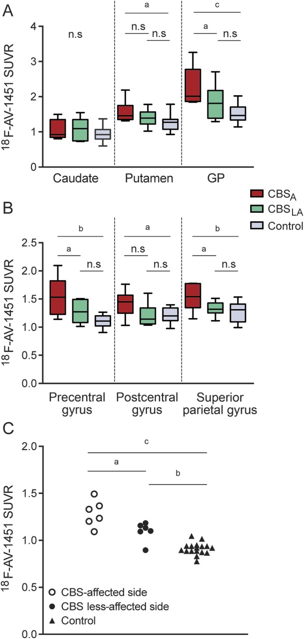
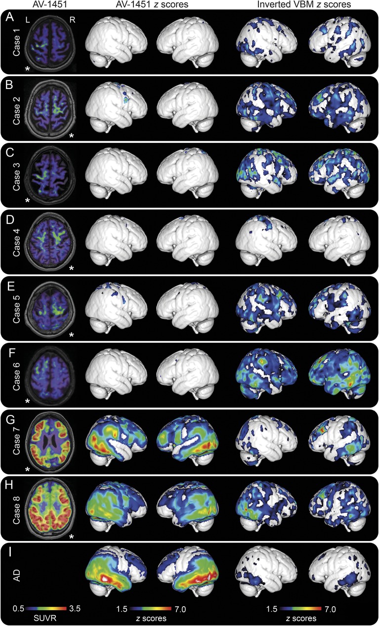
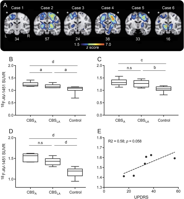
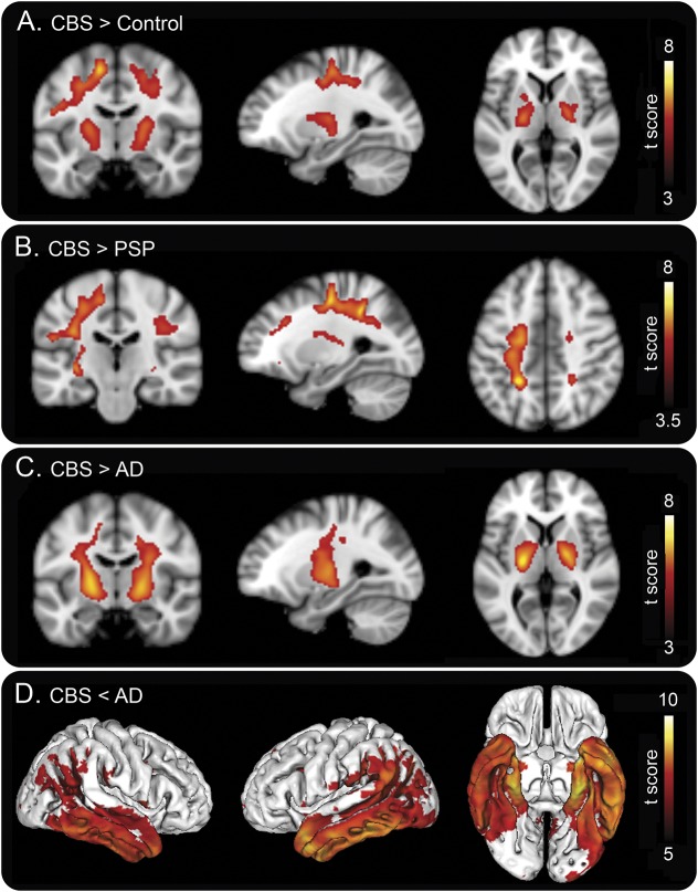
Verification
Claims the paper makes
The claims and statistics of this paper haven’t been checked yet.
Run the check — free, no account needed. It reads the open-access text and checks the sources it cites.
Claims other papers make about this one
Each is checked against this paper’s own text: does it say what the citing paper says it does?
The source backs part of the claim, not all of it — or backs it for a different population, condition or comparison.
In the citing paper
“Elevated in vivo binding is seen in tauopathies characterized by straight filaments (Bevan-Jones et al., 2016; Passamonti et al., 2018; Smith et al., 2017; Jones et al., 2018), albeit with generally lower binding affinity than in Alzheimer’s disease”
From Neuroinflammation and protein aggregation co-localize across the frontotemporal dementia spectrum (2020) · cites this paper as Smith et al., 2017
Experimental — accuracy not yet measured
Self-check
Percentages, sums and reported differences re-computed from the paper’s own numbers.
The self-check needs the paper’s full text.
Citations
Cites
McKhann, G., Drachman, D., Folstein, M., Katzman, R., Price, D., & Stadlan, E. M. (1984) · Neurology · doi:10.1212/wnl.34.7.939 · Cited by 27,915
McKhann, G. M., Knopman, D. S., Chertkow, H., Hyman, B. T., Jack, C. R., Kawas, C. H., Klunk, W. E., Koroshetz, W. J., Manly, J. J., Mayeux, R., et al. (2011) · Alzheimer's & Dementia · doi:10.1016/j.jalz.2011.03.005 · Cited by 18,657
Desikan, R. S., Ségonne, F., Fischl, B., Quinn, B. T., Dickerson, B. C., Blacker, D., Buckner, R. L., Dale, A. M., Maguire, R. P., Hyman, B. T., et al. (2006) · NeuroImage · doi:10.1016/j.neuroimage.2006.01.021 · Cited by 13,324
Parkinsonism: onset, progression, and mortality
Hoehn, M. M., & Yahr, M. D. (1967) · Neurology · doi:10.1212/wnl.17.5.427 · Cited by 12,107
Overdependence on degraded gist memory in Alzheimer's disease.
Gallo, D. A., Shahid, K. R., Olson, M. A., Solomon, T. M., Schacter, D. L., & Budson, A. E. (2006) · Neuropsychology · doi:10.1037/0894-4105.20.6.625 · Cited by 4,757
Automatically Parcellating the Human Cerebral Cortex
Fischl, B. R. (2003) · Cerebral Cortex · doi:10.1093/cercor/bhg087 · Cited by 4,208
Litvan, I., Agid, Y., Calne, D., Campbell, G., Dubois, B., Duvoisin, R. C., Goetz, C. G., Golbe, L. I., Grafman, J., Growdon, J. H., et al. (1996) · Neurology · doi:10.1212/wnl.47.1.1 · Cited by 2,538
Criteria for the diagnosis of corticobasal degeneration
Armstrong, M. J., Litvan, I., Lang, A. E., Bak, T. H., Bhatia, K. P., Borroni, B., Boxer, A. L., Dickson, D. W., Grossman, M., Hallett, M., et al. (2013) · Neurology · doi:10.1212/wnl.0b013e31827f0fd1 · Cited by 1,904
Tau pathology and neurodegeneration
Spillantini, M. G., & Goedert, M. (2013) · The Lancet Neurology · doi:10.1016/s1474-4422(13)70090-5 · Cited by 1,133
Progressive supranuclear palsy: clinicopathological concepts and diagnostic challenges
Williams, D. R., & Lees, A. J. (2009) · The Lancet Neurology · doi:10.1016/s1474-4422(09)70042-0 · Cited by 650
Cited by
Sorted by citations
Tau PET imaging in neurodegenerative tauopathies—still a challenge
Leuzy, A., Chiotis, K., Lemoine, L., Gillberg, P.-G., Almkvist, O., Rodriguez-Vieitez, E., & Nordberg, A. (2019) · Molecular Psychiatry · doi:10.1038/s41380-018-0342-8 · Cited by 628
Schöll, M., Maaß, A., Mattsson, N., Ashton, N. J., Blennow, K., Zetterberg, H., & Jagust, W. J. (2018) · Molecular and Cellular Neuroscience · doi:10.1016/j.mcn.2018.12.001 · Cited by 248
Self-citationThe development and validation of tau PET tracers: current status and future directions
Okamura, N., Harada, R., Ishiki, A., Kikuchi, A., Nakamura, T., & Kudo, Y. (2018) · Clinical and Translational Imaging · doi:10.1007/s40336-018-0290-y · Cited by 193
Soleimani‐Meigooni, D. N., Iaccarino, L., Joie, R. L., Baker, S. L., Bourakova, V., Boxer, A. L., Edwards, L., Eser, R. A., Gorno‐Tempini, M., Jagust, W. J., et al. (2020) · Brain · doi:10.1093/brain/awaa276 · Cited by 186
18F-flortaucipir (AV-1451) tau PET in frontotemporal dementia syndromes
Tsai, R. M., Bejanin, A., Lesman-Segev, O., LaJoie, R., Visani, A., Bourakova, V., O’Neil, J. P., Janabi, M., Baker, S., Lee, S. E., et al. (2019) · Alzheimer's Research & Therapy · doi:10.1186/s13195-019-0470-7 · Cited by 164
Metadata
- Authors
- Smith, R., Schöll, M., Widner, H., van Westen, D., Svenningsson, P., Hägerström, D., Ohlsson, T., Jögi, J., Nilsson, C., & Hansson, O.
- Affiliations
- Lund University (SE)
- Corresponding author
- Smith, Ruben
- Hansson, Oskar
- ORCID
- Venue
- Neurology
- Published
- 2017-08-22
- Volume
- 89
- Issue
- 8
- Pages
- 845-853
- ISSN
- 0028-3878 (ISSN-L)
- 1526-632X
- PMC
- PMC5580862
- OpenAlex
- W2739600095
- Cited by
- 117
- References
- 34
- Citations by year
- 2026: 3 · 2025: 8 · 2024: 4 · 2023: 6 · 2022: 17 · 2021: 21 · 2020: 16 · 2019: 18 · 2018: 19 · 2017: 4
- FWCI
- 12.81
- Type
- journal article
- Language
- en
- Open access
- hybrid
- doi.org
- Licence
- cc-by
- Locations
- Neurology (publishedVersion, cc-by)
- PubMed Central (publishedVersion, cc-by)
- PubMed (publishedVersion)
- Lund University Publications (Lund University) (submittedVersion)
- Funders
- Australian Government
- Vetenskapsrådet
- Topics
- Health Sciences › Medicine › Psychiatry and Mental health › Dementia and Cognitive Impairment Research
- Life Sciences › Neuroscience › Cellular and Molecular Neuroscience › Alzheimer's disease research and treatments
- Health Sciences › Medicine › Clinical Neurology › Parkinson's Disease Mechanisms and Treatments
- Keywords
- corticobasal syndrome, 18F-AV-1451, corticobasal degeneration, amyloid PET, Alzheimer's disease, progressive supranuclear palsy, differential diagnosis, cortical atrophy, corticospinal tract, basal ganglia, motor cortex, cortical thickness
- MeSH terms
- Aged, Alzheimer Disease, Basal Ganglia Diseases, Brain, Brain Mapping, Carbolines, Diagnosis, Differential, Humans, Magnetic Resonance Imaging, Middle Aged, Pyramidal Tracts, Supranuclear Palsy, Progressive, Sweden, Radiopharmaceuticals, Positron-Emission Tomography
About these checks
Registry, retraction, Cites and statistics results are computed, not judged, with one exception: a printed reference that no index lists is read once by a model before it counts as a citation error. Claims are extracted by a model and kept only when their quote appears verbatim in the text; each cited source is then checked, with a model’s help, for whether it says what the claim relies on. Those verdicts can be wrong — read the claim’s quote and the cited source before relying on one.
Statistics and arithmetic are recomputed, not judged.
Confidence bands are shown only for fields calibrated on our annotated test set. None is calibrated yet, so no band is shown.
What counts as a claim ›How we measure confidence ›How we check this ›
Are you an author, or do you see a problem with this page? Request a review
