

The DOI matches its Crossref record; 34 of the 34 references it cites were found in a registry or an index.
In vivo retention of 18 F-AV-1451 in corticobasal syndrome
Open accessAbstract
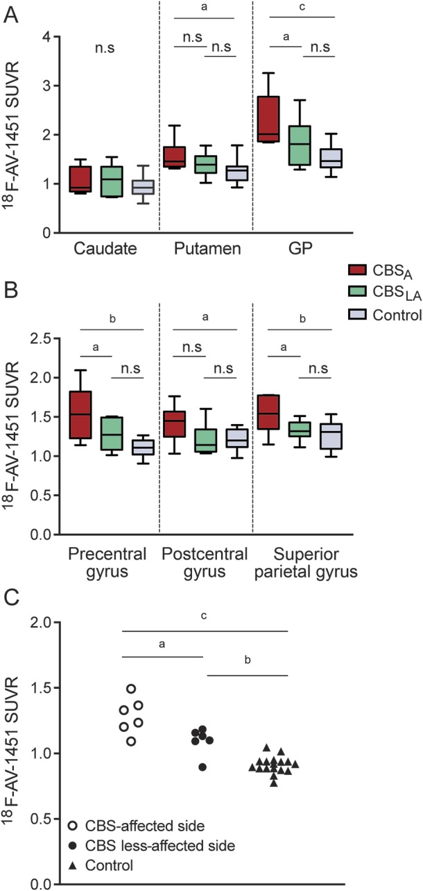
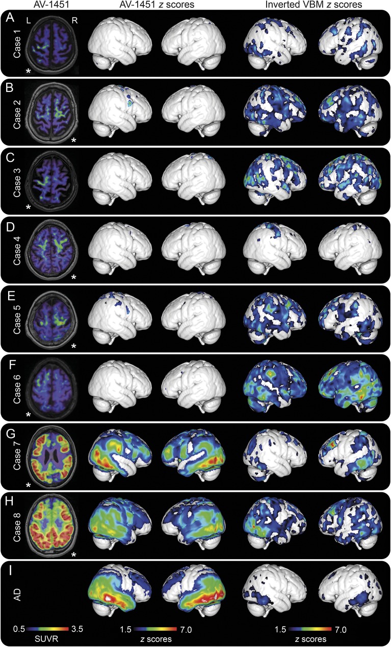
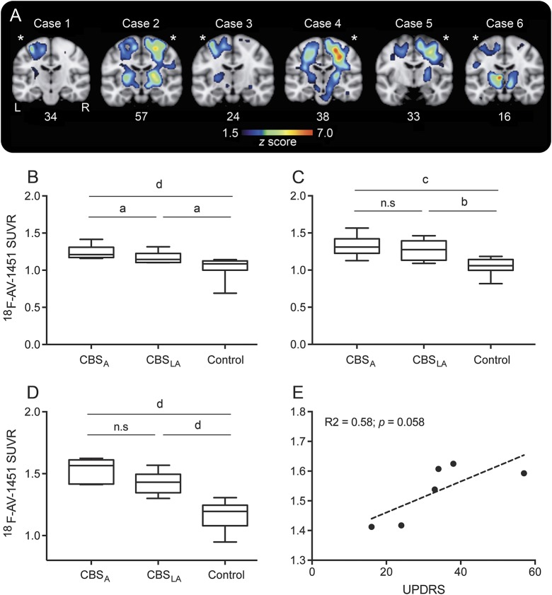
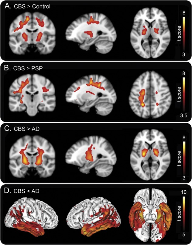
Objective: To study the usefulness of 18F-AV-1451 PET in patients with corticobasal syndrome (CBS). Methods: We recruited 8 patients with CBS, 17 controls, 31 patients with Alzheimer disease (AD), and 11 patients with progressive supranuclear palsy (PSP) from the Swedish BioFINDER study. All patients underwent clinical assessment, 18F-AV-1451 PET, MRI, and quantification of β-amyloid pathology. A subset of participants also underwent 18F-FDG-PET. Results: In the 8 patients with CBS, 6 had imaging findings compatible with the corticobasal degeneration pathology and 2 with typical AD pathology. In the 6 patients with CBS without typical AD pathology, there were substantial retentions of 18F-AV-1451 in the motor cortex, corticospinal tract, and basal ganglia contralateral to the most affected body side. These patients could be clearly distinguished from patients with AD dementia or PSP using 18F-AV-1451. However, cortical atrophy was more widespread than the cortical retention of 18F-AV1451 in these CBS cases, and cortical AV-1451 uptake did not correlate with cortical thickness or glucose hypometabolism. These results are in sharp contrast to AD dementia, where 18F-AV-1451 retention was more widespread than cortical atrophy, and correlated well with cortical thickness and hypometabolism. Conclusions: Patients with CBS without typical AD pathology exhibited AV-1451 retention in the motor cortex, corticospinal tract, and basal ganglia contralateral to the affected body side, clearly different from controls and patients with AD dementia or PSP. However, cortical atrophy measured with MRI and decreased 18F-fluorodeoxyglucose uptake were more widespread than 18F-AV-1451 uptake and probably represent earlier, yet less specific, markers of CBS. Classification of evidence: This study provides Class III evidence that 18F-AV-1451 PET distinguishes between CBS and AD or PSP.
Overview
The claims and statistics of this paper haven’t been checked yet.
Run the check — free, no account needed. It reads the open-access text and checks the sources it cites.
Integrity signals
Registry
Cites
Statistics
Figures
Verification
Claims the paper makes
The claims and statistics of this paper haven’t been checked yet.
Run the check — free, no account needed. It reads the open-access text and checks the sources it cites.
Claims other papers make about this one
Each is checked against this paper’s own text: does it say what the citing paper says it does?
The source backs part of the claim, not all of it — or backs it for a different population, condition or comparison.
In the citing paper
“Elevated in vivo binding is seen in tauopathies characterized by straight filaments (Bevan-Jones et al., 2016; Passamonti et al., 2018; Smith et al., 2017; Jones et al., 2018), albeit with generally lower binding affinity than in Alzheimer’s disease”
From Neuroinflammation and protein aggregation co-localize across the frontotemporal dementia spectrum (2020) · cites this paper as Smith et al., 2017
Experimental — accuracy not yet measured
Self-check
Percentages, sums and reported differences re-computed from the paper’s own numbers.
The self-check needs the paper’s full text.
Citations
Cites
McKhann, G., Drachman, D., Folstein, M., Katzman, R., Price, D., & Stadlan, E. M. (1984) · Neurology · doi:10.1212/wnl.34.7.939 · Cited by 27,915
McKhann, G. M., Knopman, D. S., Chertkow, H., Hyman, B. T., Jack, C. R., Kawas, C. H., Klunk, W. E., Koroshetz, W. J., Manly, J. J., Mayeux, R., et al. (2011) · Alzheimer's & Dementia · doi:10.1016/j.jalz.2011.03.005 · Cited by 18,657
Desikan, R. S., Ségonne, F., Fischl, B., Quinn, B. T., Dickerson, B. C., Blacker, D., Buckner, R. L., Dale, A. M., Maguire, R. P., Hyman, B. T., et al. (2006) · NeuroImage · doi:10.1016/j.neuroimage.2006.01.021 · Cited by 13,324
Parkinsonism: onset, progression, and mortality
Hoehn, M. M., & Yahr, M. D. (1967) · Neurology · doi:10.1212/wnl.17.5.427 · Cited by 12,107
Overdependence on degraded gist memory in Alzheimer's disease.
Gallo, D. A., Shahid, K. R., Olson, M. A., Solomon, T. M., Schacter, D. L., & Budson, A. E. (2006) · Neuropsychology · doi:10.1037/0894-4105.20.6.625 · Cited by 4,757
Automatically Parcellating the Human Cerebral Cortex
Fischl, B. R. (2003) · Cerebral Cortex · doi:10.1093/cercor/bhg087 · Cited by 4,208
Litvan, I., Agid, Y., Calne, D., Campbell, G., Dubois, B., Duvoisin, R. C., Goetz, C. G., Golbe, L. I., Grafman, J., Growdon, J. H., et al. (1996) · Neurology · doi:10.1212/wnl.47.1.1 · Cited by 2,538
Criteria for the diagnosis of corticobasal degeneration
Armstrong, M. J., Litvan, I., Lang, A. E., Bak, T. H., Bhatia, K. P., Borroni, B., Boxer, A. L., Dickson, D. W., Grossman, M., Hallett, M., et al. (2013) · Neurology · doi:10.1212/wnl.0b013e31827f0fd1 · Cited by 1,904
Tau pathology and neurodegeneration
Spillantini, M. G., & Goedert, M. (2013) · The Lancet Neurology · doi:10.1016/s1474-4422(13)70090-5 · Cited by 1,133
Progressive supranuclear palsy: clinicopathological concepts and diagnostic challenges
Williams, D. R., & Lees, A. J. (2009) · The Lancet Neurology · doi:10.1016/s1474-4422(09)70042-0 · Cited by 650
Cited by
Sorted by citations
Tau PET imaging in neurodegenerative tauopathies—still a challenge
Leuzy, A., Chiotis, K., Lemoine, L., Gillberg, P.-G., Almkvist, O., Rodriguez-Vieitez, E., & Nordberg, A. (2019) · Molecular Psychiatry · doi:10.1038/s41380-018-0342-8 · Cited by 628
Schöll, M., Maaß, A., Mattsson, N., Ashton, N. J., Blennow, K., Zetterberg, H., & Jagust, W. J. (2018) · Molecular and Cellular Neuroscience · doi:10.1016/j.mcn.2018.12.001 · Cited by 248
Self-citationThe development and validation of tau PET tracers: current status and future directions
Okamura, N., Harada, R., Ishiki, A., Kikuchi, A., Nakamura, T., & Kudo, Y. (2018) · Clinical and Translational Imaging · doi:10.1007/s40336-018-0290-y · Cited by 193
Soleimani‐Meigooni, D. N., Iaccarino, L., Joie, R. L., Baker, S. L., Bourakova, V., Boxer, A. L., Edwards, L., Eser, R. A., Gorno‐Tempini, M., Jagust, W. J., et al. (2020) · Brain · doi:10.1093/brain/awaa276 · Cited by 186
18F-flortaucipir (AV-1451) tau PET in frontotemporal dementia syndromes
Tsai, R. M., Bejanin, A., Lesman-Segev, O., LaJoie, R., Visani, A., Bourakova, V., O’Neil, J. P., Janabi, M., Baker, S., Lee, S. E., et al. (2019) · Alzheimer's Research & Therapy · doi:10.1186/s13195-019-0470-7 · Cited by 164
Neuroimaging Advances in Parkinson's Disease and Atypical Parkinsonian Syndromes
Saeed, U., Lang, A. E., & Masellis, M. (2020) · Frontiers in Neurology · doi:10.3389/fneur.2020.572976 · Cited by 156
Dissecting the clinical heterogeneity of early-onset Alzheimer’s disease
Sirkis, D. W., Bonham, L. W., Johnson, T. P., La Joie, R., & Yokoyama, J. S. (2022) · Molecular Psychiatry · doi:10.1038/s41380-022-01531-9 · Cited by 151
Evolving concepts in progressive supranuclear palsy and other 4-repeat tauopathies
Stamelou, M., Respondek, G., Giagkou, N., Whitwell, J. L., Kovács, G. G., & Höglinger, G. U. (2021) · Nature Reviews Neurology · doi:10.1038/s41582-021-00541-5 · Cited by 149
Tau PET Imaging in Neurodegenerative Disorders
Groot, C., Villeneuve, S., Smith, R., Hansson, O., & Ossenkoppele, R. (2022) · Journal of Nuclear Medicine · doi:10.2967/jnumed.121.263196 · Cited by 137
Self-citationLessons learned about [F-18]-AV-1451 off-target binding from an autopsy-confirmed Parkinson’s case.
Marquié, M., Verwer, E. E., Meltzer, A. C., Kim, S. J. W., Agüero, C., Gonzalez, J., Makaretz, S. J., Siao Tick Chong, M., Ramanan, P., Amaral, A. C., et al. (2017) · Acta Neuropathologica Communications · doi:10.1186/s40478-017-0482-0 · Cited by 128
Neuroinflammation and protein aggregation co-localize across the frontotemporal dementia spectrum
Bevan-Jones, W. R., Cope, T. E., Jones, P. S., Kaalund, S. S., Passamonti, L., Allinson, K., Green, O., Hong, Y. T., Fryer, T. D., Arnold, R., et al. (2020) · Brain · doi:10.1093/brain/awaa033 · Cited by 124
Corticobasal degeneration and corticobasal syndrome: A review
Constantinides, V. C., Paraskevas, G. P., Paraskevas, P. G., Stefanis, L., & Kapaki, E. N. (2019) · Clinical Parkinsonism & Related Disorders · doi:10.1016/j.prdoa.2019.08.005 · Cited by 113
Propagation of Tau Pathology: Integrating Insights From Postmortem and In Vivo Studies
Vogels, T., Leuzy, A., Cicognola, C., Ashton, N. J., Smolek, T., Novák, M., Blennow, K., Zetterberg, H., Hromádka, T., Žilka, N., et al. (2019) · Biological Psychiatry · doi:10.1016/j.biopsych.2019.09.019 · Cited by 99
Self-citationHead-to-head comparison of tau positron emission tomography tracers [18F]flortaucipir and [18F]RO948
Smith, R., Schöll, M., Leuzy, A., Jögi, J., Ohlsson, T., Strandberg, O., & Hansson, O. (2020) · European Journal of Nuclear Medicine and Molecular Imaging · doi:10.1007/s00259-019-04496-0 · Cited by 88
Self-citationMolecular imaging in dementia: Past, present, and future
Laforce, R. J., Soucy, J., Sellami, L., Dallaire‐Théroux, C., Brunet, F., Bergeron, D., Miller, B. L., & Ossenkoppele, R. (2018) · Alzheimer s & Dementia · doi:10.1016/j.jalz.2018.06.2855 · Cited by 88
Cortical [ 18 F ] PI ‐2620 Binding Differentiates Corticobasal Syndrome Subtypes
Palleis, C., Brendel, M., Finze, A., Weidinger, E., Bötzel, K., Danek, A., Beyer, L., Nitschmann, A., Kern, M., Biechele, G., et al. (2021) · Movement Disorders · doi:10.1002/mds.28624 · Cited by 87
Tezuka, T., Takahata, K., Seki, M., Tabuchi, H., Momota, Y., Shiraiwa, M., Suzuki, N., Morimoto, A., Nakahara, T., Iwabuchi, Y., et al. (2021) · Brain Communications · doi:10.1093/braincomms/fcab190 · Cited by 85
Tau Imaging in Neurodegenerative Diseases Using Positron Emission Tomography
Wang, Y. T., & Edison, P. (2019) · Current Neurology and Neuroscience Reports · doi:10.1007/s11910-019-0962-7 · Cited by 86
Leuzy, A., Pascoal, T. A., Strandberg, O. T., Insel, P. S., Smith, R. A., Mattsson, N., Benedet, A. L., Cho, H., Lyoo, C. H., Joie, R. L., et al. (2021) · European Journal of Nuclear Medicine and Molecular Imaging · doi:10.1007/s00259-021-05401-4 · Cited by 89
Self-citationOverview of tau PET molecular imaging
Petersen, G. C., Roytman, M., Chiang, G. C.-Y., Li, Y., Gordon, M. L., & Franceschi, A. M. (2022) · Current Opinion in Neurology · doi:10.1097/wco.0000000000001035 · Cited by 87
Tau PET and multimodal brain imaging in patients at risk for chronic traumatic encephalopathy
Lesman‐Segev, O. H., Joie, R. L., Stephens, M. L., Sonni, I., Tsai, R. M., Bourakova, V., Visani, A. V., Edwards, L., O’Neil, J. P., Baker, S. L., et al. (2019) · NeuroImage Clinical · doi:10.1016/j.nicl.2019.102025 · Cited by 83
Young, C. B., Winer, J. R., Younes, K., Cody, K. A., Betthauser, T. J., Johnson, S. C., Schultz, A., Sperling, R. A., Greicius, M. D., Cobos, I., et al. (2022) · JAMA Neurology · doi:10.1001/jamaneurol.2022.0676 · Cited by 79
Lebouvier, T., Pasquier, F., & Buée, L. (2017) · Current Opinion in Neurology · doi:10.1097/wco.0000000000000502 · Cited by 77
Malarte, M.-L., Gillberg, P.-G., Kumar, A., Bogdanovic, N., Lemoine, L., & Nordberg, A. (2023) · Molecular Psychiatry · doi:10.1038/s41380-022-01875-2 · Cited by 67
Clinical Spectrum of Tauopathies
Olfati, N., Shoeibi, A., & Litvan, I. (2022) · Frontiers in Neurology · doi:10.3389/fneur.2022.944806 · Cited by 66
Groot, C., Smith, R. A., Collij, L. E., Mastenbroek, S. E., Stomrud, E., Binette, A. P., Leuzy, A., Palmqvist, S., Mattsson, N., Strandberg, O. T., et al. (2024) · JAMA Neurology · doi:10.1001/jamaneurol.2024.1612 · Cited by 70
Self-citationTau-PET imaging with [18F]AV-1451 in primary progressive apraxia of speech
Utianski, R. L., Whitwell, J. L., Schwarz, C. G., Senjem, M. L., Tosakulwong, N., Duffy, J. R., Clark, H. M., Machulda, M. M., Petersen, R. C., Jack, C. R., et al. (2018) · Cortex · doi:10.1016/j.cortex.2017.12.021 · Cited by 54
Saranza, G., Whitwell, J. L., Kovács, G. G., & Lang, A. E. (2019) · International review of neurobiology · doi:10.1016/bs.irn.2019.10.014 · Cited by 52
Dementia spectrum disorders: lessons learnt from decades with PET research
Wilson, H., Pagano, G., & Politis, M. (2019) · Journal of Neural Transmission · doi:10.1007/s00702-019-01975-4 · Cited by 50
Corticobasal syndrome: neuroimaging and neurophysiological advances
Stasio, F. D., Suppa, A., Marsili, L., Upadhyay, N., Asci, F., Bologna, M., Colosimo, C., Fabbrini, G., Pantano, P., & Berardelli, A. (2019) · European Journal of Neurology · doi:10.1111/ene.13928 · Cited by 50
FDG‐PET Patterns Predict Amyloid Deposition and Clinical Profile in Corticobasal Syndrome
Parmera, J. B., Coutinho, A. M., Aranha, M. R., Studart‐Neto, A., Carneiro, C. D. G., de Almeida, I. J., Solla, D. J. F., Ono, C. R., Barbosa, E. R., Nitríni, R., et al. (2020) · Movement Disorders · doi:10.1002/mds.28373 · Cited by 46
Diagnostic biomarkers in Alzheimer’s disease
Andersen, E., Casteigne, B., Chapman, W. D., Creed, A., Foster, F., Lapins, A., Shatz, R., & Sawyer, R. P. (2021) · Biomarkers in Neuropsychiatry · doi:10.1016/j.bionps.2021.100041 · Cited by 42
Diffuse Lewy body disease manifesting as corticobasal syndrome
Kasanuki, K., Josephs, K. A., Ferman, T. J., Murray, M. E., Koga, S., Konno, T., Sakae, N., Parks, A. C., Uitti, R. J., van Gerpen, J. A., et al. (2018) · Neurology · doi:10.1212/wnl.0000000000005828 · Cited by 41
Imaging of Tau Pathology in Neurodegenerative Diseases: An Update
Beyer, L., & Brendel, M. (2020) · Seminars in Nuclear Medicine · doi:10.1053/j.semnuclmed.2020.12.004 · Cited by 44
Uses of Human MR and PET Imaging in Research of Neurodegenerative Brain Diseases
Schwarz, C. G. (2021) · Neurotherapeutics · doi:10.1007/s13311-021-01030-9 · Cited by 40
Murugan, N. A., Nordberg, A. K., & Ågren, H. (2021) · ACS Chemical Neuroscience · doi:10.1021/acschemneuro.0c00340 · Cited by 41
Corticobasal degeneration: key emerging issues
Ali, F., & Josephs, K. A. (2017) · Journal of Neurology · doi:10.1007/s00415-017-8644-3 · Cited by 40
Imaging Protein Misfolding in the Brain Using β-Sheet Ligands
Harada, R., Okamura, N., Furumoto, S., & Yanai, K. (2018) · Frontiers in Neuroscience · doi:10.3389/fnins.2018.00585 · Cited by 41
Amyloid and Tau PET Imaging of Alzheimer Disease and Other Neurodegenerative Conditions
Rowley, P. A., Samsonov, A. A., Betthauser, T. J., Pirasteh, A., Johnson, S. C., & Eisenmenger, L. B. (2020) · Seminars in Ultrasound CT and MRI · doi:10.1053/j.sult.2020.08.011 · Cited by 40
Ali, F., Whitwell, J. L., Martin, P. R., Senjem, M. L., Knopman, D. S., Jack, C. R., Lowe, V. J., Petersen, R. C., Boeve, B. F., & Josephs, K. A. (2018) · Journal of Neurology · doi:10.1007/s00415-018-8815-x · Cited by 38
The Role of Tau beyond Alzheimer’s Disease: A Narrative Review
Virgilio, E., Marchi, F. D., Contaldi, E., Dianzani, U., Cantello, R. M., Mazzini, L., & Comi, C. (2022) · Biomedicines · doi:10.3390/biomedicines10040760 · Cited by 32
Beyer, L., Meyer-Wilmes, J., Schönecker, S., Schnabel, J., Brendel, E., Prix, C., Nübling, G., Unterrainer, M., Albert, N. L., Pogarell, O. M., et al. (2018) · Frontiers in Neurology · doi:10.3389/fneur.2018.00483 · Cited by 34
Novel PET Biomarkers to Disentangle Molecular Pathways across Age-Related Neurodegenerative Diseases
Wilson, H., Politis, M., Rabiner, E. A., & Middleton, L. T. (2020) · Cells · doi:10.3390/cells9122581 · Cited by 34
Disease-related patterns of in vivo pathology in Corticobasal syndrome
Initiative, A. D. N., Niccolini, F., Wilson, H., Hirschbichler, S. T., Yousaf, T., Pagano, G., Whittington, A., Caminiti, S. P., Erro, R., Holton, J. L., et al. (2018) · European Journal of Nuclear Medicine and Molecular Imaging · doi:10.1007/s00259-018-4104-2 · Cited by 35
Ricci, M., Cimini, A., Chiaravalloti, A., Filippi, L., & Schillaci, O. (2020) · International Journal of Molecular Sciences · doi:10.3390/ijms21207481 · Cited by 33
Multimodal imaging evidence of pathology-mediated disease distribution in corticobasal syndrome
McMillan, C. T., Boyd, C. D., Gross, R. G., Weinstein, J., Firn, K., Toledo, J. B., Rascovsky, K., Shaw, L. M. J., Wolk, D. A., Irwin, D. J., et al. (2016) · Neurology · doi:10.1212/wnl.0000000000003119 · Cited by 31
Burnham, S. C., Iaccarino, L., Pontecorvo, M. J., Fleisher, A., Lu, M., Collins, E. C., & Devous, M. D. (2023) · Brain Communications · doi:10.1093/braincomms/fcad305 · Cited by 30
Alosco, M. L., Su, Y., Stein, T. D., Protas, H. D., Cherry, J. D., Adler, C. H., Balcer, L. J., Bernick, C. B., Pulukuri, S. V., Abdolmohammadi, B., et al. (2022) · European Journal of Nuclear Medicine and Molecular Imaging · doi:10.1007/s00259-022-05963-x · Cited by 28
Corticobasal degeneration: advances in clinicopathology and biomarkers
Svenningsson, P. (2019) · Current Opinion in Neurology · doi:10.1097/wco.0000000000000707 · Cited by 27
Self-citationIn vivo [ 18 F]-AV-1451 tau-PET imaging in sporadic Creutzfeldt-Jakob disease
Day, G. S., Gordon, B. A., Perrin, R. J., Cairns, N. J., Beaumont, H., Schwetye, K., Ferguson, C., Sinha, N., Bucelli, R., Musiek, E. S., et al. (2018) · Neurology · doi:10.1212/wnl.0000000000005064 · Cited by 26
Perani, D., Iaccarino, L., Lammertsma, A. A., Windhorst, A. D., Edison, P., Boellaard, R., Hansson, O., Nordberg, A. K., Jacobs, A. H., & IMBI Project (2019) · Alzheimer s & Dementia · doi:10.1016/j.jalz.2019.02.004 · Cited by 25
Self-citationIntegrated 18F-T807 Tau PET, Structural MRI, and Plasma Tau in Tauopathy Neurodegenerative Disorders
Li, C., Chen, T., Chiu, M., Yen, R., Shih, M., & Lin, C. (2021) · Frontiers in Aging Neuroscience · doi:10.3389/fnagi.2021.646440 · Cited by 24
18F-THK5351 Positron Emission Tomography Imaging in Neurodegenerative Tauopathies
Ezura, M., Kikuchi, A., Okamura, N., Ishiki, A., Hasegawa, T., Harada, R., Watanuki, S., Funaki, Y., Hiraoka, K., Baba, T., et al. (2021) · Frontiers in Aging Neuroscience · doi:10.3389/fnagi.2021.761010 · Cited by 25
Blazhenets, G., Soleimani‐Meigooni, D. N., Thomas, W. P., Mundada, N. S., Brendel, M., Vento, S. A., VandeVrede, L. A., Heuer, H. W., Ljubenkov, P. A., Rojas, J. C., et al. (2023) · Journal of Nuclear Medicine · doi:10.2967/jnumed.123.265856 · Cited by 23
Alster, P., Madetko‐Alster, N., Koziorowski, D., Królicki, L., Budrewicz, S. P., & Friedman, A. (2019) · Frontiers in Neurology · doi:10.3389/fneur.2019.00101 · Cited by 22
Saito, Y., Kamagata, K., Andica, C., Uchida, W., Takabayashi, K., Yoshida, S., Nakaya, M., Tanaka, Y., Kamio, S., Sato, K., et al. (2023) · Japanese Journal of Radiology · doi:10.1007/s11604-023-01454-7 · Cited by 20
PET Neuroimaging in Dementia Conditions
Perani, D., Caminiti, S. P., Carli, G., & Tondo, G. (2020) · Springer eBooks · doi:10.1007/978-3-030-53168-3_9 · Cited by 20
Tau Imaging in Parkinsonism: What Have We Learned So Far?
Whitwell, J. L. (2017) · Movement Disorders Clinical Practice · doi:10.1002/mdc3.12584 · Cited by 19
Neuroimaging in Frontotemporal Lobar Degeneration: Research and Clinical Utility
Dev, S. I., Dickerson, B. C., & Touroutoglou, A. (2021) · Advances in experimental medicine and biology · doi:10.1007/978-3-030-51140-1_7 · Cited by 16
Tau Imaging in the 4-Repeat-Tauopathies Progressive Supranuclear Palsy and Corticobasal Syndrome
Schröter, N., Blazhenets, G., Frings, L., Barkhausen, C., Jost, W. H., Weiller, C., Rijntjes, M., & Meyer, P. T. (2020) · Clinical Nuclear Medicine · doi:10.1097/rlu.0000000000002949 · Cited by 18
PET-based classification of corticobasal syndrome
Nakano, Y., Shimada, H., Shinotoh, H., Hirano, S., Tagai, K., Sano, Y., Yamamoto, Y., Endo, H., Matsuoka, K., Takahata, K., et al. (2022) · Parkinsonism & Related Disorders · doi:10.1016/j.parkreldis.2022.04.015 · Cited by 19
The Role of Tau Imaging in Parkinsonian Disorders
Hammes, J., Drzezga, A. E., & van Eimeren, T. (2018) · Current Neurology and Neuroscience Reports · doi:10.1007/s11910-018-0898-3 · Cited by 18
Disentangling brain functional network remodeling in corticobasal syndrome – A multimodal MRI study
Ballarini, T., Albrecht, F., Mueller, K., Jech, R., Diehl‐Schmid, J., Fließbach, K., Kassubek, J., Lauer, M., Fassbender, K. C., Schneider, A., et al. (2019) · NeuroImage Clinical · doi:10.1016/j.nicl.2019.102112 · Cited by 17
Barć, K., & Kuźma‐Kozakiewicz, M. (2019) · Neurologia i Neurochirurgia Polska · doi:10.5603/pjnns.a2019.0013 · Cited by 15
Longitudinal changes in 18F‐ THK 5351 positron emission tomography in corticobasal syndrome
Ezura, M., Kikuchi, A., Ishiki, A., Okamura, N., Hasegawa, T., Harada, R., Watanuki, S., Funaki, Y., Hiraoka, K., Baba, T., et al. (2019) · European Journal of Neurology · doi:10.1111/ene.13966 · Cited by 15
18F-AV-1451 positron emission tomography in neuropathological substrates of corticobasal syndrome
Goodheart, A. E., Locascio, J. J., Samore, W. R., Collins, J. A., Brickhouse, M. J., Schultz, A. P., Touroutoglou, A., Johnson, K. A., Frosch, M. P., Growdon, J. H., et al. (2020) · Brain · doi:10.1093/brain/awaa383 · Cited by 16
Carlos, A. F., Tosakulwong, N., Weigand, S. D., Buciuc, M., Ali, F., Clark, H. M., Botha, H., Utianski, R. L., Machulda, M. M., Schwarz, C. G., et al. (2022) · Brain Communications · doi:10.1093/braincomms/fcac108 · Cited by 14
Vasilevskaya, A., Taghdiri, F., Multani, N., Anor, C. J., Misquitta, K., Houle, S., Burke, C., Tang‐Wai, D. F., Lang, A. E., Fox, S. H., et al. (2020) · Frontiers in Neurology · doi:10.3389/fneur.2020.00574 · Cited by 17
Schweyer, K., Busche, M. A., Hammes, J., Zwergal, A., Buhmann, C., van Eimeren, T., & Höglinger, G. U. (2018) · Neurology · doi:10.1212/wnl.0000000000005069 · Cited by 13
Ding, J., Shen, C., Wang, Z., Yang, Y., Fakhri, G. E., Lu, J., Liang, D., Zheng, H., Zhou, Y., Sun, T., et al. (2023) · Cerebral Cortex · doi:10.1093/cercor/bhad312 · Cited by 13
Impairment of intermediate somatosensory function in corticobasal syndrome
Matsuda, K., Satoh, M., Tabei, K., Ueda, Y., Taniguchi, A., Matsuura, K., Asahi, M., Ii, Y., Niwa, A., & Tomimoto, H. (2020) · Scientific Reports · doi:10.1038/s41598-020-67991-7 · Cited by 12
Chronic Traumatic Encephalopathy: A Comparison with Alzheimer's Disease and Frontotemporal Dementia
Lesman‐Segev, O. H., Edwards, L., & Rabinovici, G. D. (2020) · Seminars in Neurology · doi:10.1055/s-0040-1715134 · Cited by 13
Tau Positron Emission Tomography Imaging in Degenerative Parkinsonisms
Lyoo, C. H., Cho, H., Choi, J. Y., Ryu, Y. H., & Lee, M. S. (2018) · Journal of Movement Disorders · doi:10.14802/jmd.17071 · Cited by 12
PET Molecular Imaging in Atypical Parkinsonism
Xu, Z., Arbizu, J., & Pavese, N. (2018) · International review of neurobiology · doi:10.1016/bs.irn.2018.09.001 · Cited by 12
Is longitudinal tau PET ready for use in Alzheimer’s disease clinical trials?
Hansson, O., & Mormino, E. C. (2018) · Brain · doi:10.1093/brain/awy065 · Cited by 10
Self-citationSantillo, A. F., Leuzy, A., Honer, M., Waldö, M. L., Tideman, P., Harper, L., Ohlsson, T. G., Moes, S., Giannini, L., Jögi, J., et al. (2022) · European Journal of Nuclear Medicine and Molecular Imaging · doi:10.1007/s00259-022-06065-4 · Cited by 11
Self-citationRelationships between PET and blood plasma biomarkers in corticobasal syndrome
Singh, N. A., Alnobani, A., Graff‐Radford, J., Machulda, M. M., Mielke, M. M., Schwarz, C. G., Senjem, M. L., Jack, C. R., Lowe, V. J., Kanekiyo, T., et al. (2024) · Alzheimer s & Dementia · doi:10.1002/alz.13914 · Cited by 9
Clear‐headed into old age: Resilience and resistance against brain aging—A PET imaging perspective
Hoenig, M. C., & Drzezga, A. E. (2022) · Journal of Neurochemistry · doi:10.1111/jnc.15598 · Cited by 9
Recent development in selective Tau tracers for PET imaging in the brain
Li, Y., Liu, T., & Cui, M. (2022) · Chinese Chemical Letters · doi:10.1016/j.cclet.2022.03.024 · Cited by 10
Zhang, W., Xu, S., Yu, H., Li, X., Jin, Z., Li, Y., & He, Z. (2021) · Annals of Nuclear Medicine · doi:10.1007/s12149-020-01566-4 · Cited by 8
The Adult Neurogenesis Theory of Alzheimer’s Disease
Abbate, C. (2023) · Journal of Alzheimer s Disease · doi:10.3233/jad-221279 · Cited by 11
A 5-year longitudinal evaluation in patients with mild cognitive impairment by 11C-PIB PET/CT
Jiménez-Bonilla, J. F., Quirce, R., de Arcocha-Torres, M., Martínez‐Rodríguez, I., Martínez‐Amador, N., Sánchez-Salmón, A., la Fuente-Gómez, F. D., Rodríguez‐Rodríguez, E., Sánchez‐Juan, P., & Banzo, I. (2019) · Nuclear Medicine Communications · doi:10.1097/mnm.0000000000001004 · Cited by 8
Molecular Imaging of Extrapyramidal Movement Disorders With Dementia: The 4R Tauopathies
Frey, K. A. (2021) · Seminars in Nuclear Medicine · doi:10.1053/j.semnuclmed.2020.12.003 · Cited by 8
PBB3 binding in a patient with corticobasal syndrome
Pérez‐Soriano, A., Matarazzo, M., Vafai, N. M., Shahinfard, E., Miao, Q., Higuchi, M., Sossi, V., & Stoessl, A. J. (2018) · Movement Disorders · doi:10.1002/mds.27405 · Cited by 7
Imaging Familial and Sporadic Neurodegenerative Disorders Associated with Parkinsonism
Brooks, D. J. (2021) · Neurotherapeutics · doi:10.1007/s13311-020-00994-4 · Cited by 7
An Update on the State of Tau Radiotracer Development: a Brief Review
Rigney, G. H., Ayubcha, C., Werner, T. J., & Alavi, A. A. (2021) · Molecular Imaging and Biology · doi:10.1007/s11307-021-01612-1 · Cited by 7
The tauopathies: Neuroimaging characteristics and emerging experimental therapies
Riley, K. J., Graner, B. D., & Veronesi, M. C. (2022) · Journal of Neuroimaging · doi:10.1111/jon.13001 · Cited by 8
The Current Role of Tau PET Imaging in Neurodegeneration
Vanderlinden, G., Vandenberghe, R. R., Vandenbulcke, M., & Laere, K. V. (2025) · Seminars in Nuclear Medicine · doi:10.1053/j.semnuclmed.2025.03.002 · Cited by 7
Role of [18F]-FDG PET in patients with atypical parkinsonism associated with dementia
Raffa, S., Donegani, M. I., Borra, A., Miceli, A., Balma, M., Bauckneht, M., & Morbelli, S. D. (2020) · Clinical and Translational Imaging · doi:10.1007/s40336-020-00360-6 · Cited by 5
PET and SPECT Imaging of Neurodegenerative Diseases
Drzezga, A. E., Bischof, G. N., Giehl, K., & van Eimeren, T. (2021) · Elsevier eBooks · doi:10.1016/b978-0-12-816386-3.00085-5 · Cited by 4
Tau positron emission tomography in tauopathies: A narrative review
Lee, H., Gu, Y., Seo, S. W., & Moon, S. H. (2023) · Precision and Future Medicine · doi:10.23838/pfm.2023.00016 · Cited by 4
Tau PET imaging evidence in patients with cognitive impairment: preparing for clinical use
Noirot, C., Mainta, I., Mendes, A., Andryszak, P., Visvaratnam, H., Unschuld, P. G., Frisoni, G. B., & Garibotto, V. (2018) · Clinical and Translational Imaging · doi:10.1007/s40336-018-0297-4 · Cited by 3
Abbate, C., & Gallucci, A. (2025) · Journal of Alzheimer s Disease · doi:10.1177/13872877251374319 · Cited by 2
Niethammer, M. (2021) · Springer eBooks · doi:10.1007/978-3-030-82367-2_32 · Cited by 2
The biological diagnosis of Alzheimer’s disease using blood-based biomarkers: A Canadian prospective
Kumar, P., Mousavi, A., & Frykman, H. (2025) · Clinical Biochemistry · doi:10.1016/j.clinbiochem.2025.110980 · Cited by 2
Dinkelbach, L., Südmeyer, M., Hartmann, C. J., Roeber, S., Arzberger, T., Felsberg, J., Ferrea, S., Moldovan, A., Amunts, K., Schnitzler, A., et al. (2020) · Neurobiology of Aging · doi:10.1016/j.neurobiolaging.2020.05.009 · Cited by 2
Imaging biomarkers for the diagnosis of corticobasal ganglia syndrome: DaTSCAN and [18F]FDG PET
Missir, E., & Cornwell, M. (2025) · NeuroMarkers. · doi:10.1016/j.neumar.2025.100079 · Cited by 1
Relationship between tau-PET and quantitative susceptibility mapping in atypical Alzheimer’s disease
Singh, N. A., Satoh, R., Graff‐Radford, J., Machulda, M. M., Lowe, V. J., Josephs, K. A., & Whitwell, J. L. (2025) · Frontiers in Aging Neuroscience · doi:10.3389/fnagi.2025.1615718 · Cited by 3
Molecular Imaging of Neurodegenerative Parkinsonism
Frey, K. A., & Bohnen, N. I. (2021) · PET Clinics · doi:10.1016/j.cpet.2020.12.002 · Cited by 1
Alster, P., Madetko‐Alster, N., Migda, B., Nieciecki, M., Kutyłowski, M., Królicki, L., & Friedman, A. (2022) · Diagnostics · doi:10.3390/diagnostics12071671 · Cited by 1
Metadata
- Authors
- Smith, R., Schöll, M., Widner, H., van Westen, D., Svenningsson, P., Hägerström, D., Ohlsson, T., Jögi, J., Nilsson, C., & Hansson, O.
- Affiliations
- Lund University (SE)
- Corresponding author
- Smith, Ruben
- Hansson, Oskar
- ORCID
- Venue
- Neurology
- Published
- 2017-08-22
- Volume
- 89
- Issue
- 8
- Pages
- 845-853
- ISSN
- 0028-3878 (ISSN-L)
- 1526-632X
- PMC
- PMC5580862
- OpenAlex
- W2739600095
- Cited by
- 117
- References
- 34
- Citations by year
- 2026: 3 · 2025: 8 · 2024: 4 · 2023: 6 · 2022: 17 · 2021: 21 · 2020: 16 · 2019: 18 · 2018: 19 · 2017: 4
- FWCI
- 12.81
- Type
- journal article
- Language
- en
- Open access
- hybrid
- doi.org
- Licence
- cc-by
- Locations
- Neurology (publishedVersion, cc-by)
- PubMed Central (publishedVersion, cc-by)
- PubMed (publishedVersion)
- Lund University Publications (Lund University) (submittedVersion)
- Funders
- Australian Government
- Vetenskapsrådet
- Topics
- Health Sciences › Medicine › Psychiatry and Mental health › Dementia and Cognitive Impairment Research
- Life Sciences › Neuroscience › Cellular and Molecular Neuroscience › Alzheimer's disease research and treatments
- Health Sciences › Medicine › Clinical Neurology › Parkinson's Disease Mechanisms and Treatments
- Keywords
- corticobasal syndrome, 18F-AV-1451, corticobasal degeneration, amyloid PET, Alzheimer's disease, progressive supranuclear palsy, differential diagnosis, cortical atrophy, corticospinal tract, basal ganglia, motor cortex, cortical thickness
- MeSH terms
- Aged, Alzheimer Disease, Basal Ganglia Diseases, Brain, Brain Mapping, Carbolines, Diagnosis, Differential, Humans, Magnetic Resonance Imaging, Middle Aged, Pyramidal Tracts, Supranuclear Palsy, Progressive, Sweden, Radiopharmaceuticals, Positron-Emission Tomography
About these checks
Registry, retraction, Cites and statistics results are computed, not judged, with one exception: a printed reference that no index lists is read once by a model before it counts as a citation error. Claims are extracted by a model and kept only when their quote appears verbatim in the text; each cited source is then checked, with a model’s help, for whether it says what the claim relies on. Those verdicts can be wrong — read the claim’s quote and the cited source before relying on one.
Statistics and arithmetic are recomputed, not judged.
Confidence bands are shown only for fields calibrated on our annotated test set. None is calibrated yet, so no band is shown.
What counts as a claim ›How we measure confidence ›How we check this ›
Are you an author, or do you see a problem with this page? Request a review
