

The DOI matches its Crossref record; 53 of the 53 references it cites were found in a registry or an index.
Frequency of Biologically Defined Alzheimer Disease in Relation to Age, Sex, APOE ε4, and Cognitive Impairment
Open accessAbstract
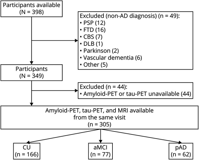
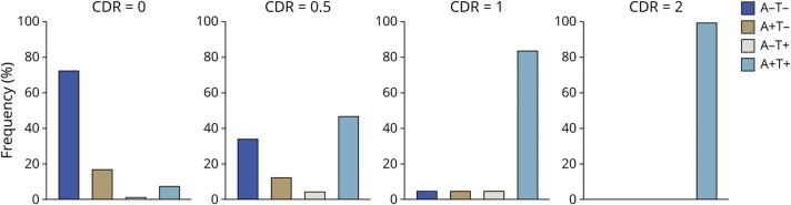
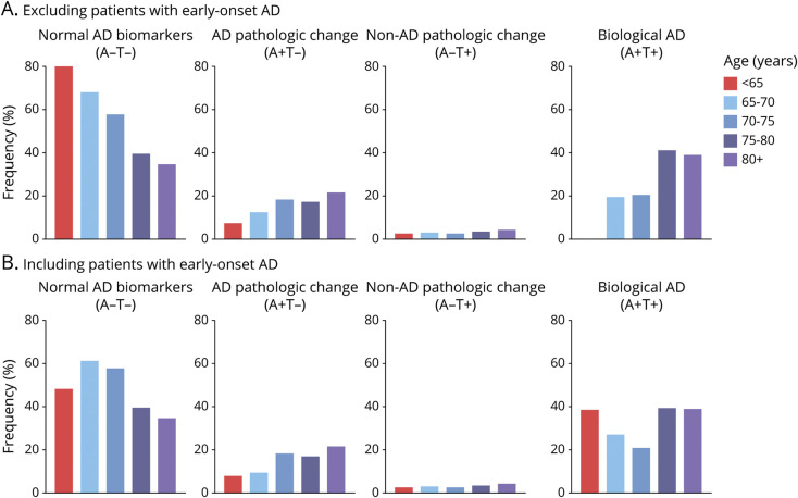
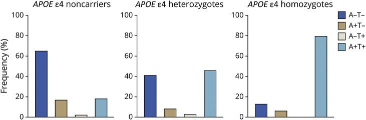
Objective To assess the frequency of biologically defined Alzheimer disease (AD) in relation to age, sex, APOE ε4, and clinical diagnosis in a prospective cohort study evaluated with amyloid-PET and tau-PET. Methods We assessed cognitively unimpaired (CU) elderly (n = 166), patients with amnestic mild cognitive impairment (n = 77), and patients with probable AD dementia (n = 62) who underwent evaluation by dementia specialists and neuropsychologists in addition to amyloid-PET with [18F]AZD4694 and tau-PET with [18F]MK6240. Individuals were grouped according to their AD biomarker profile. Positive predictive value for biologically defined AD was assessed in relation to clinical diagnosis. Frequency of AD biomarker profiles was assessed using logistic regressions with odds ratios (ORs) and 95% confidence intervals (CIs). Results The clinical diagnosis of probable AD dementia demonstrated good agreement with biologically defined AD (positive predictive value 85.2%). A total of 7.88% of CU were positive for both amyloid-PET and tau-PET. Frequency of biologically defined AD increased with age (OR 1.14; p < 0.0001) and frequency of APOE ε4 allele carriers (single ε4: OR 3.82; p < 0.0001; double ε4: OR 17.55, p < 0.0001). Conclusion Whereas we observed strong, but not complete, agreement between clinically defined probable AD dementia and biomarker positivity for both β-amyloid and tau, we also observed that biologically defined AD was not rare in CU elderly. Abnormal tau-PET was almost exclusively observed in individuals with abnormal amyloid-PET. Our results highlight that even in tertiary care memory clinics, detailed evaluation by dementia specialists systematically underestimates the frequency of biologically defined AD and related entities. Classification of Evidence This study provides Class I evidence that biologically defined AD (abnormal amyloid PET and tau PET) was observed in 85.2% of people with clinically defined AD and 7.88% of CU elderly.
Overview
The claims and statistics of this paper haven’t been checked yet.
Run the check — free, no account needed. It reads the open-access text and checks the sources it cites.
Integrity signals
Registry
Cites
Statistics
Figures
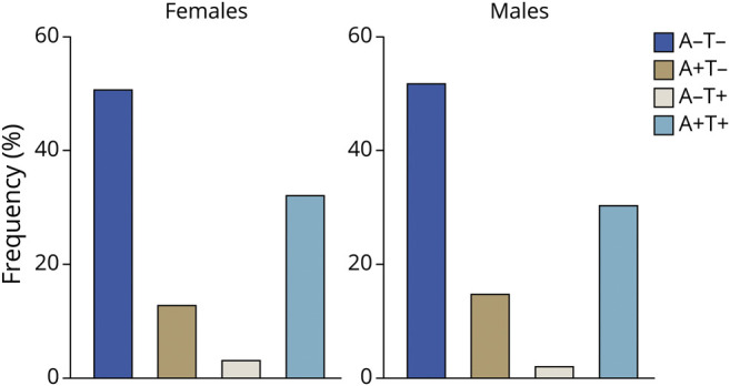
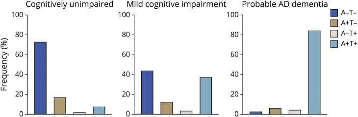
Verification
Claims the paper makes
The claims and statistics of this paper haven’t been checked yet.
Run the check — free, no account needed. It reads the open-access text and checks the sources it cites.
Claims other papers make about this one
Each is checked against this paper’s own text: does it say what the citing paper says it does?
The source backs part of the claim, not all of it — or backs it for a different population, condition or comparison.
In the citing paper
“The clinical syndrome of probable AD dementia is more closely associated with amyloid-β pathology than MCI at all ages19,20,25”
From Diagnosis of Alzheimer's disease using plasma biomarkers adjusted to clinical probability (2024) · cites this paper as [25]
Experimental — accuracy not yet measured
Self-check
Percentages, sums and reported differences re-computed from the paper’s own numbers.
The self-check needs the paper’s full text.
Citations
Cites
McKhann, G., Drachman, D., Folstein, M., Katzman, R., Price, D., & Stadlan, E. M. (1984) · Neurology · doi:10.1212/wnl.34.7.939 · Cited by 27,915
McKhann, G. M., Knopman, D. S., Chertkow, H., Hyman, B. T., Jack, C. R., Kawas, C. H., Klunk, W. E., Koroshetz, W. J., Manly, J. J., Mayeux, R., et al. (2011) · Alzheimer's & Dementia · doi:10.1016/j.jalz.2011.03.005 · Cited by 18,657
Neuropathological stageing of Alzheimer-related changes.
Braak, H., & Braak, E. (1991) · Acta Neuropathologica · doi:10.1007/bf00308809 · Cited by 16,123
NIA‐AA Research Framework: Toward a biological definition of Alzheimer's disease
Jack, C. R., Bennett, D. A., Blennow, K., Carrillo, M. C., Dunn, B., Haeberlein, S. B., Holtzman, D. M., Jagust, W., Jessen, F., Karlawish, J., et al. (2018) · Alzheimer's & Dementia · doi:10.1016/j.jalz.2018.02.018 · Cited by 10,537
Mild cognitive impairment as a diagnostic entity
Petersen, R. C. (2004) · Journal of Internal Medicine · doi:10.1111/j.1365-2796.2004.01388.x · Cited by 7,864
Jack, C. R., Knopman, D. S., Jagust, W. J., Petersen, R. C., Weiner, M. W., Aisen, P. S., Shaw, L. M., Vemuri, P., Wiste, H. J., Weigand, S. D., et al. (2013) · The Lancet Neurology · doi:10.1016/s1474-4422(12)70291-0 · Cited by 4,396
Clinical and biomarker changes in dominantly inherited Alzheimer's disease
Bateman, R. J., Xiong, C., Benzinger, T. L. S., Fagan, A. M., Goate, A., Fox, N. C., Marcus, D. S., Cairns, N. J., Xie, X., Blazey, T. M., et al. (2012) · New England Journal of Medicine · doi:10.1056/nejmoa1202753 · Cited by 3,814
Hyman, B. T., Phelps, C. H., Beach, T. G., Bigio, E. H., Cairns, N. J., Carrillo, M. C., Dickson, D. W., Duyckaerts, C., Frosch, M. P., Masliah, E., et al. (2012) · Alzheimer's & Dementia · doi:10.1016/j.jalz.2011.10.007 · Cited by 3,092
Frequency of Stages of Alzheimer-Related Lesions in Different Age Categories
Braak, H., & Braak, E. (1997) · Neurobiology of Aging · doi:10.1016/s0197-4580(97)00056-0 · Cited by 1,656
Limbic-predominant age-related TDP-43 encephalopathy (LATE): consensus working group report
Nelson, P. T., Dickson, D. W., Trojanowski, J. Q., Jack, C. R., Boyle, P. A., Arfanakis, K., Rademakers, R., Alafuzoff, I., Attems, J., Brayne, C., et al. (2019) · Brain · doi:10.1093/brain/awz099 · Cited by 1,547
Cited by
Sorted by citations
Diagnostic Accuracy of a Plasma Phosphorylated Tau 217 Immunoassay for Alzheimer Disease Pathology
Ashton, N. J., Brum, W. S., Di Molfetta, G., Benedet, A. L., Arslan, B., Jonaitis, E., Langhough, R. E., Cody, K., Wilson, R., Carlsson, C. M., et al. (2024) · JAMA Neurology · doi:10.1001/jamaneurol.2023.5319 · Cited by 884
Self-citationAPOE4 homozygosity represents a distinct genetic form of Alzheimer’s disease
Fortea, J., Pegueroles, J., Alcolea, D., Belbin, O., Dols-Icardo, O., Vaqué-Alcázar, L., Videla, L., Gispert, J. D., Suárez-Calvet, M., Johnson, S. C., et al. (2024) · Nature Medicine · doi:10.1038/s41591-024-02931-w · Cited by 369
Biomarker modeling of Alzheimer’s disease using PET-based Braak staging
Therriault, J., Pascoal, T. A., Lussier, F. Z., Tissot, C., Chamoun, M., Bezgin, G., Servaes, S., Benedet, A. L., Ashton, N. J., Karikari, T. K., et al. (2022) · Nature Aging · doi:10.1038/s43587-022-00204-0 · Cited by 308
Self-citationBrum, W. S., Cullen, N. C., Janelidze, S., Ashton, N. J., Zimmer, E. R., Therriault, J., Benedet, A. L., Rahmouni, N., Tissot, C., Stevenson, J., et al. (2023) · Nature Aging · doi:10.1038/s43587-023-00471-5 · Cited by 248
Self-citationBiomarker-based staging of Alzheimer disease: rationale and clinical applications
Therriault, J., Schindler, S. E., Salvadó, G., Pascoal, T. A., Benedet, A. L., Ashton, N. J., Karikari, T. K., Apostolova, L. G., Murray, M. E., Verberk, I. M. W., et al. (2024) · Nature Reviews Neurology · doi:10.1038/s41582-024-00942-2 · Cited by 225
Self-citationTREM2 dependent and independent functions of microglia in Alzheimer’s disease
Hou, J., Chen, Y., Grajales-Reyes, G., & Colonna, M. (2022) · Molecular Neurodegeneration · doi:10.1186/s13024-022-00588-y · Cited by 191
Equivalence of plasma p‐tau217 with cerebrospinal fluid in the diagnosis of Alzheimer's disease
Therriault, J., Servaes, S., Tissot, C., Rahmouni, N., Ashton, N. J., Benedet, A. L., Karikari, T. K., Macedo, A. C., Lussier, F. Z., Stevenson, J., et al. (2023) · Alzheimer s & Dementia · doi:10.1002/alz.13026 · Cited by 180
Self-citationAstrocyte biomarker signatures of amyloid-β and tau pathologies in Alzheimer’s disease
Ferrari‐Souza, J. P., Ferreira, P. C. L., Bellaver, B., Tissot, C., Wang, Y.-T., Leffa, D. T., Brum, W. S., Benedet, A. L., Ashton, N. J., Bastiani, M. A. D., et al. (2022) · Molecular Psychiatry · doi:10.1038/s41380-022-01716-2 · Cited by 150
Self-citationMontoliu-Gaya, L., Benedet, A. L., Tissot, C., Vrillon, A., Ashton, N. J., Brum, W. S., Lantero-Rodriguez, J., Stevenson, J., Nilsson, J., Sauer, M., et al. (2023) · Nature Aging · doi:10.1038/s43587-023-00405-1 · Cited by 122
Self-citationStaging of Alzheimer’s disease: past, present, and future perspectives
Therriault, J., Zimmer, E. R., Benedet, A. L., Pascoal, T. A., Gauthier, S. G., & Rosa‐Neto, P. (2022) · Trends in Molecular Medicine · doi:10.1016/j.molmed.2022.05.008 · Cited by 123
Self-citationDiagnosis of Alzheimer's disease using plasma biomarkers adjusted to clinical probability
Therriault, J., Janelidze, S., Benedet, A. L., Ashton, N. J., Arranz Martínez, J., Gonzalez-Escalante, A., Bellaver, B., Alcolea, D., Vrillon, A., Karim, H., et al. (2024) · Nature Aging · doi:10.1038/s43587-024-00731-y · Cited by 131
Self-citationAntiamyloid Monoclonal Antibody Therapy for Alzheimer Disease
Ramanan, V. K., Armstrong, M. J., Choudhury, P., Coerver, K. A., Hamilton, R. H., Klein, B. C., Wolk, D. A., Wessels, S. R., & Jones, L. K. (2023) · Neurology · doi:10.1212/wnl.0000000000207757 · Cited by 120
Therriault, J., Brum, W. S., Trudel, L., Macedo, A. C., Bitencourt, F. V., Martins, C. C., Nakouzi, M., Pola, I., Wong, M. W. K., Kac, P. R., et al. (2025) · The Lancet Neurology · doi:10.1016/s1474-4422(25)00227-3 · Cited by 118
Self-citationCassidy, C., Therriault, J., Pascoal, T. A., Cheung, V., Savard, M., Tuominen, L. J., Chamoun, M., McCall, A., Celebi, S., Lussier, F. Z., et al. (2022) · Neuropsychopharmacology · doi:10.1038/s41386-022-01293-6 · Cited by 99
Self-citationComparison of two plasma p-tau217 assays to detect and monitor Alzheimer’s pathology
Therriault, J., Ashton, N. J., Pola, I., Triana‐Baltzer, G., Brum, W. S., Molfetta, G. D., Arslan, B., Rahmouni, N., Tissot, C., Servaes, S., et al. (2024) · EBioMedicine · doi:10.1016/j.ebiom.2024.105046 · Cited by 83
Self-citationComparing tau status determined via plasma pTau181, pTau231 and [18F]MK6240 tau-PET
Tissot, C., Therriault, J., Kunach, P., Benedet, A. L., Pascoal, T. A., Ashton, N. J., Karikari, T. K., Servaes, S., Lussier, F. Z., Chamoun, M., et al. (2022) · EBioMedicine · doi:10.1016/j.ebiom.2022.103837 · Cited by 78
Self-citationCSF p-tau205: a biomarker of tau pathology in Alzheimer’s disease
Lantero‐Rodriguez, J., Montoliu‐Gaya, L., Benedet, A. L., Vrillon, A., Dumurgier, J., Cognat, E., Brum, W. S., Rahmouni, N., Stevenson, J., Servaes, S., et al. (2024) · Acta Neuropathologica · doi:10.1007/s00401-023-02659-w · Cited by 74
Self-citationIslam, T., Hill, E., Abrahamson, E. E., Servaes, S., Smirnov, D. S., Zeng, X., Sehrawat, A., Chen, Y., Kac, P. R., Kvartsberg, H., et al. (2025) · Nature Medicine · doi:10.1038/s41591-024-03400-0 · Cited by 76
Self-citationTherriault, J., Pascoal, T. A., Savard, M., Mathotaarachchi, S. S., Benedet, A. L., Chamoun, M., Tissot, C., Lussier, F. Z., Rahmouni, N., Stevenson, J., et al. (2022) · Science Translational Medicine · doi:10.1126/scitranslmed.abc8693 · Cited by 58
Self-citationNeuropsychiatric Symptoms and Microglial Activation in Patients with Alzheimer Disease
Aguzzoli, C. S., Ferreira, P. C. L., Povala, G., Ferrari‐Souza, J. P., Bellaver, B., Soares, C., Zalzale, H., Lussier, F. Z., Rohden, F., Abbas, S., et al. (2023) · JAMA Network Open · doi:10.1001/jamanetworkopen.2023.45175 · Cited by 54
Self-citationWang, Y., Therriault, J., Servaes, S., Tissot, C., Rahmouni, N., Macedo, A. C., Arias, J. F., Mathotaarachchi, S. S., Benedet, A. L., Stevenson, J., et al. (2023) · Brain · doi:10.1093/brain/awad397 · Cited by 54
Self-citationAshton, N. J., Brum, W. S., Molfetta, G. D., Benedet, A. L., Arslan, B., Jonaitis, E. M., Langhough, R. E., Cody, K. A., Wilson, R. H., Carlsson, C. M., et al. (2023) · medRxiv · doi:10.1101/2023.07.11.23292493 · Cited by 50
Self-citationPlasma tau biomarkers for biological staging of Alzheimer’s disease
Montoliu‐Gaya, L., Salvadó, G., Therriault, J., Nilsson, J., Janelidze, S., Weiner, S., Ashton, N. J., Benedet, A. L., Rahmouni, N., Lantero‐Rodriguez, J., et al. (2025) · Nature Aging · doi:10.1038/s43587-025-00951-w · Cited by 48
Self-citationAPOEε4 potentiates amyloid β effects on longitudinal tau pathology
Ferrari-Souza, J. P., Bellaver, B., Ferreira, P. C. L., Benedet, A. L., Povala, G., Lussier, F. Z., Leffa, D. T., Therriault, J., Tissot, C., Soares, C., et al. (2023) · Nature Aging · doi:10.1038/s43587-023-00490-2 · Cited by 43
Self-citationWoo, M. S., Tissot, C., Lantero‐Rodriguez, J., Snellman, A., Therriault, J., Rahmouni, N., Macedo, A. C., Servaes, S., Wang, Y., Arias, J. F., et al. (2023) · Alzheimer s & Dementia · doi:10.1002/alz.13528 · Cited by 37
Self-citationMicroglia modulate Aβ-dependent astrocyte reactivity in Alzheimer’s disease
Ferrari‐Souza, J. P., Povala, G., Rahmouni, N., Bellaver, B., Ferreira, P. C. L., Bastiani, M. A. D., Leffa, D. T., Lussier, F. Z., Aguzzoli, C. S., Brum, W. S., et al. (2025) · Nature Neuroscience · doi:10.1038/s41593-025-02103-0 · Cited by 46
Self-citationBellaver, B., Puig‐Pijoan, A., Ferrari‐Souza, J. P., Leffa, D. T., Lussier, F. Z., Ferreira, P. C. L., Tissot, C., Povala, G., Therriault, J., Benedet, A. L., et al. (2023) · Alzheimer s & Dementia · doi:10.1002/alz.13014 · Cited by 36
Self-citationTissot, C., Servaes, S., Lussier, F. Z., Ferrari‐Souza, J. P., Therriault, J., Ferreira, P. C. L., Bezgin, G., Bellaver, B., Leffa, D. T., Mathotaarachchi, S. S., et al. (2022) · Journal of Nuclear Medicine · doi:10.2967/jnumed.122.264434 · Cited by 30
Self-citationMohammadi, Z., Alizadeh, H., Marton, J., & Cumming, P. D. (2023) · Biomolecules · doi:10.3390/biom13020290 · Cited by 28
Antonioni, A., Raho, E. M., Lorenzo, F. D., Manzoli, L., Flacco, M. E., & Koch, G. (2025) · Journal of Neurology · doi:10.1007/s00415-025-12996-3 · Cited by 34
Development, initial validation, and application of a visual read method for [ 18 F]MK‐6240 tau PET
Shuping, J., Matthews, D. C., Adamczuk, K., Scott, D., Rowe, C. C., Kreisl, W. C., Johnson, S. C., Lukic, A. S., Johnson, K. A., Rosa‐Neto, P., et al. (2023) · Alzheimer s & Dementia Translational Research & Clinical Interventions · doi:10.1002/trc2.12372 · Cited by 26
Self-citationTemporal Dynamics and Biological Variability of Alzheimer Biomarkers
Yun, J., Shin, D., Lee, E. H., Kim, J. P., Ham, H., Gu, Y., Chun, M. Y., Kang, S. H., Kim, Y. J., Na, D. L., et al. (2025) · JAMA Neurology · doi:10.1001/jamaneurol.2024.5263 · Cited by 32
Self-citationClinical Correlates of the PET-based Braak Staging Framework in Alzheimer's Disease
Macedo, A. C., Durço, D. F. P. A., Tissot, C., Therriault, J., Vilela de Faria, A. O., Aumont, É., Servaes, S., Rahmouni, N., Fernandez-Arias, J., Wang, Y.-T., et al. (2024) · The Journal of Prevention of Alzheimer's Disease · doi:10.14283/jpad.2024.15 · Cited by 26
Self-citationTherriault, J., Woo, M. S., Salvadó, G., Gobom, J., Karikari, T. K., Janelidze, S., Servaes, S., Rahmouni, N., Tissot, C., Ashton, N. J., et al. (2024) · Molecular Neurodegeneration · doi:10.1186/s13024-023-00689-2 · Cited by 25
Self-citationAPOE genotype and sex modulate Alzheimer’s disease pathology in aged EFAD transgenic mice
Balu, D., Valencia‐Olvera, A. C., Islam, Z., Mielczarek, C., Hansen, A. L., Ramos, T. M. P., York, J. M., LaDu, M. J., & Tai, L. M. (2023) · Frontiers in Aging Neuroscience · doi:10.3389/fnagi.2023.1279343 · Cited by 20
Nabizadeh, F. (2023) · Brain Communications · doi:10.1093/braincomms/fcad286 · Cited by 19
Fu, J. F., Lois, C., Sanchez, J. S., Becker, J. A., Rubinstein, Z. B., Thibault, E. G., Salvatore, A. N., Sari, H., Farrell, M. E., Guehl, N. J., et al. (2022) · Journal of Cerebral Blood Flow & Metabolism · doi:10.1177/0271678x221142139 · Cited by 17
Gunter, N. B., Gebre, R. K., Graff‐Radford, J., Heckman, M. G., Jack, C. R., Lowe, V. J., Knopman, D. S., Petersen, R. C., Ross, O. A., Vemuri, P., et al. (2024) · Neurology Genetics · doi:10.1212/nxg.0000000000200120 · Cited by 18
Quispialaya, K. M., Therriault, J., Aliaga, A., Benedet, A. L., Ashton, N. J., Karikari, T. K., Macedo, A. C., Rahmouni, N., Tissot, C., Arias, J. F., et al. (2024) · Neurology · doi:10.1212/wnl.0000000000210211 · Cited by 17
Self-citationMattsson, N., Palmqvist, S., Horie, K., Barthélemy, N. R., Salvadó, G., Binette, A. P., Tideman, P., Morris, J. C., Benzinger, T. L. S., Perrin, R. J., et al. (2026) · The Lancet Neurology · doi:10.1016/s1474-4422(26)00029-3 · Cited by 16
Cataldo, A., Criscuolo, S., Benedetto, E. D., Masciullo, A., Pesola, M., & Schiavoni, R. (2024) · Bioengineering · doi:10.3390/bioengineering11040324 · Cited by 15
Michael, H. U., Youbi, E., Ohadoma, S. C., Ramlall, S., Oosthuizen, F., & Polyakova, M. (2021) · Neuropsychology Review · doi:10.1007/s11065-021-09527-y · Cited by 15
Identify biological Alzheimer’s disease using a novel nucleic acid–linked protein immunoassay
Wang, Y., Ashton, N. J., Therriault, J., Benedet, A. L., Macedo, A. C., Pola, I., Aumont, É., Molfetta, G. D., Arias, J. F., Tan, K., et al. (2024) · Brain Communications · doi:10.1093/braincomms/fcaf004 · Cited by 14
Self-citationPola, I., Ashton, N. J., Bastiani, M. A. D., Brum, W. S., Rahmouni, N., Tan, K., Machado, L. S., Servaes, S., Stevenson, J., Tissot, C., et al. (2025) · Alzheimer s & Dementia · doi:10.1002/alz.70171 · Cited by 15
Self-citationBiofluid-based staging of Alzheimer’s disease
Lantero‐Rodriguez, J., Montoliu‐Gaya, L., Ashton, N. J., Pola, I., Therriault, J., Rahmouni, N., Brum, W. S., Servaes, S., Stevenson, J., Molfetta, G. D., et al. (2025) · Acta Neuropathologica · doi:10.1007/s00401-025-02863-w · Cited by 13
Self-citationTau polygenic risk scoring: a cost-effective aid for prognostic counseling in Alzheimer’s disease
Ramanan, V. K., Heckman, M. G., Lesnick, T. G., Przybelski, S. A., Cahn, E. J., Kosel, M., Murray, M. E., Mielke, M. M., Botha, H., Graff‐Radford, J., et al. (2022) · Acta Neuropathologica · doi:10.1007/s00401-022-02419-2 · Cited by 10
Benedet, A. L., Arslan, B., Tan, K., Huber, H., Pola, I., Molfetta, G. D., Kvartsberg, H., Dolado, A. O., Janelidze, S., Blennow, K., et al. (2025) · Clinical Chemistry · doi:10.1093/clinchem/hvaf162 · Cited by 14
Self-citationQuispialaya, K. M., Therriault, J., Aliaga, A., Tissot, C., Servaes, S., Rahmouni, N., Karikari, T. K., Benedet, A. L., Ashton, N. J., Macedo, A. C., et al. (2024) · European Journal of Neurology · doi:10.1111/ene.16255 · Cited by 12
Self-citationTrudel, L., Therriault, J., Macedo, A. C., Servaes, S., Hosseini, S. A., Bezgin, G., Aumont, É., Rahmouni, N., Arias, J. F., Zheng, Y., et al. (2025) · Neurology · doi:10.1212/wnl.0000000000214111 · Cited by 13
Self-citationDreyer, A. J., Nightingale, S., Heaps‐Woodruff, J. M., Henry, M., Gouse, H., Paul, R. H., Thomas, K. G. F., & Joska, J. A. (2021) · Journal of NeuroVirology · doi:10.1007/s13365-021-00993-x · Cited by 10
Rates of clinical progression according to biological Alzheimer's disease stages
Trudel, L., Therriault, J., Macedo, A. C., Servaes, S., Hosseini, S. A., Bezgin, G., Rahmouni, N., Chan, T., Arias, J. F., Aumont, É., et al. (2025) · Alzheimer s & Dementia · doi:10.1002/alz.70624 · Cited by 13
Self-citationBodryzlova, Y., Mehrabi, F., Bosson, A., Maïano, C., André, C., Bélanger, E., & Moullec, G. (2023) · Journal of Aging & Social Policy · doi:10.1080/08959420.2023.2245672 · Cited by 8
Amyloid beta plaque accumulation with longitudinal [18F]AZD4694 PET
Therriault, J., Lussier, F. Z., Tissot, C., Chamoun, M., Stevenson, J., Rahmouni, N., Pallen, V., Bezgin, G., Servaes, S., Kunach, P., et al. (2023) · Alzheimer s & Dementia Diagnosis Assessment & Disease Monitoring · doi:10.1002/dad2.12391 · Cited by 6
Self-citationKnopman, D. S., & Jagust, W. J. (2020) · Neurology · doi:10.1212/wnl.0000000000011415 · Cited by 7
Clinical–biological Alzheimer's disease stage concordance: insights from cohorts and autopsy data
Trudel, L., Therriault, J., Macedo, A. C., Hosseini, S. A., Arias, J. F., Chan, T., Rahmouni, N., Bezgin, G., Tissot, C., Woo, M. S., et al. (2026) · Brain · doi:10.1093/brain/awag018 · Cited by 8
Self-citationBelasso, C. J., Cai, Z.-C., Bezgin, G., Pascoal, T., Stevenson, J., Rahmouni, N., Tissot, C., Lussier, F., Rosa-Neto, P., Soucy, J., et al. (2023) · Frontiers in Aging Neuroscience · doi:10.3389/fnagi.2023.1225816 · Cited by 5
Self-citationAstrocyte biomarker signatures of amyloid-β and tau pathologies in Alzheimer’s diseasePreprint
Ferrari‐Souza, J. P., Ferreira, P. C. L., Bellaver, B., Tissot, C., Wang, Y.-T., Leffa, D. T., Brum, W. S., Benedet, A. L., Ashton, N. J., Bastiani, M. A. D., et al. (2022) · medRxiv · doi:10.1101/2022.01.25.22269841 · Cited by 6
Self-citationWeiner, S., Sauer, M., Montoliu‐Gaya, L., Benedet, A. L., Ashton, N. J., González‐Ortiz, F., Simrén, J., Rahmouni, N., Tissot, C., Therriault, J., et al. (2025) · Molecular Neurodegeneration · doi:10.1186/s13024-025-00841-0 · Cited by 8
Self-citationClassifying Alzheimer’s Disease Neuropathology Using Clinical and MRI Measurements
Zhuang, X., Cordes, D., Bender, A. R., Nandy, R. R., Oh, E. C. T., Kinney, J. W., Caldwell, J., Cummings, J. L., & Miller, J. B. (2024) · Journal of Alzheimer s Disease · doi:10.3233/jad-231321 · Cited by 7
The Evolution of Randomized Clinical Trial Designs to Assess Therapeutics in Alzheimer Disease
Aumont, É., Therriault, J., Kwan, A. T. H., Carello‐Collar, G., Arfaie, S., Hall, B. J., Ferrari‐Souza, J. P., Woo, M. S., Macedo, A. C., Tissot, C., et al. (2025) · JAMA Network Open · doi:10.1001/jamanetworkopen.2025.29665 · Cited by 5
Self-citationTherriault, J., Gauthier, S. G., & Rosa‐Neto, P. (2023) · The Journal of Prevention of Alzheimer s Disease · doi:10.14283/jpad.2023.52 · Cited by 4
Self-citationWei, B., Xu, Y., Du, Y., Zhou, J., Zhong, F., Wu, C., & Lou, Y. (2024) · Actas Españolas de Psiquiatría · doi:10.62641/aep.v52i2.1552 · Cited by 4
Body Composition and Alzheimer’s Disease: A Holistic Review
Frank, G., Gualtieri, P., Cianci, R., Caldarelli, M., Palma, R., Santis, G. D. D., Porfilio, C., Nicoletti, F., Bigioni, G., & di Renzo, L. (2024) · International Journal of Molecular Sciences · doi:10.3390/ijms25179573 · Cited by 3
Labuza, A., Pidikiti, H., Alldred, M. J., Ibrahim, K. W., Peng, K. Y., Pasato, J., Heguy, A., Mathews, P. M., & Ginsberg, S. D. (2025) · Brain Sciences · doi:10.3390/brainsci15101117 · Cited by 4
Stiver, J., Wang, X., Lui, K. K., Dratva, M. A., Lifset, E. T., Heyworth, N. C., Rivera, C. S., Malhotra, A., Thomas, M. L., Sundermann, E. E., et al. (2025) · Alzheimer s & Dementia · doi:10.1002/alz.71051 · Cited by 3
Plasma proteomic profiles of Alzheimer’s disease and neurodegeneration in African cohorts
Pola, I., Akinyemi, T. O., Tan, K., Olalusi, O., Traichel, W., Giacomucci, G., Yaria, J. O., Rahmouni, N., Ogunde, G. O., Oguntiloye, O., et al. (2026) · Nature Communications · doi:10.1038/s41467-026-74971-4 · Cited by 4
Self-citationLATE, Hippocampal Sclerosis, and Primary Age-related Tauopathy
Ramanan, V. K., & Graff‐Radford, J. (2024) · CONTINUUM Lifelong Learning in Neurology · doi:10.1212/con.0000000000001499 · Cited by 4
LoBue, C., Stopschinski, B. E., Saez-Calveras, N., Douglas, P. M., Huebinger, R. M., Cullum, C. M., Hart, J., & Gonzales, M. M. (2023) · Journal of Alzheimer s Disease · doi:10.3233/jad-231027 · Cited by 4
AT(N) predicts near-term development of Alzheimer’s disease symptoms in unimpaired older adultsPreprint
Strikwerda‐Brown, C., Gonneaud, J., Hobbs, D. A., St‐Onge, F., Binette, A. P., Ozlen, H., Provost, K., Soucy, J., Buckley, R. F., Benzinger, T. L. S., et al. (2022) · medRxiv · doi:10.1101/2022.05.09.22274638 · Cited by 3
Self-citationThe Effect of the Tau Protein on D. melanogaster Lifespan Depends on GSK3 Expression and Sex
Veselkina, E. R., Trostnikov, M. V., Roshina, N. V., & PASYUKOVA, E. G. (2023) · International Journal of Molecular Sciences · doi:10.3390/ijms24032166 · Cited by 3
Chen, X., Huang, T., Shi, C., Xu, S., & Fan, S. (2026) · Frontiers in Aging Neuroscience · doi:10.3389/fnagi.2026.1834591 · Cited by 2
Dementia-Specific Nursing Care Competencies for Nursing Education and Long-Term Care Practice
Mayo, A. M. (2022) · Nursing Clinics of North America · doi:10.1016/j.cnur.2022.02.004 · Cited by 2
Therriault, J., Pascoal, T. A., Gauthier, S. G., & Rosa‐Neto, P. (2021) · Neurology · doi:10.1212/wnl.0000000000012586 · Cited by 2
Self-citationApplications of Alzheimer’s disease staging to clinical trials
Therriault, J., Gauthier, S. G., & Rosa‐Neto, P. (2023) · Aging · doi:10.18632/aging.204482 · Cited by 2
Self-citationHuang, S., Liu, Y., Zhang, J., & Wang, Y. (2025) · Artificial Cells Nanomedicine and Biotechnology · doi:10.1080/21691401.2025.2506591 · Cited by 2
Long-Term Forecasting of Alzheimer’s Disease Progression Using Time-Aware Models
Solak, N., Sabanovic, A., Dedovic, H., Hubana, T., Hodžić, M. I., Fojnica, A., & Mesalic, A. (2025) · International Conference on Information, Communication and Automation Technologies (ICAT) · doi:10.1109/icat66432.2025.11189283 · Cited by 1
Arslan, B., Gobom, J., Andréasson, U., Montoliu‐Gaya, L., Benedet, A. L., Dittrich, A., Kern, S., Skoog, I. G., Ashton, N. J., Chan, T., et al. (2026) · Alzheimer s & Dementia · doi:10.1002/alz.71374 · Cited by 3
Self-citationTau extent outperforms tau load as a predictor of neurodegeneration in Alzheimer’s disease
Macedo, A. C., Trudel, L., Hosseini, S. A., Therriault, J., Aumont, É., Bezgin, G., Rahmouni, N., Stevenson, J., Tissot, C., Woo, M. S., et al. (2026) · Molecular Neurodegeneration · doi:10.1186/s13024-026-00945-1 · Cited by 3
Self-citationChai, L., Zhan, Y., Huang, Y., Tian, M., Zhao, G., Ni, J., Xiong, X., Huang, J., & Lin, B. (2026) · Alzheimer s Research & Therapy · doi:10.1186/s13195-026-02104-6 · Cited by 2
Macedo, A. C., Trudel, L., Hosseini, S. A., Bezgin, G., Quispialaya-Socualaya, K., Therriault, J., Rahmouni, N., Tissot, C., Woo, M. S., Limberger, C., et al. (2026) · EBioMedicine · doi:10.1016/j.ebiom.2026.106460 · Cited by 2
Self-citationGarnier‐Crussard, A., & Krolak‐Salmon, P. (2021) · Neurology · doi:10.1212/wnl.0000000000012585
Fluid and Imaging Markers of Amyloid Pathology
West, T., Meyer, M. R., Kirmess, K. M., Yarasheski, K. E., Braunstein, J. B., & Verghese, P. B. (2025) · Springer eBooks · doi:10.1007/978-3-031-80849-4_1
Akinyemi, T. O., Pola, I., Tan, K., Olalusi, O., Yaria, J. O., Ogunde, G. O., Traichel, W., Rahmouni, N., Oguntiloye, O., Fagbemi, A., et al. (2026) · npj Dementia · doi:10.1038/s44400-026-00136-1 · Cited by 2
Self-citationZhang, H., Wu, J., Bian, Z., Xiaoli Li, A., Guo, X., Gu, G., & Cui, D. Y. (2025) · Journal of Neural Engineering · doi:10.1088/1741-2552/ae220e
Therriault, J., Woo, M. S., Salvadó, G., Gobom, J., Karikari, T. K., Janelidze, S., Servaes, S., Rahmouni, N., Tissot, C., Ashton, N. J., et al. (2024) · Figshare · doi:10.6084/m9.figshare.26664625
Self-citationTherriault, J., Woo, M. S., Salvadó, G., Gobom, J., Karikari, T. K., Janelidze, S., Servaes, S., Rahmouni, N., Tissot, C., Ashton, N. J., et al. (2024) · Figshare · doi:10.6084/m9.figshare.26664619
Self-citationThe Dementia Cascade: From the Preclinical Stage
Chen, Q., Xu, J., Gao, H., & Cai, H. (2026) · Springer eBooks · doi:10.1007/978-981-95-5222-1_1
Rousset, R. Z., den Braber, A., Bongers, B., Boonkamp, L., Wilson, D., Ligthart, L., Pool, R., Visser, P. J., van der Landen, S. M., de Geus, E. J. C., et al. (2026) · Research Square · doi:10.21203/rs.3.rs-8426026/v1
Hemispheric lateralization of Tau associations with visual and verbal memory in Alzheimer’s disease
Arias, J. F., Aumont, É., Therriault, J., Trudel, L., Poltronetti, N. M., Thomas, E. M., Bezgin, G., Servaes, S., Wang, Y., Rahmouni, N., et al. (2026) · European Journal of Nuclear Medicine and Molecular Imaging · doi:10.1007/s00259-026-07898-z
Self-citationLife’s Essential 8, Polygenic Risk for Type 2 Diabetes, and Dementia: Evidence from the UK Biobank
Li, Y.-J., Ekblom, Ö., Bucha, B., Cojocari, A., Wang, H.-X., & Wang, R. (2026) · Nutrients · doi:10.3390/nu18132080
Concordance between plasma biomarkers of phosphorylated tau and tau-PET: A narrative review
van Houwelingen, A., Vernooij, M. W., Vermeiren, M. R., Ossenkoppele, R., van de Giessen, E., Seelaar, H., & Neitzel, J. (2026) · Journal of Alzheimer s Disease · doi:10.1177/13872877261468708
Metadata
- Authors
- Therriault, J., Pascoal, T. A., Benedet, A. L., Tissot, C., Savard, M., Chamoun, M., Lussier, F., Kang, M. S., Berzgin, G., Wang, T., Fernandes-Arias, J., Massarweh, G., Soucy, J.-P., Vitali, P., Saha-Chaudhuri, P., Gauthier, S., & Rosa-Neto, P.
- Affiliations
- Montreal Neurological Institute and Hospital (CA)
- From the Translational Neuroimaging Laboratory, The McGill University Research Centre for Studies in Aging, Douglas Hospital (J.T., T.A.P., A.L.B., C.T., M.S., M.C., F.L., M.S.K., G.B., T.W., J.F.-A., S.G., P.R.-N.), and the Departments of Neurology and Neurosurgery (J.T., T.A.P., A.L.B., C.T., M.C., F.L., M.S.K., G.B., T.W., J.F.-A., J.-P.S., P.V., S.G., P.R.-N.), Psychiatry (S.G., P.R.-N.), Radiochemistry (G.M.), and Epidemiology and Biostatistics (P.S.-C.), McGill University, Montreal; and...
- ORCID
- Venue
- Neurology
- Published
- 2021-02-16
- Volume
- 96
- Issue
- 7
- Pages
- e975-e985
- ISSN
- 0028-3878 (ISSN-L)
- 1526-632X
- PMC
- PMC8055338
- OpenAlex
- W3118160302
- Cited by
- 91
- References
- 53
- Citations by year
- 2026: 14 · 2025: 19 · 2024: 19 · 2023: 21 · 2022: 13 · 2021: 4
- FWCI
- 7.11
- Type
- journal article
- Language
- en
- Open access
- hybrid
- doi.org
- Licence
- cc-by-nc-nd
- Locations
- Neurology (publishedVersion, cc-by-nc-nd)
- PubMed Central (publishedVersion, cc-by-nc-nd)
- PubMed (publishedVersion)
- Funders
- McGill University
- Canadian Institutes of Health Research
- Topics
- Health Sciences › Medicine › Psychiatry and Mental health › Dementia and Cognitive Impairment Research
- Life Sciences › Neuroscience › Cellular and Molecular Neuroscience › Alzheimer's disease research and treatments
- Health Sciences › Medicine › Oncology › Cancer-related cognitive impairment studies
- Keywords
- amyloid PET, tau PET, APOE ε4, amnestic mild cognitive impairment, positive predictive value, sex differences, [18F]MK-6240
- MeSH terms
- Age Factors, Aged, Alzheimer Disease, Brain, Cognition, Female, Humans, Male, Middle Aged, Neuropsychological Tests, Sex Factors, Amyloid beta-Peptides, tau Proteins, Positron-Emission Tomography, Apolipoprotein E4, Cognitive Dysfunction
About these checks
Registry, retraction, Cites and statistics results are computed, not judged, with one exception: a printed reference that no index lists is read once by a model before it counts as a citation error. Claims are extracted by a model and kept only when their quote appears verbatim in the text; each cited source is then checked, with a model’s help, for whether it says what the claim relies on. Those verdicts can be wrong — read the claim’s quote and the cited source before relying on one.
Statistics and arithmetic are recomputed, not judged.
Confidence bands are shown only for fields calibrated on our annotated test set. None is calibrated yet, so no band is shown.
What counts as a claim ›How we measure confidence ›How we check this ›
Are you an author, or do you see a problem with this page? Request a review
