

The DOI matches its Crossref record; 1 of the 1 references it cites were found in a registry or an index.
Barrier Capacity of Human Placenta for Nanosized Materials
Abstract
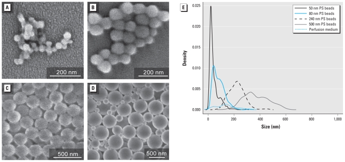
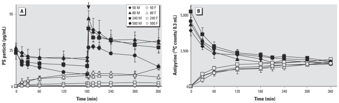
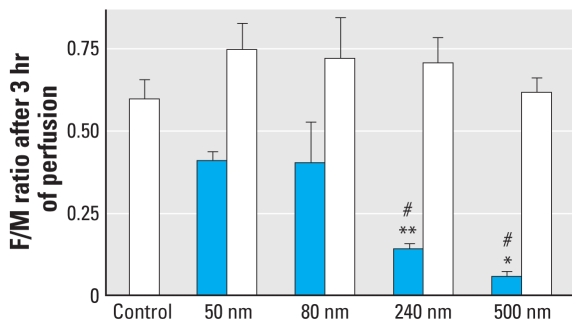
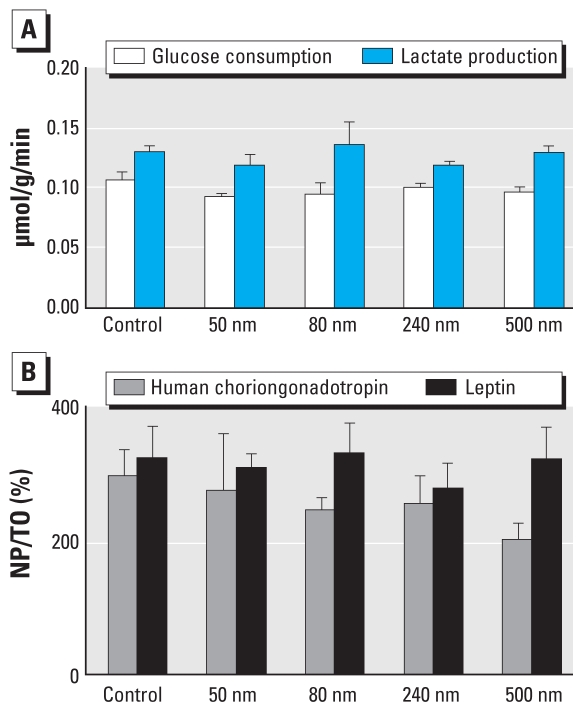
Background: Humans have been exposed to fine and ultrafine particles throughout their history. Since the Industrial Revolution, sources, doses, and types of nanoparticles have changed dramatically. In the last decade, the rapidly developing field of nanotechnology has led to an increase of engineered nanoparticles with novel physical and chemical properties. Regardless of whether this exposure is unintended or not, a careful assessment of possible adverse effects is needed. A large number of projects have been carried out to assess the consequences of combustion-derived or engineered nanoparticle exposure on human health. In recent years there has been a growing concern about the possible health influence of exposure to air pollutants during pregnancy, hence an implicit concern about potential risk for nanoparticle exposure in utero. Previous work has not addressed the question of whether nanoparticles may cross the placenta. Objective: In this study we investigated whether particles can cross the placental barrier and affect the fetus. Methods: We used the ex vivo human placental perfusion model to investigate whether nanoparticles can cross this barrier and whether this process is size dependent. Fluorescently labeled polystyrene beads with diameters of 50, 80, 240, and 500 nm were chosen as model particles. Results: We showed that fluorescent polystyrene particles with diameter up to 240 nm were taken up by the placenta and were able to cross the placental barrier without affecting the viability of the placental explant. Conclusions: The findings suggest that nanomaterials have the potential for transplacental transfer and underscore the need for further nanotoxicologic studies on this important organ system.
Overview
The claims and statistics of this paper haven’t been checked yet.
Run the check — free, no account needed. It reads the open-access text and checks the sources it cites.
Integrity signals
Registry
Cites
Statistics
Figures
Verification
Claims the paper makes
The claims and statistics of this paper haven’t been checked yet.
Run the check — free, no account needed. It reads the open-access text and checks the sources it cites.
Claims other papers make about this one
Each is checked against this paper’s own text: does it say what the citing paper says it does?
In the citing paper
“Experiments with an ex vivo placental perfusion model showed an inverse correlation between the translocation of nano- and submicro-particles (in this case, polystyrene beads) and particle size [192].”
From Particle toxicology and health - where are we? (2019) · cites this paper as [192]
Self-check
Percentages, sums and reported differences re-computed from the paper’s own numbers.
The self-check needs the paper’s full text.
Citations
Cites
Ultrafine Particles in the Urban Air: To the Respiratory Tract--And Beyond?
Oberdörster, G., & Utell, M. J. (2002) · Environmental Health Perspectives · doi:10.1289/ehp.110-a440 · Cited by 304
Research works printed but not linked: 31
- Alfaro-Moreno, 2007, Particulate matter in the environment: pulmonary and cardiovascular effects, Curr Opin Pulm Med, 13, 98, 10.1097/MCP.0b013e328013f47e · DOI
- Challier, 1985, Criteria for evaluating perfusion experiments and presentation of results, Contrib Gynecol Obstet, 13, 32, 10.1159/000410667 · DOI
- Conner, 2003, Regulated portals of entry into the cell, Nature, 422, 37, 10.1038/nature01451 · DOI
- Enders, 1999, Comparative placental structure, Adv Drug Deliv Rev, 38, 3, 10.1016/S0169-409X(99)00003-4 · DOI
- Ganapathy, 2000, Placental transporters relevant to drug distribution across the maternal-fetal interface, J Pharmacol Exp Ther, 294, 413, 10.1016/S0022-3565(24)39087-1 · DOI
- Geiser, 2005, Ultrafine particles cross cellular membranes by nonphagocytic mechanisms in lungs and in cultured cells, Environ Health Perspect, 113, 1555, 10.1289/ehp.8006 · DOI
- Hillaireau, 2009, Nanocarrier’s entry into the cell: relevance to drug delivery, Cell Mol Life Sci, 66, 2873, 10.1007/s00018-009-0053-z · DOI
- Kanno, 2007, A murine scavenger receptor MARCO recognizes polystyrene nanoparticles, Toxicol Sci, 97, 398, 10.1093/toxsci/kfm050 · DOI
- Kertschanska, 1997, Pressure dependence of so-called transtrophoblastic channels during fetal perfusion of human placental villi, Microsc Res Tech, 38, 52, 10.1002/(SICI)1097-0029(19970701/15)38:1/2<52::AID-JEMT7>3.0.CO;2-W · DOI
- Kreyling, 2004, Dosimetry and toxicology of ultrafine particles, J Aerosol Med, 17, 140, 10.1089/0894268041457147 · DOI
- Lacasana, 2005, Exposure to ambient air pollution and prenatal and early childhood health effects, Eur J Epidemiol, 20, 183, 10.1007/s10654-004-3005-9 · DOI
- Latzin, 2008, Air pollution during pregnancy and lung function in newborns: a birth cohort study, Eur Respir J, 33, 594, 10.1183/09031936.00084008 · DOI
- Malek, 2009, The impact of cocaine and heroin on the placental transfer of methadone, Reprod Biol Endocrinol, 7, 61, 10.1186/1477-7827-7-61 · DOI
- Malek, 1994, Maternal-fetal transport of immunoglobulin G and its subclasses during the third trimester of human pregnancy, Am J Reprod Immunol, 32, 8, 10.1111/j.1600-0897.1994.tb00873.x · DOI · Paper page
- Malek, 2000, Production of protein hormones by cultured trophoblast cells isolated from term and early placentae, Am J Reprod Immunol, 43, 278, 10.1111/j.8755-8920.2000.430506.x · DOI
- Malek, 2001, Capacity for hormone production of cultured trophoblast cells obtained from placentae at term and in early pregnancy, J Assist Reprod Genet, 18, 299, 10.1023/A:1016674503298 · DOI
- Myllynen, 2008, Kinetics of gold nanoparticles in the human placenta, Reprod Toxicol, 26, 130, 10.1016/j.reprotox.2008.06.008 · DOI · Paper page
- Nemmar, 2007, Cardiovascular and lung inflammatory effects induced by systemically administered diesel exhaust particles in rats, Am J Physiol Lung Cell Mol Physiol, 292, L664, 10.1152/ajplung.00240.2006 · DOI
- Nemmar, 2005, Evaluation of particle translocation across the alveolo-capillary barrier in isolated perfused rabbit lung model, Toxicology, 208, 105, 10.1016/j.tox.2004.11.012 · DOI
- Nemmar, 2008, The acute proinflammatory and prothrombotic effects of pulmonary exposure to rutile TiO2 nanorods in rats, Exp Biol Med (Maywood), 233, 610, 10.3181/0706-RM-165 · DOI
- Panigel, 1967, Radioangiographic study of circulation in the villi and intervillous space of isolated human placental cotyledon kept viable by perfusion [Letter] [in French], J Physiol (Paris), 59, 277
- Pietzonka, 2002, Transfer of lipophilic markers from PLGA and polystyrene nanoparticles to caco-2 monolayers mimics particle uptake, Pharm Res, 19, 595, 10.1023/A:1015393710253 · DOI
- Reddy, 2007, Exploiting lymphatic transport and complement activation in nanoparticle vaccines, Nat Biotechnol, 25, 1159, 10.1038/nbt1332 · DOI
- Rothen-Rutishauser, 2006, Interaction of fine particles and nanoparticles with red blood cells visualized with advanced microscopic techniques, Environ Sci Technol, 40, 4353, 10.1021/es0522635 · DOI
- Rudin, 2003, Molecular imaging in drug discovery and development, Nat Rev Drug Discov, 2, 123, 10.1038/nrd1007 · DOI
- Schneider, 1972, Transfer across the perfused human placenta of antipyrine, sodium and leucine, Am J Obstet Gynecol, 114, 822, 10.1016/0002-9378(72)90909-X · DOI
- Semmler-Behnke, 2007, Uptake of 1.4 nm versus 18 nm gold nanoparticles in secondary target organs is size dependent in control and pregnant rats after intratrecheal or intravenous application, 102
- Stoeger, 2006, Instillation of six different ultrafine carbon particles indicates a surface area threshold dose for acute lung inflammation in mice, Environ Health Perspect, 114, 328, 10.1289/ehp.8266 · DOI
- Syme, 2004, Drug transfer and metabolism by the human placenta, Clin Pharmacokinet, 43, 487, 10.2165/00003088-200443080-00001 · DOI
- Takata, 1997, Ultrastructure of the rodent placental labyrinth: a site of barrier and transport, J Reprod Dev, 43, 13, 10.1262/jrd.43.13 · DOI
- Thiesen, 2008, Clinical applications of magnetic nanoparticles for hyperthermia, Int J Hyperthermia, 24, 467, 10.1080/02656730802104757 · DOI · Paper page
Cited by
Sorted by citations
Discovery and quantification of plastic particle pollution in human blood
Leslie, H. A., van Velzen, M. J. M., Brandsma, S. H., Vethaak, A. D., Garcia-Vallejo, J. J., & Lamoree, M. H. (2022) · Environment International · doi:10.1016/j.envint.2022.107199 · Cited by 3,906
Environmental exposure to microplastics: An overview on possible human health effects
Prata, J. C., da Costa, J. P., Lopes, I., Duarte, A. C., & Rocha-Santos, T. (2020) · Science of The Total Environment · doi:10.1016/j.scitotenv.2019.134455 · Cited by 2,690
Diverse Applications of Nanomedicine
Pelaz, B., Alexiou, C., Alvarez-Puebla, R. A., Alves, F., Andrews, A. M., Ashraf, S., Balogh, L. P., Ballerini, L., Bestetti, A., Brendel, C., et al. (2017) · ACS Nano · doi:10.1021/acsnano.6b06040 · Cited by 1,350
Self-citationPresence of microplastics and nanoplastics in food, with particular focus on seafood
(CONTAM), E. P. O. C. I. T. F. C. (2016) · EFSA Journal · doi:10.2903/j.efsa.2016.4501 · Cited by 958
Ambient black carbon particles reach the fetal side of human placenta
Bové, H., Bongaerts, E., Slenders, E., Bijnens, E. M., Saenen, N. D., Gyselaers, W., Van Eyken, P., Plusquin, M., Roeffaers, M. B. J., Ameloot, M., et al. (2019) · Nature Communications · doi:10.1038/s41467-019-11654-3 · Cited by 709
Metadata
- Authors
- Wick, P., Malek, A., Manser, P., Meili, D., Maeder-Althaus, X., Diener, L., Diener, P.-A., Zisch, A., Krug, H. F., & von Mandach, U.
- Venue
- Environmental Health Perspectives
- Publisher
- American Chemical Society (ACS)
- Published
- 2009-11-12
- Volume
- 118
- Issue
- 3
- Pages
- 432-436
- ISSN
- 0091-6765
- 1552-9924
- PMC
- PMC2854775
- Cited by
- 584
- References
- 1
- Language
- en
About these checks
Registry, retraction, Cites and statistics results are computed, not judged, with one exception: a printed reference that no index lists is read once by a model before it counts as a citation error. Claims are extracted by a model and kept only when their quote appears verbatim in the text; each cited source is then checked, with a model’s help, for whether it says what the claim relies on. Those verdicts can be wrong — read the claim’s quote and the cited source before relying on one.
Statistics and arithmetic are recomputed, not judged.
Confidence bands are shown only for fields calibrated on our annotated test set. None is calibrated yet, so no band is shown.
What counts as a claim ›How we measure confidence ›How we check this ›
Are you an author, or do you see a problem with this page? Request a review
How to cite
Cite CoChat for the checks on this page, and the paper for its own findings.
- This page
- CoChat. (2026). Citation check: Barrier Capacity of Human Placenta for Nanosized Materials. https://app.cochat.ai/papers/10.1289/ehp.0901200
- The paper
- Wick, P., Malek, A., Manser, P., Meili, D., Maeder-Althaus, X., Diener, L., Diener, P.-A., Zisch, A., Krug, H. F., & von Mandach, U. (2010). Barrier Capacity of Human Placenta for Nanosized Materials. Environmental Health Perspectives. https://doi.org/10.1289/ehp.0901200
