Cited by 103Cancer-related Molecular PathwaysGenomics and Chromatin DynamicsProtein Structure and Dynamics

The DOI matches its Crossref record; 112 of the 113 references it cites were found in a registry or an index.

Noncanonical DNA Motifs as Transactivation Targets by Wild Type and Mutant p53

Open access
Jordan, Jennifer J., Menendez, Daniel, Inga, Alberto, Nourredine, Maher, Bell, Douglas, Resnick, Michael A.·2008·PLoS Genetics

Abstract

Sequence-specific binding by the human p53 master regulator is critical to its tumor suppressor activity in response to environmental stresses. p53 binds as a tetramer to two decameric half-sites separated by 0-13 nucleotides (nt), originally defined by the consensus RRRCWWGYYY (n = 0-13) RRRCWWGYYY. To better understand the role of sequence, organization, and level of p53 on transactivation at target response elements (REs) by wild type (WT) and mutant p53, we deconstructed the functional p53 canonical consensus sequence using budding yeast and human cell systems. Contrary to early reports on binding in vitro, small increases in distance between decamer half-sites greatly reduces p53 transactivation, as demonstrated for the natural TIGER RE. This was confirmed with human cell extracts using a newly developed, semi-in vitro microsphere binding assay. These results contrast with the synergistic increase in transactivation from a pair of weak, full-site REs in the MDM2 promoter that are separated by an evolutionary conserved 17 bp spacer. Surprisingly, there can be substantial transactivation at noncanonical (1/2)-(a single decamer) and (3/4)-sites, some of which were originally classified as biologically relevant canonical consensus sequences including PIDD and Apaf-1. p53 family members p63 and p73 yielded similar results. Efficient transactivation from noncanonical elements requires tetrameric p53, and the presence of the carboxy terminal, non-specific DNA binding domain enhanced transactivation from noncanonical sequences. Our findings demonstrate that RE sequence, organization, and level of p53 can strongly impact p53-mediated transactivation, thereby changing the view of what constitutes a functional p53 target. Importantly, inclusion of (1/2)- and (3/4)-site REs greatly expands the p53 master regulatory network.

Overview

The claims and statistics of this paper haven’t been checked yet.

Run the check — free, no account needed. It reads the open-access text and checks the sources it cites.

A few free checks a day · papers already verified open instantly · results are public

Integrity signals

Registry

Crossref · 10.1371/journal.pgen.1000104

Cites

112 of 112 references verified0 citation errors · 0 not checkable

Statistics

Figures

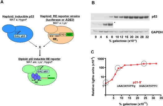
Figure 1 Isogenomic diploid yeast system to investigate transcriptional capacity of p53 towards many REs at various p53 levels. (A) Transactivation capacities of p53 (WT or mutant) from cognate REs were determined using diploid strains derived from haploids where p53 and reporters with upstream REs were integrated into different chromosomal loci. Within the p53-host strains, p53 (WT or mutant) was controlled by a “rheostatable” GAL1 promoter (blue cells) (previously described [109]) which allows for controlled, inducible expression of p53. Reporter strains contained promoter REs upstream of the minimal CYC1 promoter and either the ADE2 color reporter (REP) or the firefly luciferase reporter (tan cells) [28]. Mating of the REP and p53-host strains results in isogenomic diploid yeast that provide for rapid assessment of the transactivation potential for WT or mutant p53 proteins towards many individual REs in the p53 transcriptional network [14]. (B) Inducible expression of p53 under the rheostatable GAL1 promoter. The GAL1 promoter allows for controlled expression of p53 depending on the level of galactose in the media. Raffinose was added to provide a basal level of expression, as the GAL1 promoter is derepressed, but not induced in media containing this carbon source. Presented is a Western blot analysis of p53 expression 24 hours post-inoculation with raffinose or raffinose plus increasing amounts of galactose (0.004–0.032%). Increases in galactose from 0–0.032% correlated with an increase in p53 over a 100-fold range. The p53 protein was detected with DO-1 and pAb1801 antibodies. The asterisk (*) depicts a longer exposure to reveal protein at basal and lower galactose levels. GAPDH, identified by immunodetection provided a standard loading control. (C) Quantitative assessment of p53-induced transactivation from the p21-5′ RE in vivo. The diploid yeast strain containing GAL1::WT p53 crossed with the p21-5′ RE-luciferase REP was grown overnight in complete medium, diluted, washed and inoculated into selective medium containing either raffinose or raffinose plus increasing galactose (0–0.032%) for 24 hours. Protein lysates were obtained and a luciferase assay was used to determine the transactivation capacity of p53 from the p21-5′ RE. The strength of transactivation was calculated as relative light units/µg protein. Circled are the basal, linear-increase, and plateau phases of the transactivation response as a function of galactose concentration and are referred to as basal, moderate and high levels of p53 expression.
Figure 1 Isogenomic diploid yeast system to investigate transcriptional capacity of p53 towards many REs at various p53 levels. (A) Transactivation capacities of p53 (WT or mutant) from cognate REs were determined using diploid strains derived from haploids where p53 and reporters with upstream REs were integrated into different chromosomal loci. Within the p53-host strains, p53 (WT or mutant) was controlled by a “rheostatable” GAL1 promoter (blue cells) (previously described [109]) which allows for controlled, inducible expression of p53. Reporter strains contained promoter REs upstream of the minimal CYC1 promoter and either the ADE2 color reporter (REP) or the firefly luciferase reporter (tan cells) [28]. Mating of the REP and p53-host strains results in isogenomic diploid yeast that provide for rapid assessment of the transactivation potential for WT or mutant p53 proteins towards many individual REs in the p53 transcriptional network [14]. (B) Inducible expression of p53 under the rheostatable GAL1 promoter. The GAL1 promoter allows for controlled expression of p53 depending on the level of galactose in the media. Raffinose was added to provide a basal level of expression, as the GAL1 promoter is derepressed, but not induced in media containing this carbon source. Presented is a Western blot analysis of p53 expression 24 hours post-inoculation with raffinose or raffinose plus increasing amounts of galactose (0.004–0.032%). Increases in galactose from 0–0.032% correlated with an increase in p53 over a 100-fold range. The p53 protein was detected with DO-1 and pAb1801 antibodies. The asterisk (*) depicts a longer exposure to reveal protein at basal and lower galactose levels. GAPDH, identified by immunodetection provided a standard loading control. (C) Quantitative assessment of p53-induced transactivation from the p21-5′ RE in vivo. The diploid yeast strain containing GAL1::WT p53 crossed with the p21-5′ RE-luciferase REP was grown overnight in complete medium, diluted, washed and inoculated into selective medium containing either raffinose or raffinose plus increasing galactose (0–0.032%) for 24 hours. Protein lysates were obtained and a luciferase assay was used to determine the transactivation capacity of p53 from the p21-5′ RE. The strength of transactivation was calculated as relative light units/µg protein. Circled are the basal, linear-increase, and plateau phases of the transactivation response as a function of galactose concentration and are referred to as basal, moderate and high levels of p53 expression.CC0 · PubMed Central
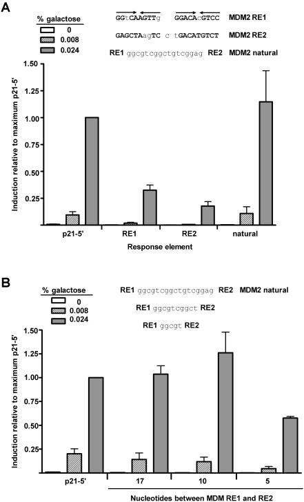
Figure 2 Weak REs can function synergistically when separated by a spacer. (A) To ascertain if p53 functions from the two full-site REs of MDM2 independently or if the REs interact, p53 transactivation from the isolated REs, as well as the natural MDM2 RE containing a 17 nucleotide spacer were evaluated. Isogenomic diploid yeast strains containing the p21-5′ and MDM2 REs, as indicated, were grown in increasing concentrations of galactose to induce p53 protein to basal, moderate and high levels of expression. The ability of WT p53 to transactivate from RE sequences was measured by a luciferase assay 24 hours after inoculation into the galactose supplemented media. Induction from each RE was compared relative to the ability of p53 to transactivate from the p21-5′ RE at 0.024% galactose and is depicted as the mean and standard error of measurement (SEM) of 6 independent experiments. The average light units/µg protein from p21-5′ at 0.024% galactose was 2.1 million. Solid arrows over the sequences indicate a ¼-binding site. (B) Impact of reducing the spacer to 10 and 5 nucleotides. The average light units/µg protein for p21-5′ at 0.024% galactose was 2,800,000.
Figure 2 Weak REs can function synergistically when separated by a spacer. (A) To ascertain if p53 functions from the two full-site REs of MDM2 independently or if the REs interact, p53 transactivation from the isolated REs, as well as the natural MDM2 RE containing a 17 nucleotide spacer were evaluated. Isogenomic diploid yeast strains containing the p21-5′ and MDM2 REs, as indicated, were grown in increasing concentrations of galactose to induce p53 protein to basal, moderate and high levels of expression. The ability of WT p53 to transactivate from RE sequences was measured by a luciferase assay 24 hours after inoculation into the galactose supplemented media. Induction from each RE was compared relative to the ability of p53 to transactivate from the p21-5′ RE at 0.024% galactose and is depicted as the mean and standard error of measurement (SEM) of 6 independent experiments. The average light units/µg protein from p21-5′ at 0.024% galactose was 2.1 million. Solid arrows over the sequences indicate a ¼-binding site. (B) Impact of reducing the spacer to 10 and 5 nucleotides. The average light units/µg protein for p21-5′ at 0.024% galactose was 2,800,000.CC0 · PubMed Central
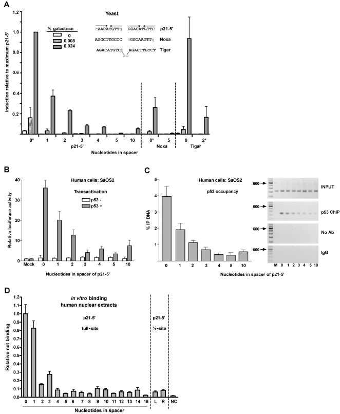
Figure 3 Spacer decreases p53 transactivation, promoter occupancy and binding in yeast and mammalian cells and in vitro. (A) Transactivation in yeast. The ability of p53 to transactivate from REs containing spacers of variable nucleotide length sequences was measured 24 hours after p53 induction with a quantitative luciferase assay. Induction from each RE at various p53 expression levels were compared to the induction from the p21-5′ RE at 0.024% galactose. The average light units/µg protein for WT p53 towards p21-5′ at 0.024% galactose for a minimum of 6 biological repeats was 1.26 million. *indicates the number of nucleotides in the spacer of the natural RE. Solid arrows identify a ¼-binding site. (B) Human SaOS2 cells were transfected with p21-5′::luciferase reporter constructs containing spacers of increasing length between decamer half-sites in the presence (solid bars) or absence (open bars) of the high expressing pCMV-p53 WT vector. At 48 hours post transfection, induction of the luciferase reporter was assessed. Relative luciferase activity was compared to the pGL3-P plasmid lacking the p53 RE (mock) and is represented by the average and standard deviations of three independent experiments, each containing three replicates. (C) Occupancy of p53 at p21-5′ REs in human cells. The p21-5′ promoter constructs containing the increasing spacers between half-sites were co-transfected with p53 into SaOS2 cells (as described in (B)). Twenty-four hours later, ChIP analysis was performed. Presented are the average and standard deviation from 4 independent experiments (left). PCR products of the Input DNA (input) and ChIP DNA (p53, IgG, or no antibody, Ab) are shown (right). The “M” corresponds to a pGL3-P plasmid control lacking the p53 RE. No bands were observed above the 600 bp markers. (D) In vitro fluorescent microsphere binding assay to evaluate sequence-specific p53-DNA binding interactions (see Materials and Methods). Fluorescent microspheres bearing double stranded DNA fragments were multiplexed and incubated in the presence of nuclear extracts from non-treated (NT) or Doxo-treated (0.6 ug/mL [1 mM] Doxorubicin for 18 hours at 37°C) lymphoblastoid cells. The DNA fragments contained the p21-5′ RE, p21-5′ RE with spacers of increasing length (0–15), p21-5′ half-site RE (left or right), or a scramble sequence.
Figure 3 Spacer decreases p53 transactivation, promoter occupancy and binding in yeast and mammalian cells and in vitro. (A) Transactivation in yeast. The ability of p53 to transactivate from REs containing spacers of variable nucleotide length sequences was measured 24 hours after p53 induction with a quantitative luciferase assay. Induction from each RE at various p53 expression levels were compared to the induction from the p21-5′ RE at 0.024% galactose. The average light units/µg protein for WT p53 towards p21-5′ at 0.024% galactose for a minimum of 6 biological repeats was 1.26 million. *indicates the number of nucleotides in the spacer of the natural RE. Solid arrows identify a ¼-binding site. (B) Human SaOS2 cells were transfected with p21-5′::luciferase reporter constructs containing spacers of increasing length between decamer half-sites in the presence (solid bars) or absence (open bars) of the high expressing pCMV-p53 WT vector. At 48 hours post transfection, induction of the luciferase reporter was assessed. Relative luciferase activity was compared to the pGL3-P plasmid lacking the p53 RE (mock) and is represented by the average and standard deviations of three independent experiments, each containing three replicates. (C) Occupancy of p53 at p21-5′ REs in human cells. The p21-5′ promoter constructs containing the increasing spacers between half-sites were co-transfected with p53 into SaOS2 cells (as described in (B)). Twenty-four hours later, ChIP analysis was performed. Presented are the average and standard deviation from 4 independent experiments (left). PCR products of the Input DNA (input) and ChIP DNA (p53, IgG, or no antibody, Ab) are shown (right). The “M” corresponds to a pGL3-P plasmid control lacking the p53 RE. No bands were observed above the 600 bp markers. (D) In vitro fluorescent microsphere binding assay to evaluate sequence-specific p53-DNA binding interactions (see Materials and Methods). Fluorescent microspheres bearing double stranded DNA fragments were multiplexed and incubated in the presence of nuclear extracts from non-treated (NT) or Doxo-treated (0.6 ug/mL [1 mM] Doxorubicin for 18 hours at 37°C) lymphoblastoid cells. The DNA fragments contained the p21-5′ RE, p21-5′ RE with spacers of increasing length (0–15), p21-5′ half-site RE (left or right), or a scramble sequence.CC0 · PubMed Central
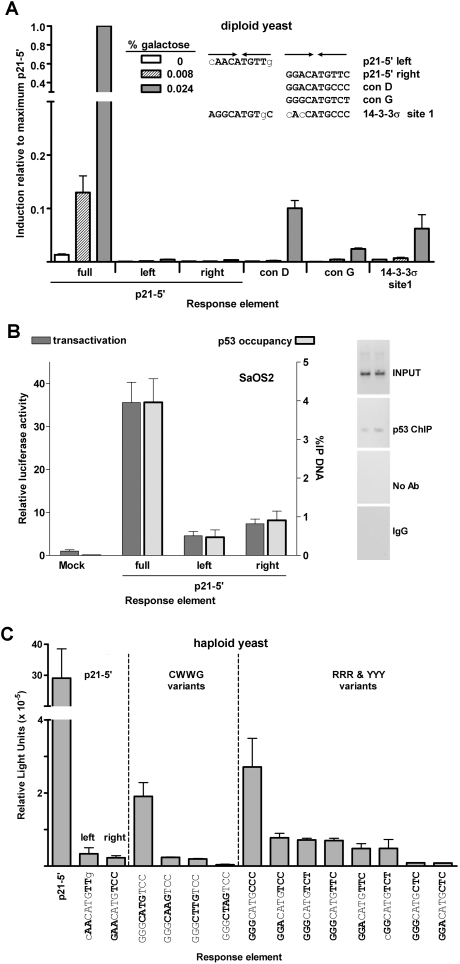
Figure 4 Half-sites function as noncanonical REs for transactivation in a sequence-dependent manner in yeast and human cells. (A) Transactivation from decamer REs in the diploid yeast was quantified with a luciferase assay. Protein lysates were obtained 24 hours post inoculation into galactose supplemented media. The average light units/µg protein for WT p53 towards p21-5′ at 0.024% galactose for six biological repeats was 1.8 million. Solid arrows indicate ¼-binding sites. Comparisons were made with transactivation from the p21-5′ full site at high protein levels. (B) Transactivation in human SaOS2 cells. The cells were co-transfected with WT p53 along with either the full, left or right half-sites of the p21-5′ RE containing reporter construct. Transactivation was assessed with the luciferase assay 48 hours later. Relative luciferase activity was compared to a mock transfection containing the promoter-less pGL3 plasmid. Presented are the averages and standard deviations of 3 independent experiments that were each done in triplicate. PCR products of the input DNA (input) and ChIP DNA (p53, IgG or no antibody, Ab) are shown (right). Input and ChIP PCR products for the mock and p21-5′ full-site are shown in Figure 3C. No bands were observed above the 600 bp markers. (C) Sequence dependence of p53 transactivation from decamer half-sites. The extent to which p53 transactivation from half-sites is sequence dependent at high expression levels (2% galactose) was determined with a plasmid-based haploid yeast system [30]. Relative light units/µg protein from ½-sites were compared to transactivation from the yLFM strain containing the p21-5′ full-site RE. The average light units/µg protein from p21-5′ at 2% galactose was 2.9 million.
Figure 4 Half-sites function as noncanonical REs for transactivation in a sequence-dependent manner in yeast and human cells. (A) Transactivation from decamer REs in the diploid yeast was quantified with a luciferase assay. Protein lysates were obtained 24 hours post inoculation into galactose supplemented media. The average light units/µg protein for WT p53 towards p21-5′ at 0.024% galactose for six biological repeats was 1.8 million. Solid arrows indicate ¼-binding sites. Comparisons were made with transactivation from the p21-5′ full site at high protein levels. (B) Transactivation in human SaOS2 cells. The cells were co-transfected with WT p53 along with either the full, left or right half-sites of the p21-5′ RE containing reporter construct. Transactivation was assessed with the luciferase assay 48 hours later. Relative luciferase activity was compared to a mock transfection containing the promoter-less pGL3 plasmid. Presented are the averages and standard deviations of 3 independent experiments that were each done in triplicate. PCR products of the input DNA (input) and ChIP DNA (p53, IgG or no antibody, Ab) are shown (right). Input and ChIP PCR products for the mock and p21-5′ full-site are shown in Figure 3C. No bands were observed above the 600 bp markers. (C) Sequence dependence of p53 transactivation from decamer half-sites. The extent to which p53 transactivation from half-sites is sequence dependent at high expression levels (2% galactose) was determined with a plasmid-based haploid yeast system [30]. Relative light units/µg protein from ½-sites were compared to transactivation from the yLFM strain containing the p21-5′ full-site RE. The average light units/µg protein from p21-5′ at 2% galactose was 2.9 million.CC0 · PubMed Central
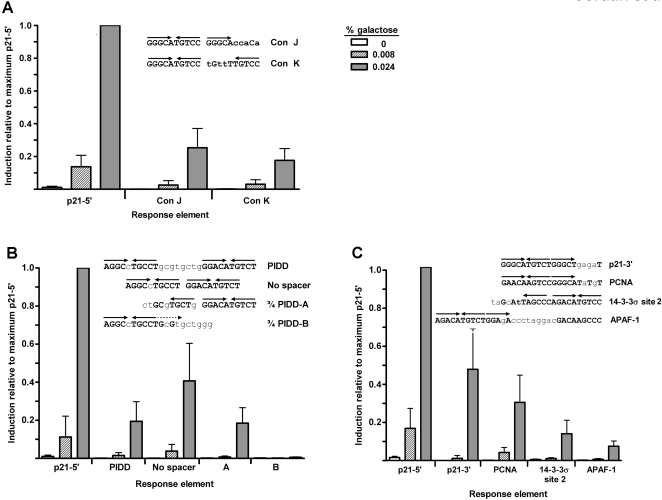
Figure 5 A ¾-site can function as a noncanonical RE in p53 transactivation. (A) Transactivation was assessed from ¾ REs Con J and Con K. The ability of WT p53 to transactivate was measured with the diploid yeast luciferase assay 24 hours post inoculation into the galactose-supplemented media. Induction from each RE was compared relative to the ability of p53 to transactivate from the p21-5′ RE at 0.024% galactose and is depicted as the mean and SEM of 6 independent experiments. The average light units/µg protein from p21-5′ at 0.024% galactose was 2.1 million. Solid arrows indicate a ¼-binding site. (B) p53 functions from a ¾-site RE in the PIDD RE. Removal of the natural 8 bp spacer increased transactivation as expected; however, p53 was also able to transactivate from the natural PIDD RE containing the spacer. To determine the true functional p53 binding element, the canonical PIDD RE was broken into two noncanonical sites, ¾ PIDD-A and ¾ PIDD-B, which incorporated the spacer sequence into the RE and assessed for transactivation capacity with the luciferase assay. Solid arrows indicate ¼-binding site; dashed arrows indicate putative ¼-binding sites. (C) Analysis of p53 transactivation from various p53 targets in the genome that may be functioning ¾-sites. Shown are the identified p53 target sequences for p21-3′, PCNA, 14-3-3σ site 2 and APAF1 with the noncanonical ¾-site RE contained in these sites (identified by solid arrows).
Figure 5 A ¾-site can function as a noncanonical RE in p53 transactivation. (A) Transactivation was assessed from ¾ REs Con J and Con K. The ability of WT p53 to transactivate was measured with the diploid yeast luciferase assay 24 hours post inoculation into the galactose-supplemented media. Induction from each RE was compared relative to the ability of p53 to transactivate from the p21-5′ RE at 0.024% galactose and is depicted as the mean and SEM of 6 independent experiments. The average light units/µg protein from p21-5′ at 0.024% galactose was 2.1 million. Solid arrows indicate a ¼-binding site. (B) p53 functions from a ¾-site RE in the PIDD RE. Removal of the natural 8 bp spacer increased transactivation as expected; however, p53 was also able to transactivate from the natural PIDD RE containing the spacer. To determine the true functional p53 binding element, the canonical PIDD RE was broken into two noncanonical sites, ¾ PIDD-A and ¾ PIDD-B, which incorporated the spacer sequence into the RE and assessed for transactivation capacity with the luciferase assay. Solid arrows indicate ¼-binding site; dashed arrows indicate putative ¼-binding sites. (C) Analysis of p53 transactivation from various p53 targets in the genome that may be functioning ¾-sites. Shown are the identified p53 target sequences for p21-3′, PCNA, 14-3-3σ site 2 and APAF1 with the noncanonical ¾-site RE contained in these sites (identified by solid arrows).CC0 · PubMed Central

Verification

Claims the paper makes

The claims and statistics of this paper haven’t been checked yet.

Run the check — free, no account needed. It reads the open-access text and checks the sources it cites.

A few free checks a day · papers already verified open instantly · results are public

Claims other papers make about this one

Each is checked against this paper’s own text: does it say what the citing paper says it does?

Self-check

Percentages, sums and reported differences re-computed from the paper’s own numbers.

The self-check needs the paper’s full text.

Citations

Cites

Show all 113 references

Cited by

Sorted by citations

See all 103

Metadata

Authors
Jordan, J. J., Menendez, D., Inga, A., Nourredine, M., Bell, D., & Resnick, M. A.
Affiliations
  • University of North Carolina at Chapel Hill (US)
  • National Institutes of Health (US)
Corresponding author
Resnick, Michael A.
Venue
PLoS Genetics
Published
2008-06-27
Volume
4
Issue
6
Pages
e1000104
ISSN
  • 1553-7390 (ISSN-L)
  • 1553-7404
OpenAlex
W2049945082
Cited by
103
References
113
Citations by year
2026: 1 · 2025: 1 · 2024: 1 · 2023: 6 · 2022: 3 · 2021: 3 · 2020: 4 · 2019: 3 · 2018: 3 · 2017: 3 · 2016: 7 · 2015: 6 · 2014: 4 · 2013: 10 · 2012: 13
FWCI
6.16
Type
journal article
Language
en
Open access
Locations
  • PLoS Genetics (publishedVersion, public-domain)
  • PubMed Central (publishedVersion, public-domain)
  • PubMed (publishedVersion)
  • DOAJ (DOAJ: Directory of Open Access Journals) (submittedVersion, cc-by-sa)
  • Figshare (submittedVersion, cc-by)
Funders
  • U.S. Department of Defense
  • Associazione Italiana per la Ricerca sul Cancro
  • National Institute of Environmental Health Sciences
Topics
  • Health Sciences › Medicine › Oncology › Cancer-related Molecular Pathways
  • Life Sciences › Biochemistry, Genetics and Molecular Biology › Molecular Biology › Genomics and Chromatin Dynamics
  • Life Sciences › Biochemistry, Genetics and Molecular Biology › Molecular Biology › Protein Structure and Dynamics
Keywords
p53, transcriptional activation, response elements, p63, p73, p53 tetramer, tumor suppressor regulation
MeSH terms
Base Sequence, Binding Sites, Biological Assay, Diploidy, DNA, Humans, Molecular Sequence Data, Promoter Regions, Genetic, Saccharomyces cerevisiae, Transfection, Transcriptional Activation, Tumor Suppressor Protein p53, Consensus Sequence, Point Mutation, Protein Structure, Tertiary, Genes, Reporter, Dimerization, Response Elements, DNA, Intergenic, Cell Line, Tumor, Luciferases, Renilla, Proto-Oncogene Proteins c-mdm2

About these checks

Registry, retraction, Cites and statistics results are computed, not judged, with one exception: a printed reference that no index lists is read once by a model before it counts as a citation error. Claims are extracted by a model and kept only when their quote appears verbatim in the text; each cited source is then checked, with a model’s help, for whether it says what the claim relies on. Those verdicts can be wrong — read the claim’s quote and the cited source before relying on one.

Statistics and arithmetic are recomputed, not judged.

Confidence bands are shown only for fields calibrated on our annotated test set. None is calibrated yet, so no band is shown.

What counts as a claim ›How we measure confidence ›How we check this ›

Are you an author, or do you see a problem with this page? Request a review