

The DOI matches its Crossref record; 61 of the 61 references it cites were found in a registry or an index.
Cdk2 Is Required for p53-Independent G2/M Checkpoint Control
Open accessAbstract
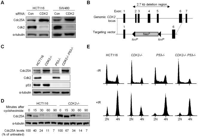
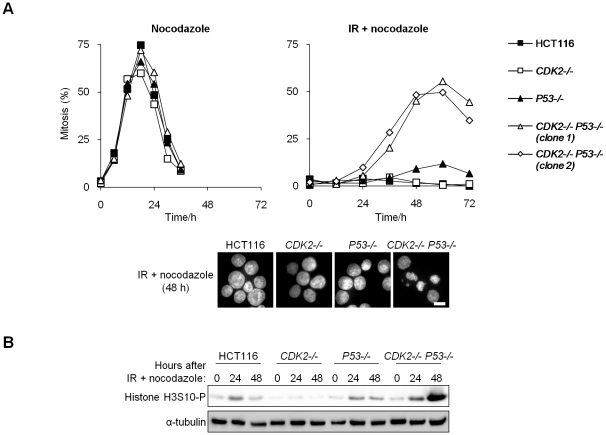
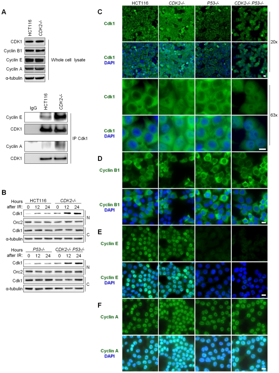
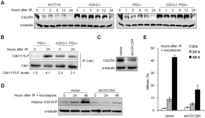
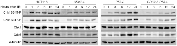
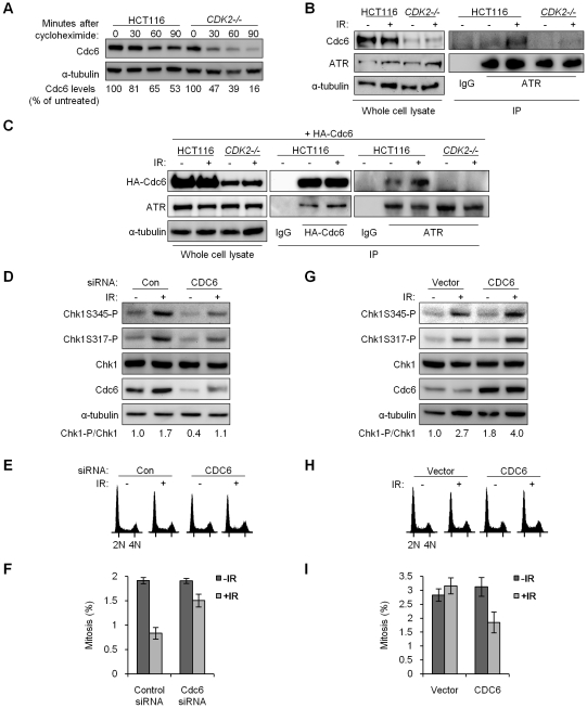
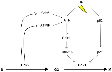
The activation of phase-specific cyclin-dependent kinases (Cdks) is associated with ordered cell cycle transitions. Among the mammalian Cdks, only Cdk1 is essential for somatic cell proliferation. Cdk1 can apparently substitute for Cdk2, Cdk4, and Cdk6, which are individually dispensable in mice. It is unclear if all functions of non-essential Cdks are fully redundant with Cdk1. Using a genetic approach, we show that Cdk2, the S-phase Cdk, uniquely controls the G(2)/M checkpoint that prevents cells with damaged DNA from initiating mitosis. CDK2-nullizygous human cells exposed to ionizing radiation failed to exclude Cdk1 from the nucleus and exhibited a marked defect in G(2)/M arrest that was unmasked by the disruption of P53. The DNA replication licensing protein Cdc6, which is normally stabilized by Cdk2, was physically associated with the checkpoint regulator ATR and was required for efficient ATR-Chk1-Cdc25A signaling. These findings demonstrate that Cdk2 maintains a balance of S-phase regulatory proteins and thereby coordinates subsequent p53-independent G(2)/M checkpoint activation.
Overview
The claims and statistics of this paper haven’t been checked yet.
Run the check — free, no account needed. It reads the open-access text and checks the sources it cites.
Integrity signals
Registry
Cites
Statistics
Figures
Verification
Claims the paper makes
The claims and statistics of this paper haven’t been checked yet.
Run the check — free, no account needed. It reads the open-access text and checks the sources it cites.
Claims other papers make about this one
Each is checked against this paper’s own text: does it say what the citing paper says it does?
In the citing paper
“Recently, Chung and Bunz have shown that Cdk2 is required for a p53-independent, but Chk1- and Chk2-dependent cell cycle arrest [60]”
From NER initiation factors, DDB2 and XPC, regulate UV radiation response by recruiting ATR and ATM kinases to DNA damage sites (2013) · cites this paper as [60]
Self-check
Percentages, sums and reported differences re-computed from the paper’s own numbers.
The self-check needs the paper’s full text.
Citations
Cites
Cell cycle, CDKs and cancer: a changing paradigm
Malumbres, M., & Barbacid, M. (2009) · Nature Reviews Cancer · doi:10.1038/nrc2602 · Cited by 3,957
Cell-cycle checkpoints and cancer
Kastan, M. B., & Bartek, J. (2004) · Nature · doi:10.1038/nature03097 · Cited by 2,836
Requirement for p53 and p21 to Sustain G 2 Arrest After DNA Damage
Bunz, F., Dutriaux, A., Lengauer, C., Waldman, T., Zhou, S., Brown, J. P., Sedivy, J. M., Kinzler, K. W., & Vogelstein, B. (1998) · Science · doi:10.1126/science.282.5393.1497 · Cited by 2,803
Self-citationAn Oncogene-Induced DNA Damage Model for Cancer Development
Halazonetis, T. D., Gorgoulis, V. G., & Bartek, J. (2008) · Science · doi:10.1126/science.1140735 · Cited by 1,851
ATR: an essential regulator of genome integrity
Cimprich, K. A., & Cortez, D. (2008) · Nature Reviews Molecular Cell Biology · doi:10.1038/nrm2450 · Cited by 1,798
The DNA Damage Response: Ten Years After
Harper, J. W., & Elledge, S. J. (2007) · Molecular Cell · doi:10.1016/j.molcel.2007.11.015 · Cited by 1,705
Liu, Q., Guntuku, S., Cui, X.-S., Matsuoka, S., Cortez, D., Tamai, K., Luo, G., Carattini-Rivera, S., DeMayo, F., Bradley, A., et al. (2000) · Genes & Development · doi:10.1101/gad.14.12.1448 · Cited by 1,625
Mammalian cyclin-dependent kinases
Malumbres, M., & Barbacid, M. (2005) · Trends in Biochemical Sciences · doi:10.1016/j.tibs.2005.09.005 · Cited by 1,333
p21 is necessary for the p53-mediated G1 arrest in human cancer cells.
Waldman, T. A., Kinzler, K. W., & Vogelstein, B. (1995) · PubMed · Cited by 1,188
Cdk1 is sufficient to drive the mammalian cell cycle
Santamarı́a, D., Barrière, C., Cerqueira, A., Hunt, S. L., Tardy, C., Newton, K., Cáceres, J. F., Dubus, P. R., Malumbres, M., & Barbacid, M. (2007) · Nature · doi:10.1038/nature06046 · Cited by 1,096
Cited by
Sorted by citations
Tripathi, V., Shen, Z., Chakraborty, A., Giri, S., Freier, S. M., Wu, X., Zhang, Y., Gorospe, M., Prasanth, S. G., Lal, A., et al. (2013) · PLoS Genetics · doi:10.1371/journal.pgen.1003368 · Cited by 763
Chk1 inhibits replication factory activation but allows dormant origin firing in existing factories
Ge, X. Q., & Blow, J. J. (2010) · The Journal of Cell Biology · doi:10.1083/jcb.201007074 · Cited by 219
Physiological Relevance of Cell Cycle Kinases
Malumbres, M. (2011) · Physiological Reviews · doi:10.1152/physrev.00025.2010 · Cited by 225
Spatial competition constrains resistance to targeted cancer therapy
Bačević, K., Noble, R., Soffar, A. A., Ammar, O., Boszonyik, B., Prieto, S., Vincent, C., Hochberg, M., Krasińska, L., & Fisher, D. (2017) · Nature Communications · doi:10.1038/s41467-017-01516-1 · Cited by 115
The CDK Network: Linking Cycles of Cell Division and Gene Expression
Fisher, R. P. (2012) · Genes & Cancer · doi:10.1177/1947601912473308 · Cited by 118
Ray, A., Milum, K., Battu, A., Wani, G., & Wani, A. A. (2013) · DNA Repair · doi:10.1016/j.dnarep.2013.01.003 · Cited by 85
“Recently, Chung and Bunz have shown that Cdk2 is required for a p53-independent, but Chk1- and Chk2dependent cell cycle arrest [60], raising the possibility that DDB2 and XPC might influence this axis of checkpoint signaling pathway.”
BackgroundCdk2 strengthens the intra-S checkpoint and counteracts cell cycle exit induced by DNA damage
Bačević, K., Lossaint, G., Achour, T. N., Georget, V., Fisher, D., & Dulić, V. (2017) · Scientific Reports · doi:10.1038/s41598-017-12868-5 · Cited by 91
Brill, E., Yokoyama, T., Nair, J. R., Yu, M., Ahn, Y.-R., & Lee, J. (2017) · Oncotarget · doi:10.18632/oncotarget.22195 · Cited by 79
Lin, F., Zhang, G., Yang, X., Wang, M., Wang, R., Wan, M., Wang, J., Wu, B. M., Yan, T., & Jia, Y. (2022) · Journal of Ethnopharmacology · doi:10.1016/j.jep.2022.115933 · Cited by 77
El‐Naggar, A. M., El-Hashash, M. A. A., & Elkaeed, E. B. (2021) · Bioorganic Chemistry · doi:10.1016/j.bioorg.2020.104615 · Cited by 68
ATR mediates cisplatin resistance in a p53 genotype-specific manner
Sangster‐Guity, N., Conrad, B., Papadopoulos, N., & Bunz, F. (2011) · Oncogene · doi:10.1038/onc.2010.624 · Cited by 65
Self-citationTo control or to be controlled? Dual roles of CDK2 in DNA damage and DNA damage response
Liu, Q., Gao, J., Zhao, C., Guo, Y., Wang, S., Shen, F., Xing, X., & Luo, Y. (2019) · DNA repair · doi:10.1016/j.dnarep.2019.102702 · Cited by 63
Hassan, G. S., Georgey, H. H., Mohammed, E. Z., George, R. F., Mahmoud, W. R., & Omar, F. A. (2021) · European Journal of Medicinal Chemistry · doi:10.1016/j.ejmech.2021.113389 · Cited by 62
Wohlbold, L., Merrick, K. A., De, S., Amat, R., Kim, J. H., Larochelle, S., Allen, J. J., Zhang, C., Shokat, K. M., Petrini, J. H. J., et al. (2012) · PLoS Genetics · doi:10.1371/journal.pgen.1002935 · Cited by 65
Lapek, J. D., Lewinski, M. K., Wozniak, J. M., Guatelli, J., & Gonzalez, D. J. (2017) · Molecular & Cellular Proteomics · doi:10.1074/mcp.m116.066019 · Cited by 59
Nian, W., Xu-jun, A., Wu, Y., Huang, Y., Shao, J., Wang, Y., Chen, Z., CHEN, F., & Wang, D. (2013) · Oncology Letters · doi:10.3892/ol.2013.1375 · Cited by 59
Gatti-Mays, M. E., Karzai, F. H., Soltani, S. N., Zimmer, A. S., Green, J. E., Lee, M.-J., Trepel, J. B., Yuno, A., Lipkowitz, S., Nair, J. R., et al. (2020) · The Oncologist · doi:10.1634/theoncologist.2020-0491 · Cited by 58
Molecular chemotherapeutic potential of butein: A concise review
Jayasooriya, R. G. P. T., Molagoda, I. M. N., Park, C., Jeong, J.-W., Choi, Y. H., Moon, D., Kim, M.-O., & Kim, G. (2017) · Food and Chemical Toxicology · doi:10.1016/j.fct.2017.12.028 · Cited by 56
Lou, X., Zhang, J., Liu, S., Lou, X., & Liao, D. J. (2014) · Cell Cycle · doi:10.4161/cc.29082 · Cited by 50
A Comparative Analysis of Constitutive Promoters Located in Adeno-Associated Viral Vectors
Damdindorj, L., Karnan, S., Ota, A., Hossain, E., Konishi, Y., Hosokawa, Y., & Konishi, H. (2014) · PLoS ONE · doi:10.1371/journal.pone.0106472 · Cited by 54
Xiang, Y., Song, Y., Li, Y., Zhao, D., Ma, L., & Tan, L. (2016) · Medical Science Monitor · doi:10.12659/msm.897301 · Cited by 46
Kandala, P. K., & Srivastava, S. (2010) · Molecular Pharmacology · doi:10.1124/mol.110.063750 · Cited by 45
Echalier, A., Cot, E., Camasses, A., Hodimont, E., Hoh, F., Jay, P., Sheinerman, F. B., Krasińska, L., & Fisher, D. (2012) · Chemistry & Biology · doi:10.1016/j.chembiol.2012.06.015 · Cited by 46
CyclinD-CDK4/6 complexes phosphorylate CDC25A and regulate its stability
Dozier, C., Mazzolini, L., Cénac, C., Froment, C., Burlet‐Schiltz, O., Besson, A., & Manenti, S. (2017) · Oncogene · doi:10.1038/onc.2016.506 · Cited by 43
Wang, A., Wang, S., Zhou, F., Li, P., Wang, Y., Gan, L., & Lin, L. (2018) · Biomedicine & Pharmacotherapy · doi:10.1016/j.biopha.2018.02.094 · Cited by 42
Characterization of CDK(5) inhibitor, 20-223 (aka CP668863) for colorectal cancer therapy
Robb, C. M., Kour, S., Contreras, J. I., Agarwal, E., Barger, C. J., Rana, S., Sonawane, Y. A., Neilsen, B. K., Taylor, M., Kizhake, S., et al. (2017) · Oncotarget · doi:10.18632/oncotarget.23749 · Cited by 41
Li, T., Gu, M., Deng, A., & Qian, C. (2020) · Cancer Cell International · doi:10.1186/s12935-020-01309-5 · Cited by 42
An, S. S. A., Kim, J.-E., Kim, H., Meang, E.-H., Song, Y., & Kim, M.-K. (2014) · International Journal of Nanomedicine · doi:10.2147/ijn.s57936 · Cited by 40
Lo, P., Lee, J. S., & Sukumar, S. V. (2011) · Cellular Signalling · doi:10.1016/j.cellsig.2011.09.017 · Cited by 36
Promotion of HepG2 cell apoptosis by flower of Allium atroviolaceum and the mechanism of action
Khazaei, S., Hamid, R. A., Esa, N., Ramachandran, V., Aalam, F., Etemad, A., & Ismail, P. (2017) · BMC Complementary and Alternative Medicine · doi:10.1186/s12906-017-1594-6 · Cited by 36
Mohammed, E. Z., Mahmoud, W. R., George, R. F., Hassan, G. S., Omar, F. A., & Georgey, H. H. (2021) · Bioorganic Chemistry · doi:10.1016/j.bioorg.2021.105347 · Cited by 35
Three independent mechanisms for arrest in G2after ionizing radiation
Landsverk, K. S., Patzke, S., Rein, I. D., Stokke, C., Lyng, H., Angelis, P. M. D., & Stokke, T. (2011) · Cell Cycle · doi:10.4161/cc.10.5.14968 · Cited by 35
Spetz, J. K. E., Langen, B., Rudqvist, N.-P., Parris, T. Z., Helou, K., Nilsson, O., & Forssell-Aronsson, E. B. (2017) · BMC Cancer · doi:10.1186/s12885-017-3524-x · Cited by 32
Neri, L., Lasa, M., Elósegui-Artola, A., D’Avola, D., Carte, B., Gázquez, C., Alve, S., Roca‐Cusachs, P., Iñarrairaegui, M., Herrero, J. I., et al. (2017) · Oncotarget · doi:10.18632/oncotarget.17332 · Cited by 31
Interplay Between the Cell Cycle and Double-Strand Break Response in Mammalian Cells
Beishline, K., & Azizkhan‐Clifford, J. (2014) · Methods in molecular biology · doi:10.1007/978-1-4939-0888-2_3 · Cited by 31
Discovery of AXL Degraders with Improved Potencies in Triple-Negative Breast Cancer (TNBC) Cells
He, R. R., Song, Z., Bai, Y., He, S., Huang, J., Wang, Y., Zhou, F., Huang, W., Guo, J., Wang, Z., et al. (2023) · Journal of Medicinal Chemistry · doi:10.1021/acs.jmedchem.2c01682 · Cited by 30
Phosphorylation of CDC25A on SER283 in late S/G2 by CDK/cyclin complexes accelerates mitotic entry
Mazzolini, L., Broban, A., Froment, C., Burlet‐Schiltz, O., Besson, A., Manenti, S., & Dozier, C. (2016) · Cell Cycle · doi:10.1080/15384101.2016.1220455 · Cited by 27
Hussmann, M., Janke, K., Kranz, P., Neumann, F., Mersch, E., Baumann, M., Göpelt, K., Brockmeier, U., & Metzen, E. (2015) · Oncotarget · doi:10.18632/oncotarget.5746 · Cited by 27
Novel pyrazolopyrimidines: Synthesis, in vitro cytotoxic activity and mechanistic investigation
Hassan, G. S., Rahman, D. E. A., Nissan, Y. M., Abdelmajeed, E. A., & Abdelghany, T. M. (2017) · European Journal of Medicinal Chemistry · doi:10.1016/j.ejmech.2017.07.003 · Cited by 24
Dowejko, A., Bauer, R. J., Bauer, K., Müller-Richter, U. D. A., & Reichert, T. E. (2011) · Experimental Cell Research · doi:10.1016/j.yexcr.2011.11.004 · Cited by 22
Cyclin-dependent kinases regulate apoptosis of intestinal epithelial cells
Bhattacharya, S., Ray, R. M., & Johnson, L. R. (2013) · APOPTOSIS · doi:10.1007/s10495-013-0942-3 · Cited by 22
The impact of age on oncogenic potential: tumor-initiating cells and the brain microenvironment
Stoll, E. A., Horner, P. J., & Rostomily, R. (2013) · Aging Cell · doi:10.1111/acel.12104 · Cited by 23
Chang, J.-W., Ding, Y., Qamar, M. T. U., Shen, Y., Gao, J., & Chen, L. (2019) · Carcinogenesis · doi:10.1093/carcin/bgz044 · Cited by 22
Lamberto, I., Plano, D., Moreno, E., Font, M., Palop, J. A., Sanmartín, C., & Encı́o, I. (2013) · Current Medicinal Chemistry · doi:10.2174/0929867311320120010 · Cited by 21
Liu, Q., Liu, X., Gao, J., Shi, X., Hu, X., Wang, S., & Luo, Y. (2013) · International Journal of Biological Sciences · doi:10.7150/ijbs.5763 · Cited by 21
Huang, C., Yang, S.-H., Huang, S.-M., Lin, C.-M., Chien, C., Chen, Y., Lee, C., Wu, H.-H., & Chang, C. (2011) · Journal of Translational Medicine · doi:10.1186/1479-5876-9-82 · Cited by 20
MAEL gene contributes to bovine testicular development through the m5C-mediated splicing
Liu, S., Ma, X., Wang, Z., Lin, F., Li, M., Li, Y., Yang, L., Rushdi, H. E., Riaz, H., Gao, T., et al. (2023) · iScience · doi:10.1016/j.isci.2023.105941 · Cited by 16
KIM, O.-Y., Cha, H. J., Ahn, K. J., An, I., An, S., & Bae, S. (2014) · Molecular Medicine Reports · doi:10.3892/mmr.2014.2158 · Cited by 16
Liu, L.-Y., Huang, W., Lin, R.-J., Lin, S.-Y., & Liang, Y. (2013) · Chemical Research in Toxicology · doi:10.1021/tx400271n · Cited by 16
Simple Monitoring of Gene Targeting Efficiency in Human Somatic Cell Lines Using the PIGA Gene
Karnan, S., Konishi, Y., Ota, A., Takahashi, M., Damdindorj, L., Hosokawa, Y., & Konishi, H. (2012) · PLoS ONE · doi:10.1371/journal.pone.0047389 · Cited by 16
Ezzat, M. A. F., Elmasry, G. F., El‐Mageed, M. M. A. A., Fouad, M. A., Abdel‐Aziz, H. A., & Elewa, S. I. (2023) · Drug Development Research · doi:10.1002/ddr.22079 · Cited by 15
Rivas, V., González-Muñoz, T., Albitre, Á., Lafarga, V., Delgado‐Arévalo, C., Mayor, F., & Penela, P. (2024) · Cell Death Discovery · doi:10.1038/s41420-024-02143-8 · Cited by 14
Chk1 suppresses bypass of mitosis and tetraploidization in p53-deficient cancer cells
Wilsker, D. F., Chung, J., & Bunz, F. (2012) · Cell Cycle · doi:10.4161/cc.19944 · Cited by 14
Self-citationJin, X., Wang, J., Hu, J., Liu, X., Li, S., Lu, Y., Zhen, H., Li, M., Zhao, Z., & Luo, Y. (2021) · Animals · doi:10.3390/ani11082417 · Cited by 14
Wip1 contributes to cell homeostasis maintained by the steady-state level of Wtp53
Park, H. K., Panneerselvam, J., Dudimah, F. D., Dong, G.-Z., Sebastian, S., Zhang, J., & Fei, P. (2011) · Cell Cycle · doi:10.4161/cc.10.15.15923 · Cited by 14
Xu, C., Zhang, W., Zhang, X., Zhou, D., Qu, L., Liu, J., Xiao, M., Ni, R., Jiang, F., Ni, W., et al. (2018) · Cancer Science · doi:10.1111/cas.13882 · Cited by 14
Kim, S., Hinoue, T., Kim, M. S., Sohn, K., Cho, R. C., Cole, P. D., Weisenberger, D. J., Laird, P. W., & Kim, Y. (2014) · Genes & Nutrition · doi:10.1007/s12263-014-0444-0 · Cited by 14
New Insights into Cell Cycle Regulation and DNA Damage Response in Embryonic Stem Cells
Suvorova, I. I., Katolikova, N. V., & Pospelov, V. A. (2012) · International review of cytology · doi:10.1016/b978-0-12-394310-1.00004-7 · Cited by 14
Aberrant activation of cyclin A-CDK induces G2/M-phase checkpoint in human cells
Akaike, Y., & Chibazakura, T. (2019) · Cell Cycle · doi:10.1080/15384101.2019.1693119 · Cited by 13
Non‐Cell Cycle Functions of the CDK Network in Ciliogenesis: Recycling the Cell Cycle Oscillator
Krasińska, L., & Fisher, D. (2018) · BioEssays · doi:10.1002/bies.201800016 · Cited by 12
Effect of methionine deficiency on the apoptosis and cell cycle of kidney in broilers
Song, B., Zeng, Q., Liu, Y., & Wu, B. (2019) · Research in Veterinary Science · doi:10.1016/j.rvsc.2019.09.013 · Cited by 12
Evangelisti, G., Barra, F., Moioli, M., Sala, P., Stigliani, S., Gustavino, C., Costantini, S., & Ferrero, S. (2020) · Expert Opinion on Investigational Drugs · doi:10.1080/13543784.2020.1783238 · Cited by 12
Cyclin Dependent Kinase as Significant Target for Cancer Treatment
Singh, S. K., Tripathi, S. K., Dessalew, N., & Singh, P. C. (2012) · Current Cancer Therapy Reviews · doi:10.2174/157339412802653164 · Cited by 11
Maher, M., Kassab, A. E., Zaher, A. F., & Mahmoud, Z. (2019) · Anti-Cancer Agents in Medicinal Chemistry · doi:10.2174/1871520619666190417153350 · Cited by 11
Ribociclib Inhibits P-gp-Mediated Multidrug Resistance in Human Epidermoid Carcinoma Cells
Zhang, L., Ye, B., Lin, Y., Li, Y.-D., Wang, J., Chen, Z., Ping, F., & Chen, Z.-S. (2022) · Frontiers in Pharmacology · doi:10.3389/fphar.2022.867128 · Cited by 11
CDK2 knockdown enhances head and neck cancer cell radiosensitivity
Soffar, A. A., Storch, K., Aleem, E. A., & Cordes, N. (2013) · International Journal of Radiation Biology · doi:10.3109/09553002.2013.782108 · Cited by 11
Wilsker, D. F., Chung, J., Pradilla, I. F., Petermann, E., Helleday, T., & Bunz, F. (2011) · Molecular Cancer Therapeutics · doi:10.1158/1535-7163.mct-11-0675 · Cited by 10
Self-citationTumor tissue protein signatures reflect histological grade of breast cancer
Skoog, P., Ohlsson, M., Fernö, M., Rydén, L., Borrebaeck, C. A. K., & Wingren, C. (2017) · PLoS ONE · doi:10.1371/journal.pone.0179775 · Cited by 10
Cheng, X., Li, D., Qi, T., Sun, J., Zhou, T., & Zheng, W. (2022) · Scientific Reports · doi:10.1038/s41598-021-04055-4 · Cited by 9
CtIP promotes G2/M arrest in etoposide‐treated HCT116 cells in a p53‐independent manner
Chen, H., Shan, J., Chen, D., Wang, R., Qi, W., Wang, H., Ke, Y., Liu, W., & Zeng, X. (2018) · Journal of Cellular Physiology · doi:10.1002/jcp.27824 · Cited by 10
New pyrrolidine‐carboxamide derivatives as dual antiproliferative EGFR / CDK2 inhibitors
Frejat, F. O. A., Zhao, B., Furaijit, N., Wang, L., Abou‐Zied, H. A., Fathy, H. M., Mohamed, F. A., Youssif, B. G. M., & Wu, C. (2023) · Chemical Biology & Drug Design · doi:10.1111/cbdd.14422 · Cited by 7
Zhang, Y., Liu, X., Li, Z., Wang, X., & Tang, C. (2025) · Bioorganic & Medicinal Chemistry Letters · doi:10.1016/j.bmcl.2025.130166 · Cited by 6
Interferon regulatory factor 4 mediates nonenzymatic IRE1 dependency in multiple myeloma cells
Oikonomidi, I., Kameswaran, V., Pham, V. C., Zuazo-Gaztelu, I., Gutgesell, L. M., Marsters, S. A., Dániel, B., Lill, J. R., Modrusan, Z. D., & Ashkenazi, A. (2025) · PLoS Biology · doi:10.1371/journal.pbio.3003096 · Cited by 6
DNA damage responses in murine Pre-B cells with genetic deficiencies in damage response genes
Innes, C. L., Hesse, J. E., Morales, A. J., Helmink, B. A., Schurman, S. H., Sleckman, B. P., & Paules, R. S. (2019) · Cell Cycle · doi:10.1080/15384101.2019.1693118 · Cited by 6
Wang, Y., Li, Y., Zhang, Y., Li, Y., Zhao, L., Tong, L., Zhu, X. M., Wang, J., & Dong, Y. (2025) · Chemico-Biological Interactions · doi:10.1016/j.cbi.2025.111562 · Cited by 5
Konishi, Y., Karnan, S., Takahashi, M., Ota, A., Damdindorj, L., Hosokawa, Y., & Konishi, H. (2012) · BioTechniques · doi:10.2144/0000113911 · Cited by 5
Limited Repair of Critical DNA Damage in Cells Exposed to Low Dose Radiation
Gaziev, A. I., & Shaikhaev, G. (2012) · InTech eBooks · doi:10.5772/33611 · Cited by 5
Li, E., Ho, R., Tao, R., Choi, Y. W. Y., Shin, C. Y., Chen, S. Y., Gokbayrak, B., Senz, J., Yao, B., Johnston, L., et al. (2025) · npj Precision Oncology · doi:10.1038/s41698-025-01053-x · Cited by 5
Chan, Z. C. K., Leong, K. H., Kareem, H. S., Norazit, A., Noor, S. M., & Ariffin, A. (2019) · Naunyn-Schmiedeberg s Archives of Pharmacology · doi:10.1007/s00210-019-01730-2 · Cited by 3
Alshaye, N. A. (2023) · Journal of Biomolecular Structure and Dynamics · doi:10.1080/07391102.2023.2187222 · Cited by 3
Sun, J., Liu, J., Xue, M., Zhao, T., Song, J., Zhang, W., Chang, Y., & Zhan, Y. (2025) · Fish & Shellfish Immunology · doi:10.1016/j.fsi.2025.110176 · Cited by 3
Yang, T., Gu, J., Du, C., Shan, T., Wei, T., Li, Y., & Wang, L. (2021) · Journal of Cardiovascular Pharmacology · doi:10.1097/fjc.0000000000000997 · Cited by 3
Li, Y., Lin, Y., Chen, Z., Ji, W., & Liu, H. (2024) · Cellular Signalling · doi:10.1016/j.cellsig.2024.111579 · Cited by 2
Han, X., Wang, Z., Wang, W., Bai, R., Zhao, P., & Shang, J. (2014) · Cytotechnology · doi:10.1007/s10616-013-9604-0 · Cited by 2
Hematulin, A., Meethang, S., Utapom, K., Wongkham, S., & Sagan, D. (2018) · Oncology Letters · doi:10.3892/ol.2018.7754 · Cited by 2
Kashyap, D., Kaur, T., Dutta, P. S., & Sinha, S. K. (2025) · Physical Chemistry Chemical Physics · doi:10.1039/d4cp04863f · Cited by 1
McLean, I. C., Gross, S. M., Copperman, J., Derrick, D. S., Paul, I., Emili, A., & Heiser, L. M. (2025) · iScience · doi:10.1016/j.isci.2025.113407 · Cited by 1
Patel, D. A., Prajapati, A. S., Patel, S. S., Dabhi, R. C., Kotadiya, D. D., & Patel, H. D. (2026) · Biotechnology and Applied Biochemistry · doi:10.1002/bab.70202 · Cited by 1
Zhou, M.-L., Ma, J.-N., Xu, X., Gao, X.-Y., Wang, H.-X., Shen, J., & Xue, L. (2023) · Research Square · doi:10.21203/rs.3.rs-2533161/v1 · Cited by 1
Cyclin A/CDK2 regulates multiple G2 phase pathways
Oakes, V. (2012)
Overexpression of DOC-1R Inhibits Cell Cycle G1/S Transition by Repressing CDK2 Expression and
Liu, Q., Liu, X., Gao, J., Shi, X., Hu, X., Wang, S., & Luo, Y. (2013)
(2013) · CRC Press eBooks · doi:10.1201/b14587-17
Computational studies on RNA processing in higher eukaryotes
Brüggemann, M. (2021) · Publication Server of Goethe University Frankfurt am Main (Goethe University Frankfurt) · doi:10.21248/gups.61384
MA, J., ZHOU, M., Xu, X., GAO, X., WANG, H., SHEN, J., & Xue, L. (2024) · Biocell · doi:10.32604/biocell.2023.045076
IRF4 mediates non-enzymatic dependency on IRE1 in Multiple Myeloma cellsPreprint
Oikonomidi, I., Kameswaran, V., Pham, V. C., Zuazo-Gaztelu, I., Gutgesell, L. M., Marsters, S. A., Dániel, B., Lill, J. R., Modrusan, Z. D., & Ashkenazi, A. (2024) · Research Square · doi:10.21203/rs.3.rs-4494530/v1
Zhang, R., Zhang, Y., Xue, J., Zhang, R., Xie, J., Luo, X., Yang, X., Wang, H., Liu, J., Wei, J., et al. (2025) · SSRN Electronic Journal · doi:10.2139/ssrn.5116771
Mclean, I. C., Gross, S. M., Copperman, J., Derrick, D. S., & Heiser, L. M. (2025) · bioRxiv · doi:10.1101/2025.04.03.647095
Li, T., Gu, M., 邓安梅, & Qian, C. (2020) · Figshare · doi:10.6084/m9.figshare.12486737
Li, T., Gu, M., Deng, A., & Qian, C. (2020) · Figshare · doi:10.6084/m9.figshare.12486725
Metadata
- Authors
- Chung, J. H., & Bunz, F.
- Affiliations
- Johns Hopkins University (US)
- Corresponding author
- Bunz, Fred
- Venue
- PLoS Genetics
- Publisher
- Public Library of Science (PLoS)
- Published
- 2010-02-26
- Volume
- 6
- Issue
- 2
- Pages
- e1000863
- ISSN
- 1553-7390 (ISSN-L)
- 1553-7404
- PMC
- PMC2829054
- OpenAlex
- W2093147524
- Cited by
- 105
- References
- 61
- Citations by year
- 2026: 4 · 2025: 9 · 2024: 5 · 2023: 6 · 2022: 3 · 2021: 6 · 2020: 5 · 2019: 7 · 2018: 5 · 2017: 12 · 2016: 3 · 2015: 1 · 2014: 7 · 2013: 11 · 2012: 10
- FWCI
- 5.07
- Type
- journal article
- Language
- en
- Open access
- gold
- doi.org
- Locations
- PLoS Genetics (publishedVersion, cc-by)
- PubMed Central (publishedVersion, cc-by)
- PubMed (publishedVersion)
- DOAJ (DOAJ: Directory of Open Access Journals) (submittedVersion, cc-by-sa)
- Figshare (submittedVersion, cc-by)
- Funders
- W. W. Smith Charitable Trust
- Flight Attendant Medical Research Institute
- National Cancer Institute (CA 104253, R01CA104253)
- Topics
- Health Sciences › Medicine › Oncology › Cancer-related Molecular Pathways
- Life Sciences › Biochemistry, Genetics and Molecular Biology › Molecular Biology › DNA Repair Mechanisms
- Life Sciences › Biochemistry, Genetics and Molecular Biology › Cell Biology › Microtubule and mitosis dynamics
- Keywords
- CDK2, G2/M checkpoint, CDK1, CDC6, ATR, Chk1, CDC25A, ionizing radiation, DNA damage response, DNA replication licensing
- MeSH terms
- Checkpoint Kinase 1, Animals, Cytoplasm, Enzyme Activation, Enzyme Stability, Humans, Mitosis, Models, Biological, Nuclear Proteins, Phosphorylation, Protein Kinases, Protein Processing, Post-Translational, Radiation, Ionizing, Tumor Suppressor Protein p53, G2 Phase, Protein Serine-Threonine Kinases, Cell Cycle Proteins, cdc25 Phosphatases, Protein Transport, HCT116 Cells, Cyclin-Dependent Kinase 2, Mice, Ataxia Telangiectasia Mutated Proteins
About these checks
Registry, retraction, Cites and statistics results are computed, not judged, with one exception: a printed reference that no index lists is read once by a model before it counts as a citation error. Claims are extracted by a model and kept only when their quote appears verbatim in the text; each cited source is then checked, with a model’s help, for whether it says what the claim relies on. Those verdicts can be wrong — read the claim’s quote and the cited source before relying on one.
Statistics and arithmetic are recomputed, not judged.
Confidence bands are shown only for fields calibrated on our annotated test set. None is calibrated yet, so no band is shown.
What counts as a claim ›How we measure confidence ›How we check this ›
Are you an author, or do you see a problem with this page? Request a review
