

The DOI matches its Crossref record; 35 of the 35 references it cites were found in a registry or an index.
Oncogenic Transformation by Inhibitor-Sensitive and -Resistant EGFR Mutants
Open accessAbstract
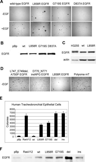
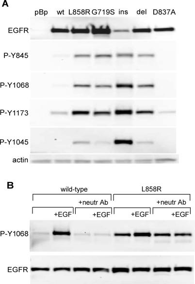
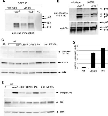
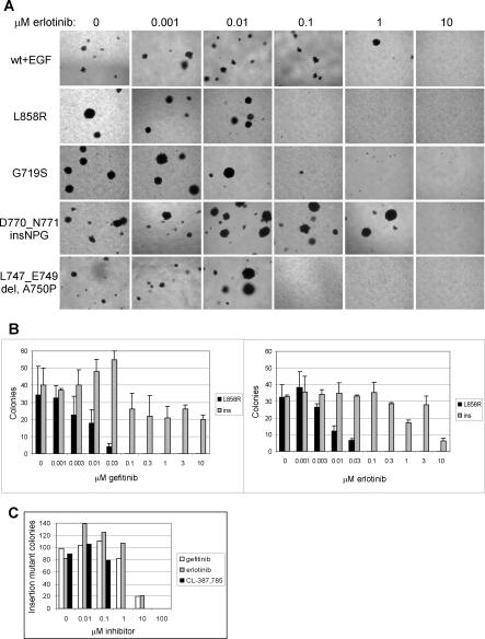
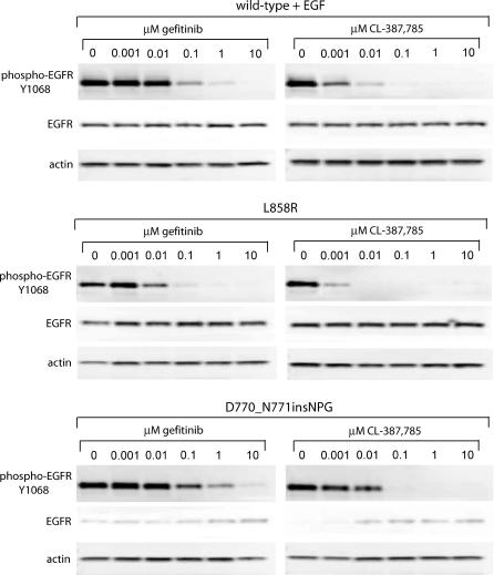
BACKGROUND: Somatic mutations in the kinase domain of the epidermal growth factor receptor tyrosine kinase gene EGFR are common in lung adenocarcinoma. The presence of mutations correlates with tumor sensitivity to the EGFR inhibitors erlotinib and gefitinib, but the transforming potential of specific mutations and their relationship to drug sensitivity have not been described. METHODS AND FINDINGS: Here, we demonstrate that EGFR active site mutants are oncogenic. Mutant EGFR can transform both fibroblasts and lung epithelial cells in the absence of exogenous epidermal growth factor, as evidenced by anchorage-independent growth, focus formation, and tumor formation in immunocompromised mice. Transformation is associated with constitutive autophosphorylation of EGFR, Shc phosphorylation, and STAT pathway activation. Whereas transformation by most EGFR mutants confers on cells sensitivity to erlotinib and gefitinib, transformation by an exon 20 insertion makes cells resistant to these inhibitors but more sensitive to the irreversible inhibitor CL-387,785. CONCLUSION: Oncogenic transformation of cells by different EGFR mutants causes differential sensitivity to gefitinib and erlotinib. Treatment of lung cancers harboring EGFR exon 20 insertions may therefore require the development of alternative kinase inhibition strategies.
Overview
The claims and statistics of this paper haven’t been checked yet.
Run the check — free, no account needed. It reads the open-access text and checks the sources it cites.
Integrity signals
Registry
Cites
Statistics
Figures
Verification
Claims the paper makes
The claims and statistics of this paper haven’t been checked yet.
Run the check — free, no account needed. It reads the open-access text and checks the sources it cites.
Claims other papers make about this one
Each is checked against this paper’s own text: does it say what the citing paper says it does?
In the citing paper
“CL-387785 has also shown potential to overcome T790M mutation-related resistance at the functional level, possibly by effectively inhibiting downstream signaling pathways.”
From Recent advances in targeting the “undruggable” proteins: from drug discovery to clinical trials (2023) · cites this paper as [201]
In the citing paper
“These mutant kinases present a reduced affinity for the ATP, accounting for the increased sensitivity to EGFR TKIs compared with the wild type counterparts, since these inhibitors compete with ATP for binding to the catalytic site [10–12].”
From A decade of EGFR inhibition in EGFR-mutated non small cell lung cancer (NSCLC): Old successes and future perspectives (2015) · cites this paper as [10]
In the citing paper
“Most EGFR exon 20 insertion mutations are 1st generation EGFR-TKIs resistant mutations [15, 18, 19].”
In the citing paper
“Cell culture and transgenic mouse model studies have shown that mutant EGFR has transforming activity (Greulich et al., 2005; Ji et al., 2006; Politi et al., 2006).”
From ERBB Receptors: From Oncogene Discovery to Basic Science to Mechanism-Based Cancer Therapeutics (2014) · cites this paper as Greulich et al., 2005
Self-check
Percentages, sums and reported differences re-computed from the paper’s own numbers.
The self-check needs the paper’s full text.
Citations
Cites
Lynch, T. J., Bell, D. W., Sordella, R., Gurubhagavatula, S., Okimoto, R. A., Brannigan, B. W., Harris, P. L., Haserlat, S. M., Supko, J. G., Haluska, F. G., et al. (2004) · New England Journal of Medicine · doi:10.1056/nejmoa040938 · Cited by 11,486
EGFR Mutations in Lung Cancer: Correlation with Clinical Response to Gefitinib Therapy
Paez, J. G., Jänne, P. A., Lee, J. C., Tracy, S., Greulich, H., Gabriel, S., Herman, P., Kaye, F. J., Lindeman, N., Boggon, T. J., et al. (2004) · Science · doi:10.1126/science.1099314 · Cited by 9,368
Self-citationPao, W., Miller, V., Zakowski, M., Doherty, J., Politi, K., Sarkaria, I., Singh, B., Heelan, R., Rusch, V., Fulton, L., et al. (2004) · Proceedings of the National Academy of Sciences · doi:10.1073/pnas.0405220101 · Cited by 4,338
EGFR Mutation and Resistance of Non–Small-Cell Lung Cancer to Gefitinib
Kobayashi, S., Boggon, T. J., Dayaram, T., Jänne, P. A., Kocher, O., Meyerson, M., Johnson, B. E., Eck, M. J., Tenen, D. G., & Halmos, B. (2005) · New England Journal of Medicine · doi:10.1056/nejmoa044238 · Cited by 3,964
Self-citationPao, W., Miller, V. A., Politi, K. A., Riely, G. J., Somwar, R., Zakowski, M. F., Kris, M. G., & Varmus, H. (2005) · PLoS Medicine · doi:10.1371/journal.pmed.0020073 · Cited by 3,527 · cited 1×
“Patients who responded to gefitinib and subsequently relapsed were found to have T790M secondary mutations, also in exon 20 [19,20].”
BackgroundThe ErbB signaling network: receptor heterodimerization in development and cancer
Olayioye, M. A., Neve, R. M., Lane, H. A., & Hynes, N. E. (2000) · The EMBO Journal · doi:10.1093/emboj/19.13.3159 · Cited by 2,489
Shigematsu, H., Lin, L., Takahashi, T., Nomura, M., Suzuki, M., Wistuba, I. I., Fong, K. M., Lee, H., Toyooka, S., Shimizu, N., et al. (2005) · JNCI Journal of the National Cancer Institute · doi:10.1093/jnci/dji055 · Cited by 2,377
Creation of human tumour cells with defined genetic elements
Hahn, W. C., Counter, C. M., Lundberg, A. S., Beijersbergen, R. L., Brooks, M. W., & Weinberg, R. A. (1999) · Nature · doi:10.1038/22780 · Cited by 2,345
Self-citationZhong, Z., Wen, Z., & Darnell, J. E. (1994) · Science · doi:10.1126/science.8140422 · Cited by 2,070
Gefitinib-Sensitizing EGFR Mutations in Lung Cancer Activate Anti-Apoptotic Pathways
Sordella, R., Bell, D. W., Haber, D. A., & Settleman, J. E. (2004) · Science · doi:10.1126/science.1101637 · Cited by 1,605
The EGFR family and its ligands in human cancer
Yarden, Y. (2001) · European Journal of Cancer · doi:10.1016/s0959-8049(01)00230-1 · Cited by 1,605
Epidermal growth factor receptor: Mechanisms of activation and signalling
Jorissen, R. (2003) · Experimental Cell Research · doi:10.1016/s0014-4827(02)00098-8 · Cited by 1,565
A novel transforming protein (SHC) with an SH2 domain is implicated in mitogenic signal transduction
Pelicci, G., Lanfrancone, L., Grignani, F., McGlade, J. C., Cavallo, F., Forni, G., Nicoletti, I., Grignani, F., Pawson, T. J., & Pelicci, P. G. (1992) · Cell · doi:10.1016/0092-8674(92)90536-l · Cited by 1,326
Mutations of the Epidermal Growth Factor Receptor Gene in Lung Cancer
Kosaka, T., Yatabe, Y., Endoh, H., Kuwano, H., Takahashi, T., & Mitsudomi, T. (2004) · Cancer Research · doi:10.1158/0008-5472.can-04-2818 · Cited by 1,220
Levkowitz, G., Waterman, H., Ettenberg, S. A., Katz, M., Tsygankov, A. Y., Alroy, I., Lavi, S., Iwaï, K., Reiss, Y., Ciechanover, A. J., et al. (1999) · Molecular Cell · doi:10.1016/s1097-2765(00)80231-2 · Cited by 1,003
Hidalgo, M., Siu, L. L., Nemunaitis, J., Rizzo, J., Hammond, L. A., Takimoto, C., Eckhardt, S. G., Tolcher, A., Britten, C. D., Denis, L., et al. (2001) · Journal of Clinical Oncology · doi:10.1200/jco.2001.19.13.3267 · Cited by 1,001
Mitsudomi, T., Kosaka, T., Endoh, H., Horio, Y., Hida, T., Mori, S., Hatooka, S., Shinoda, M., Takahashi, T., & Yatabe, Y. (2005) · Journal of Clinical Oncology · doi:10.1200/jco.2005.00.992 · Cited by 940
Frequent expression of a mutant epidermal growth factor receptor in multiple human tumors.
Moscatello, D. K., Holgado-Madruga, M., Godwin, A. K., Ramirez, G., Gunn, G. R., Zoltick, P. W., Biegel, J. A., Hayes, R. L., & Wong, A. J. (1995) · PubMed · Cited by 637
Huang, H., Nagane, M., Klingbeil, C. K., Lin, H., Nishikawa, R., Ji, X.-D., Huang, C., Gill, G. N., Wiley, H. S., & Cavenee, W. K. (1997) · Journal of Biological Chemistry · doi:10.1074/jbc.272.5.2927 · Cited by 600
Huang, S.-F. K., Liu, H.-P., Li, L.-H., Ku, Y.-C., Fu, Y.-N., Tsai, H., Chen, Y.-T., Lin, Y., Chang, W.-C., Kuo, H.-P., et al. (2004) · Clinical Cancer Research · doi:10.1158/1078-0432.ccr-04-1245 · Cited by 545
United States Food and Drug Administration Drug Approval Summary
Cohen, M. H., Williams, G. A., Sridhara, R., Chen, G., McGuinn, W. D., Morse, D. E., Abraham, S., Rahman, A., Liang, C., Lostritto, R. T., et al. (2004) · Clinical Cancer Research · doi:10.1158/1078-0432.ccr-03-0564 · Cited by 505
Epidermal-Growth-Factor-Dependent Transformation by a Human EGF Receptor Proto-Oncogene
Velu, T. J., Beguinot, L., Vass, W. C., Willingham, M. C., Merlino, G. T., Pastan, I. H., & Lowy, D. R. (1987) · Science · doi:10.1126/science.3500513 · Cited by 433
Amann, J. M., Kalyankrishna, S., Massion, P. P., Ohm, J. E., Girard, L., Shigematsu, H., Peyton, M., Juroske, D. M., Huang, Y., Salmon, J. S., et al. (2005) · Cancer Research · doi:10.1158/0008-5472.226.65.1 · Cited by 432
Gefitinib Induces Apoptosis in the EGFRL858R Non–Small-Cell Lung Cancer Cell Line H3255
Tracy, S. I., Mukohara, T., Hansen, M., Meyerson, M. M., Johnson, B. E., & Janne, P. A. (2004) · Cancer Research · doi:10.1158/0008-5472.can-04-1905 · Cited by 372
Self-citationRodrigues, G. A., Falasca, M., Zhang, Z., Ong, S. H., & Schlessinger, J. (2000) · Molecular and Cellular Biology · doi:10.1128/mcb.20.4.1448-1459.2000 · Cited by 351
Chou, T., Chiu, C., Li, L., Hsiao, C.-Y., Tzen, C., Chang, K.-T., Chen, Y., Perng, R.-P., Tsai, S., & Tsai, C. (2005) · Clinical Cancer Research · doi:10.1158/1078-0432.ccr-04-1981 · Cited by 315
Immortalization and transformation of primary human airway epithelial cells by gene transfer
Lundberg, A. S., Randell, S. H., Stewart, S. A., Elenbaas, B., Hartwell, K. A., Brooks, M. W., Fleming, M. D., Olsen, J. C., Miller, S. W., Weinberg, R. A., et al. (2002) · Oncogene · doi:10.1038/sj.onc.1205550 · Cited by 263
Self-citationExpression of mutated epidermal growth factor receptor by non-small cell lung carcinomas.
de Palazzo, I. G., Adams, G. P., Sundareshan, P., Wong, A. J., Testa, J. R., Bigner, D. D., & Weiner, L. M. (1993) · PubMed · Cited by 243
Jiang, J., Greulich, H. E., Janne, P. A., Sellers, W. R., Meyerson, M. M., & Griffin, J. D. (2005) · Cancer Research · doi:10.1158/0008-5472.can-05-1829 · Cited by 188
Self-citationBesser, D., Bromberg, J., Darnell, J. E., Jr., & Hanafusa, H. (1999) · Molecular and Cellular Biology · doi:10.1128/mcb.19.2.1401 · Cited by 123
Okabayashi, Y., Kido, Y., Okutani, T., Sugimoto, Y., Sakaguchi, K., & Kasuga, M. (1994) · Journal of Biological Chemistry · doi:10.1016/s0021-9258(17)32363-3 · Cited by 118
Discafani, C. M., Carroll, M. L., Floyd, M. B., Hollander, I. J., Husain, Z., Johnson, B. D., Kitchen, D., May, M. K., Malo, M. S., Minnick, A. A., et al. (1999) · Biochemical Pharmacology · doi:10.1016/s0006-2952(98)00356-6 · Cited by 112
Gamett, D. C., Tracy, S., & Robinson, H. L. (1986) · Proceedings of the National Academy of Sciences · doi:10.1073/pnas.83.16.6053 · Cited by 53
Ligand-independent oncogenic signaling by the epidermal growth factor receptor: v-ErbB as a paradigm
Boerner, J. L. (2003) · Experimental Cell Research · doi:10.1016/s0014-4827(02)00096-4 · Cited by 52
Danielsen, A. J., & Maihle, N. J. (2002) · Experimental Cell Research · doi:10.1006/excr.2002.5494 · Cited by 12
Research works printed but not linked: 2
- Garcia de Palazzo, 1993, Cancer Res, 53, 3217
- Moscatello, 1995, Cancer Res, 55, 5536
Cited by
Sorted by citations
Epidermal growth factor receptor mutations in lung cancer
Sharma, S. V., Bell, D. W., Settleman, J., & Haber, D. A. (2007) · Nature Reviews Cancer · doi:10.1038/nrc2088 · Cited by 3,170
Herbst, R. S., Heymach, J. V., & Lippman, S. M. (2008) · New England Journal of Medicine · doi:10.1056/nejmra0802714 · Cited by 2,435
Shigematsu, H., Lin, L., Takahashi, T., Nomura, M., Suzuki, M., Wistuba, I. I., Fong, K. M., Lee, H., Toyooka, S., Shimizu, N., et al. (2005) · JNCI Journal of the National Cancer Institute · doi:10.1093/jnci/dji055 · Cited by 2,377
Screening for Epidermal Growth Factor Receptor Mutations in Lung Cancer
Rosell, R., Moran, T., Queralt, C., Porta, R., Cardenal, F., Camps, C., Majem, M., Lopez-Vivanco, G., Isla, D., Provencio, M., et al. (2009) · New England Journal of Medicine · doi:10.1056/nejmoa0904554 · Cited by 2,373
The T790M mutation in EGFR kinase causes drug resistance by increasing the affinity for ATP
Yun, C.-H., Mengwasser, K. E., Toms, A. V., Woo, M. S., Greulich, H., Wong, K.-K., Meyerson, M., & Eck, M. J. (2008) · Proceedings of the National Academy of Sciences · doi:10.1073/pnas.0709662105 · Cited by 2,118
Self-citation
Metadata
- Authors
- Greulich, H., Chen, T.-H., Feng, W., Jänne, P. A., Alvarez, J. V., Zappaterra, M., Bulmer, S. E., Frank, D. A., Hahn, W. C., Sellers, W. R., & Meyerson, M.
- Affiliations
- Broad Institute (US)
- Brigham and Women's Hospital (US)
- Dana-Farber Cancer Institute (US)
- Corresponding author
- Greulich, Heidi
- Sellers, William R
- Meyerson, Matthew
- ORCID
- Venue
- PLoS Medicine
- Publisher
- Public Library of Science (PLoS)
- Published
- 2005-10-04
- Volume
- 2
- Issue
- 11
- Pages
- e313
- ISSN
- 1549-1277 (ISSN-L)
- 1549-1676
- PMC
- PMC1240052
- OpenAlex
- W2037873277
- Cited by
- 677
- References
- 35
- Citations by year
- 2026: 9 · 2025: 10 · 2024: 10 · 2023: 22 · 2022: 23 · 2021: 23 · 2020: 16 · 2019: 29 · 2018: 20 · 2017: 27 · 2016: 26 · 2015: 47 · 2014: 27 · 2013: 54 · 2012: 46
- FWCI
- 45.72
- Type
- journal article
- Language
- en
- Update notices
- correction (2024-09-16)
- Open access
- gold
- doi.org
- Locations
- PLoS Medicine (publishedVersion, cc-by)
- Europe PMC (PubMed Central) (publishedVersion, cc-by)
- PubMed Central (publishedVersion, cc-by)
- PubMed (publishedVersion)
- Digital Access to Scholarship at Harvard (DASH) (Harvard University) (submittedVersion, cc-by)
- DOAJ (DOAJ: Directory of Open Access Journals) (submittedVersion, cc-by-sa)
- Funders
- American Cancer Society
- Doris Duke Charitable Foundation
- Flight Attendant Medical Research Institute
- Barr Foundation
- National Institutes of Health
- National Cancer Institute
- Topics
- Health Sciences › Medicine › Pulmonary and Respiratory Medicine › Lung Cancer Treatments and Mutations
- Health Sciences › Medicine › Oncology › HER2/EGFR in Cancer Research
- Life Sciences › Biochemistry, Genetics and Molecular Biology › Cancer Research › Cancer Genomics and Diagnostics
- Keywords
- EGFR mutation, oncogenic transformation, lung adenocarcinoma, erlotinib, gefitinib, EGFR inhibitors, exon 20 insertion, autophosphorylation, Shc phosphorylation, STAT pathway, drug resistance, kinase inhibitors
- MeSH terms
- Erlotinib Hydrochloride, Src Homology 2 Domain-Containing, Transforming Protein 1, Gefitinib, Animals, Antineoplastic Agents, Cell Transformation, Neoplastic, Exons, Humans, Mice, Nude, Mutation, Neoplasms, Experimental, Quinazolines, Transfection, Tumor Stem Cell Assay, Genetic Therapy, Drug Resistance, Neoplasm, NIH 3T3 Cells, Protein Kinase Inhibitors, Adaptor Proteins, Signal Transducing, STAT3 Transcription Factor, Proto-Oncogene Proteins c-akt, Mice, Shc Signaling Adaptor Proteins, ErbB Receptors
About these checks
Registry, retraction, Cites and statistics results are computed, not judged, with one exception: a printed reference that no index lists is read once by a model before it counts as a citation error. Claims are extracted by a model and kept only when their quote appears verbatim in the text; each cited source is then checked, with a model’s help, for whether it says what the claim relies on. Those verdicts can be wrong — read the claim’s quote and the cited source before relying on one.
Statistics and arithmetic are recomputed, not judged.
Confidence bands are shown only for fields calibrated on our annotated test set. None is calibrated yet, so no band is shown.
What counts as a claim ›How we measure confidence ›How we check this ›
Are you an author, or do you see a problem with this page? Request a review
