

Decoding the similarities and differences among mycobacterial species
Open accessThe DOI matches its Crossref record; 63 of the 63 references it cites were found in a registry or an index.
Abstract
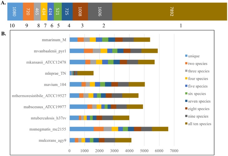
Mycobacteriaceae comprises pathogenic species such as Mycobacterium tuberculosis, M. leprae and M. abscessus, as well as non-pathogenic species, for example, M. smegmatis and M. thermoresistibile. Genome comparison and annotation studies provide insights into genome evolutionary relatedness, identify unique and pathogenicity-related genes in each species, and explore new targets that could be used for developing new diagnostics and therapeutics. Here, we present a comparative analysis of ten-mycobacterial genomes with the objective of identifying similarities and differences between pathogenic and non-pathogenic species. We identified 1080 core orthologous clusters that were enriched in proteins involved in amino acid and purine/pyrimidine biosynthetic pathways, DNA-related processes (replication, transcription, recombination and repair), RNA-methylation and modification, and cell-wall polysaccharide biosynthetic pathways. For their pathogenicity and survival in the host cell, pathogenic species have gained specific sets of genes involved in repair and protection of their genomic DNA. M. leprae is of special interest owing to its smallest genome (1600 genes and ~1300 psuedogenes), yet poor genome annotation. More than 75% of the pseudogenes were found to have a functional ortholog in the other mycobacterial genomes and belong to protein families such as transferases, oxidoreductases and hydrolases.
Overview
The claims and statistics of this paper haven’t been checked yet.
Free: it reads the open-access text and checks the first 5 claims that cite a source we can check. Create a free account to check every claim.
Integrity signals
Registry
Cites
Statistics
Figures



Verification
Claims the paper makes
The claims and statistics of this paper haven’t been checked yet.
Free: it reads the open-access text and checks the first 5 claims that cite a source we can check. Create a free account to check every claim.
Claims other papers make about this one
Each is checked against this paper’s own text: does it say what the citing paper says it does?
The source backs part of the claim, not all of it — or backs it for a different population, condition or comparison.
In the citing paper
“By contrast, M. smegmatis encodes thousands of conserved mycobacterial gene orthologs and has the same cell architecture and physiology.”
From Mycobacterium smegmatis: The Vanguard of Mycobacterial Research (2023) · cites this paper as [33]
Experimental — accuracy not yet measuredThe cited source says what the claim attributes to it.
In the citing paper
“The average protein identities between protein orthologs in M. smegmatis and their pathogenic cousins are >70% in most cases (33).”
From Mycobacterium smegmatis: The Vanguard of Mycobacterial Research (2023) · cites this paper as [33]
Self-check
Percentages, sums and reported differences re-computed from the paper’s own numbers.
The self-check needs the paper’s full text.
Citations
Cites
Deciphering the biology of Mycobacterium tuberculosis from the complete genome sequence
Cole, S. T., Brosch, R., Parkhill, J., Garnier, T., Churcher, C., Harris, D., Gordon, S. V., Eiglmeier, K., Gas, S., Barry, C. E., et al. (1998) · Nature · doi:10.1038/31159 · Cited by 7,847
UniProt: the Universal Protein knowledgebase
Apweiler, R. (2004) · Nucleic Acids Research · doi:10.1093/nar/gkh131 · Cited by 7,498
Accelerated Profile HMM Searches
Eddy, S. R. (2011) · PLoS Computational Biology · doi:10.1371/journal.pcbi.1002195 · Cited by 6,975
The Pfam protein families database: towards a more sustainable future
Finn, R. D., Coggill, P., Eberhardt, R. Y., Eddy, S. R., Mistry, J., Mitchell, A. L., Potter, S. C., Punta, M., Qureshi, M., Sangrador-Vegas, A., et al. (2016) · Nucleic Acids Research · doi:10.1093/nar/gkv1344 · Cited by 5,975
UniProt: the universal protein knowledgebase
The UniProt Consortium (2017) · Nucleic Acids Research · doi:10.1093/nar/gkw1099 · Cited by 4,436
Massive gene decay in the leprosy bacillus.
Cole, S. T., Eiglmeier, K., Parkhill, J., James, K. D., Thomson, N. R., Wheeler, P. R., Honoré, N., Garnier, T., Churcher, C., Harris, D., et al. (2001) · Nature · doi:10.1038/35059006 · Cited by 1,777
Improved method for predicting linear B-cell epitopes.
Larsen, J., Lund, O., & Nielsen, M. (2006) · Immunome Research · doi:10.1186/1745-7580-2-2 · Cited by 1,413
PATRIC, the bacterial bioinformatics database and analysis resource
Wattam, A. R., Abraham, D., Dalay, O., Disz, T. L., Driscoll, T., Gabbard, J. L., Gillespie, J. J., Gough, R., Hix, D., Kenyon, R., et al. (2014) · Nucleic Acids Research · doi:10.1093/nar/gkt1099 · Cited by 1,243
Proteinortho: Detection of (Co-)orthologs in large-scale analysis
Lechner, M., Findeiß, S., Steiner, L., Marz, M., Stadler, P. F., & Prohaska, S. J. (2011) · BMC Bioinformatics · doi:10.1186/1471-2105-12-124 · Cited by 1,221
Persister cells and tolerance to antimicrobials
Keren, I., Kaldalu, N., Spoering, A., Wang, Y., & Lewis, K. (2004) · FEMS Microbiology Letters · doi:10.1016/s0378-1097(03)00856-5 · Cited by 1,098
Research works printed but not linked: 1
- WHO | Weekly Epidemiological Record, 2 September 2016, vol. 91, 35 (pp. 405–420). WHO. World Health Organization; 2016;
Cited by
Mycobacterium smegmatis: The Vanguard of Mycobacterial Research
Sparks, I. L., Derbyshire, K. M., Jacobs, W. R., & Morita, Y. S. (2023) · Journal of Bacteriology · doi:10.1128/jb.00337-22 · Cited by 149
Drug Resistance in Nontuberculous Mycobacteria: Mechanisms and Models
Saxena, S., Spaink, H. P., & Forn‐Cuní, G. (2021) · Biology · doi:10.3390/biology10020096 · Cited by 182
“smegmatis is used for laboratory testing and modeling of NTM disease pathogenesis due to its rapid growth rate and ease of handling [138].”
MethodThe Many Lives of Nontuberculous Mycobacteria
Claeys, T. A., & Robinson, R. (2018) · Journal of Bacteriology · doi:10.1128/jb.00739-17 · Cited by 106
“Even though NTM species may share a similar number of genes, the presence of species-specific genes appears to be very high and diverse (40).”
BackgroundInfluentialBasha, R. Y., Sampath Kumar, T. S., & Doble, M. (2019) · Carbohydrate Polymers · doi:10.1016/j.carbpol.2019.04.056 · Cited by 84
Geographically dispersed zoonotic tuberculosis in pre-contact South American human populations
Vågene, Å. J., Honap, T. P., Harkins, K. M., Rosenberg, M. S., Giffin, K. L., Cárdenas-Arroyo, F., Leguizamón, L. P., Arnett, J., Buikstra, J. E., Herbig, A., et al. (2022) · Nature Communications · doi:10.1038/s41467-022-28562-8 · Cited by 66
“The enrichment of non-MTBC mycobacterial DNA during capture points to the well-known genetic similarity between environmental mycobacteria and MTBC strains 71 .”
Metadata
- Authors
- Malhotra, S., Vedithi, S. C., & Blundell, T. L.
- Affiliations
- University of Cambridge (GB)
- Corresponding author
- Malhotra, Sony
- Blundell, Tom L.
- Venue
- PLOS Neglected Tropical Diseases
- Publisher
- Public Library of Science (PLoS)
- Published
- 2017-08-30
- Volume
- 11
- Issue
- 8
- Pages
- e0005883
- ISSN
- 1935-2727 (ISSN-L)
- 1935-2735
- PMC
- PMC5595346
- OpenAlex
- W2751144760
- Cited by
- 63
- References
- 63
- Citations by year
- 2026: 3 · 2025: 10 · 2024: 5 · 2023: 6 · 2022: 6 · 2021: 17 · 2020: 8 · 2019: 5 · 2018: 3
- FWCI
- 3.42
- Type
- journal article
- Language
- en
- Update notices
- new_version (2017-09-12)
- Open access
- gold
- doi.org
- Licence
- Locations
- PLoS neglected tropical diseases (publishedVersion, cc-by)
- PubMed Central (publishedVersion, cc-by)
- PubMed (publishedVersion)
- DOAJ (DOAJ: Directory of Open Access Journals) (submittedVersion, cc-by-sa)
- Figshare (submittedVersion, cc-by)
- Apollo (University of Cambridge) (submittedVersion, cc-by)
- Apollo (cc-by)
- Funders
- Medical Research Council (RG78439)
- Cystic Fibrosis Foundation Therapeutics (US) (RG70975)
- Wellcome Trust (GB) (200814/Z/16/Z)
- American leprosy Mission (RG88726)
- Topics
- Health Sciences › Medicine › Epidemiology › Mycobacterium research and diagnosis
- Health Sciences › Medicine › Infectious Diseases › Tuberculosis Research and Epidemiology
- Life Sciences › Biochemistry, Genetics and Molecular Biology › Molecular Biology › Genomics and Phylogenetic Studies
- Keywords
- Mycobacteriaceae, comparative genomics, pathogenicity, Mycobacterium tuberculosis, Mycobacterium leprae, Mycobacterium abscessus, Mycobacterium smegmatis, Mycobacterium thermoresistibile, pseudogenes, DNA repair genes, amino acid biosynthesis, purine and pyrimidine biosynthesis, cell wall polysaccharide biosynthesis, RNA methylation, genome evolution
- MeSH terms
- Bacterial Proteins, Mycobacteriaceae, Genome, Bacterial, Virulence Factors
About these checks
Registry, retraction, Cites and statistics results are computed, not judged, with one exception: a printed reference that no index lists is read once by a model before it counts as a citation error. Claims are extracted by a model and kept only when their quote appears verbatim in the text; each cited source is then checked, with a model’s help, for whether it says what the claim relies on. Those verdicts can be wrong — read the claim’s quote and the cited source before relying on one.
Statistics and arithmetic are recomputed, not judged.
Confidence bands are shown only for fields calibrated on our annotated test set. None is calibrated yet, so no band is shown.
What counts as a claim ›How we measure confidence ›How we check this ›
Are you an author, or do you see a problem with this page? Request a review
How to cite
Cite CoChat for the checks on this page, and the paper for its own findings.
- This page
- CoChat. (2026). Citation check: Decoding the similarities and differences among mycobacterial species. https://app.cochat.ai/papers/10.1371/journal.pntd.0005883
- The paper
- Malhotra, S., Vedithi, S. C., & Blundell, T. L. (2017). Decoding the similarities and differences among mycobacterial species. PLOS Neglected Tropical Diseases. https://doi.org/10.1371/journal.pntd.0005883
Data sources
Bibliographic data: Semantic Scholar, OpenAlex, Crossref.
Contains information from the Semantic Scholar Academic Graph Datasets, made available under the ODC Attribution License (ODC-BY 1.0).
Semantic Scholar’s data is described in: Kinney, R., et al. (2023). The Semantic Scholar Open Data Platform. arXiv:2301.10140.
