

The DOI matches its Crossref record; 2 of the 2 references it cites were found in a registry or an index.
Structural similarities between the metacyclic and bloodstream form variant surface glycoproteins of the African trypanosome
Open accessPublished version of a preprint
Abstract
During infection of mammalian hosts, African trypanosomes thwart immunity using antigenic variation of the dense Variant Surface Glycoprotein (VSG) coat, accessing a large repertoire of several thousand genes and pseudogenes, and switching to antigenically distinct copies. The parasite is transferred to mammalian hosts by the tsetse fly. In the salivary glands of the fly, the pathogen adopts the metacyclic form and expresses a limited repertoire of VSG genes specific to that developmental stage. It has remained unknown whether the metacyclic VSGs possess distinct properties associated with this particular and discrete phase of the parasite life cycle. We present here three novel metacyclic form VSG N-terminal domain crystal structures (mVSG397, mVSG531, and mVSG1954) and show that they mirror closely in architecture, oligomerization, and surface diversity the known classes of bloodstream form VSGs. These data suggest that the mVSGs are unlikely to be a specialized subclass of VSG proteins, and thus could be poor candidates as the major components of prophylactic vaccines against trypanosomiasis.
Overview
The claims and statistics of this paper haven’t been checked yet.
Run the check — free, no account needed. It reads the open-access text and checks the sources it cites.
Integrity signals
Registry
Cites
Statistics
Figures
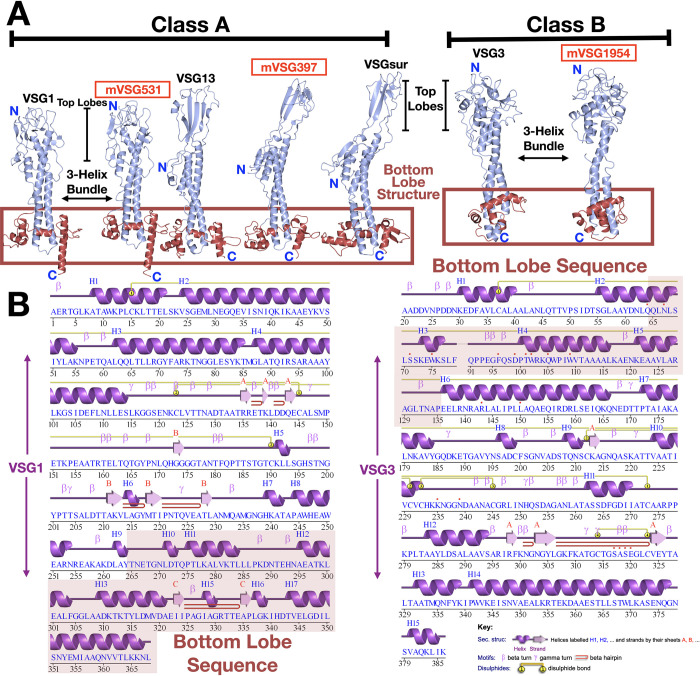
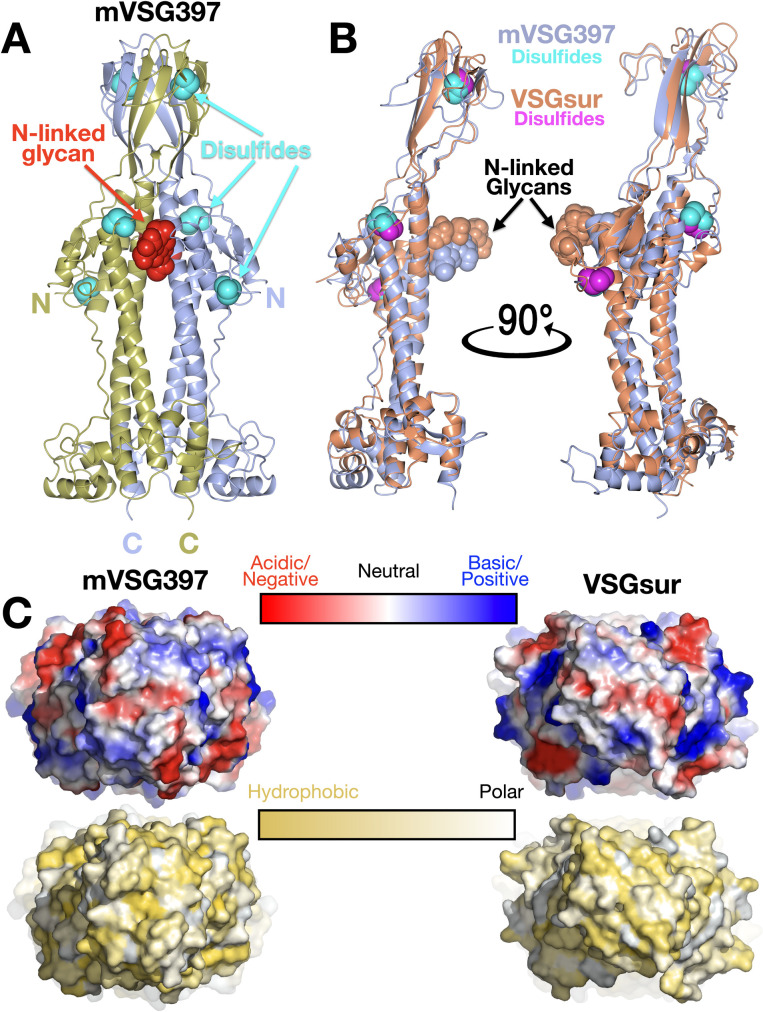
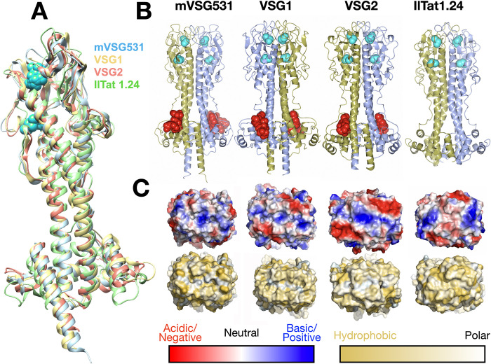
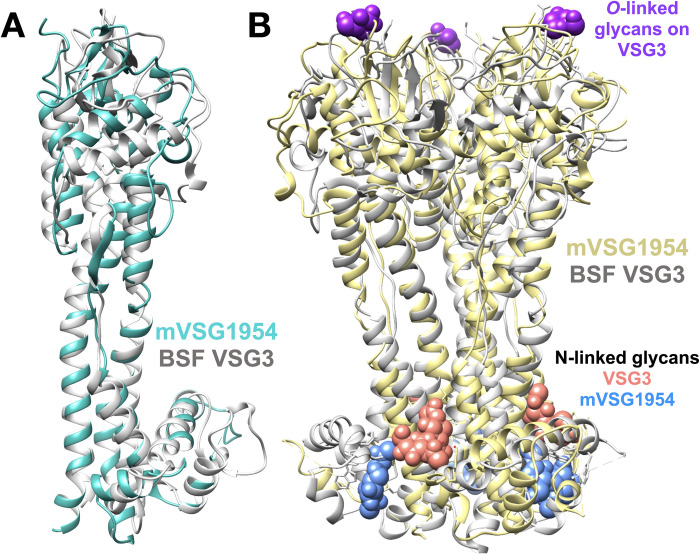
Verification
Claims the paper makes
The claims and statistics of this paper haven’t been checked yet.
Run the check — free, no account needed. It reads the open-access text and checks the sources it cites.
Self-check
Percentages, sums and reported differences re-computed from the paper’s own numbers.
The self-check needs the paper’s full text.
Citations
Cites
Highly accurate protein structure prediction with AlphaFold
Jumper, J., Evans, R., Pritzel, A., Green, T., Figurnov, M., Ronneberger, O., Tunyasuvunakool, K., Bates, R., Žídek, A., Potapenko, A., et al. (2021) · Nature · doi:10.1038/s41586-021-03819-2 · Cited by 43,764
Brun, R., Blum, J., Chappuis, F., & Burri, C. (2010) · The Lancet · doi:10.1016/s0140-6736(09)60829-1 · Cited by 926
Research works printed but not linked: 58
- J Keating, 2015, Human African trypanosomiasis prevention, treatment and control costs: A systematic review, Acta Trop, 150, 4, 10.1016/j.actatropica.2015.06.003 · DOI
- A. Ponte-Sucre, 2016, An Overview of Trypanosoma brucei Infections: An Intense Host–Parasite Interaction., Front Microbiol., 7, 10.3389/fmicb.2016.02126 · DOI
- KR Matthews, 2015, The within-host dynamics of African trypanosome infections, Philos Trans R Soc Lond B Biol Sci, 370, 10.1098/rstb.2014.0288 · DOI
- JD Bangs, 2018, Evolution of Antigenic Variation in African Trypanosomes: Variant Surface Glycoprotein Expression, Structure, and Function., BioEssays, 40, 1800181, 10.1002/bies.201800181 · DOI
- PT Manna, 2014, Life and times: synthesis, trafficking, and evolution of VSG, Trends Parasitol, 30, 251, 10.1016/j.pt.2014.03.004 · DOI
- P Borst, 1997, Mechanisms of antigenic variation in African trypanosomes, Behring Inst Mitt, 1
- D. Horn, 2014, Antigenic variation in African trypanosomes, Mol Biochem Parasitol, 195, 123, 10.1016/j.molbiopara.2014.05.001 · DOI
- MR Mugnier, 2016, Masters of Disguise: Antigenic Variation and the VSG Coat in Trypanosoma brucei., PLOS Pathog., 12, e1005784, 10.1371/journal.ppat.1005784 · DOI
- F Aresta-Branco, 2019, Mechanistic Similarities between Antigenic Variation and Antibody Diversification during Trypanosoma brucei Infection, Trends Parasitol, 35, 302, 10.1016/j.pt.2019.01.011 · DOI
- GA Cross, 2014, Capturing the variant surface glycoprotein repertoire (the VSGnome) of Trypanosoma brucei Lister 427., Mol Biochem Parasitol, 195, 59, 10.1016/j.molbiopara.2014.06.004 · DOI
- L Marcello, 2007, Analysis of the VSG gene silent archive in Trypanosoma brucei reveals that mosaic gene expression is prominent in antigenic variation and is favored by archive substructure, Genome Res, 17, 1344, 10.1101/gr.6421207 · DOI
- JL Weirather, 2012, Mapping of VSG similarities in Trypanosoma brucei, Mol Biochem Parasitol, 181, 141, 10.1016/j.molbiopara.2011.10.011 · DOI
- T Bartossek, 2017, Structural basis for the shielding function of the dynamic trypanosome variant surface glycoprotein coat., Nat Microbiol, 2, 1523, 10.1038/s41564-017-0013-6 · DOI
- M Carrington, 1991, Variant specific glycoprotein of Trypanosoma brucei consists of two domains each having an independently conserved pattern of cysteine residues, J Mol Biol, 221, 823, 10.1016/0022-2836(91)80178-W · DOI
- D Freymann, 1990, 2.9 A resolution structure of the N-terminal domain of a variant surface glycoprotein from Trypanosoma brucei, J Mol Biol, 216, 141, 10.1016/S0022-2836(05)80066-X · DOI
- J Pinger, 2018, African trypanosomes evade immune clearance by O -glycosylation of the VSG surface coat., Nat Microbiol, 3, 932, 10.1038/s41564-018-0187-6 · DOI
- J Zeelen, 2021, Structure of trypanosome coat protein VSGsur and function in suramin resistance, Nat Microbiol, 6, 392, 10.1038/s41564-020-00844-1 · DOI
- A Chattopadhyay, 2005, Structure of the C-terminal domain from Trypanosoma brucei variant surface glycoprotein MITat1.2, J Biol Chem, 280, 7228, 10.1074/jbc.M410787200 · DOI
- NG Jones, 2008, Structure of a glycosylphosphatidylinositol-anchored domain from a trypanosome variant surface glycoprotein, J Biol Chem, 283, 3584, 10.1074/jbc.M706207200 · DOI
- K Umaer, 2021, Dynamic, variable oligomerization and the trafficking of variant surface glycoproteins of Trypanosoma brucei, Traffic Cph Den, 22, 274, 10.1111/tra.12806 · DOI
- CM Alarcon, 1994, A monocistronic transcript for a trypanosome variant surface glycoprotein, Mol Cell Biol, 14, 5579
- SV Graham, 1995, Transcriptional regulation of metacyclic variant surface glycoprotein gene expression during the life cycle of Trypanosoma brucei, Mol Cell Biol, 15, 5945, 10.1128/MCB.15.11.5945 · DOI
- S Hutchinson, 2021, The establishment of variant surface glycoprotein monoallelic expression revealed by single-cell RNA-seq of Trypanosoma brucei in the tsetse fly salivary glands., PLOS Pathog., 17, e1009904, 10.1371/journal.ppat.1009904 · DOI
- LSM Müller, 2018, Genome organization and DNA accessibility control antigenic variation in trypanosomes, Nature, 563, 121, 10.1038/s41586-018-0619-8 · DOI
- P Büscher, 2017, Human African trypanosomiasis, The Lancet, 390, 2397, 10.1016/S0140-6736(17)31510-6 · DOI
- KR Matthews, 2005, The developmental cell biology of Trypanosoma brucei, J Cell Sci, 118, 283, 10.1242/jcs.01649 · DOI
- G Romero-Meza, 2020, Trypanosoma brucei., Trends Parasitol, 36, 571, 10.1016/j.pt.2019.10.007 · DOI
- OA Alfituri, 2020, To the Skin and Beyond: The Immune Response to African Trypanosomes as They Enter and Exit the Vertebrate Host., Front Immunol., 11, 1250, 10.3389/fimmu.2020.01250 · DOI
- R Christiano, 2017, The proteome and transcriptome of the infectious metacyclic form of Trypanosoma brucei define quiescent cells primed for mammalian invasion, Mol Microbiol, 106, 74, 10.1111/mmi.13754 · DOI
- S Alsford, 2005, Tagging a T. brucei RRNA locus improves stable transfection efficiency and circumvents inducible expression position effects, Mol Biochem Parasitol, 144, 142, 10.1016/j.molbiopara.2005.08.009 · DOI
- GA Cross, 1984, Release and purification of Trypanosoma brucei variant surface glycoprotein, J Cell Biochem, 24, 79, 10.1002/jcb.240240107 · DOI
- SR Ness, 2004, CRANK: new methods for automated macromolecular crystal structure solution., Struct Lond Engl 1993., 12, 1753
- K. Cowtan, 2012, Completion of autobuilt protein models using a database of protein fragments, Acta Crystallogr D Biol Crystallogr, 68, 328, 10.1107/S0907444911039655 · DOI
- PD Adams, 2010, PHENIX: a comprehensive Python-based system for macromolecular structure solution, Acta Crystallogr Biol Crystallogr, 66, 213, 10.1107/S0907444909052925 · DOI · Paper page
- D Liebschner, 2019, Macromolecular structure determination using X-rays, neutrons and electrons: recent developments in Phenix., Acta Crystallogr Sect Struct Biol, 75, 861, 10.1107/S2059798319011471 · DOI · Paper page
- P Emsley, 2004, Coot: model-building tools for molecular graphics, Acta Crystallogr D Biol Crystallogr, 60, 2126, 10.1107/S0907444904019158 · DOI · Paper page
- RP Joosten, 2014, The PDB_REDO server for macromolecular structure model optimization, IUCrJ, 1, 213, 10.1107/S2052252514009324 · DOI
- AJ McCoy, 2007, Phaser crystallographic software, J Appl Crystallogr, 40, 658, 10.1107/S0021889807021206 · DOI · Paper page
- TC Terwilliger, 2008, Iterative model building, structure refinement and density modification with the PHENIX AutoBuild wizard, Acta Crystallogr D Biol Crystallogr, 64, 61, 10.1107/S090744490705024X · DOI · Paper page
- GM Sheldrick, 2008, A short history of SHELX, Acta Crystallogr A, 64, 112, 10.1107/S0108767307043930 · DOI
- L Potterton, 2018, CCP4i2: the new graphical user interface to the CCP4 program suite, Acta Crystallogr Sect Struct Biol, 74, 68, 10.1107/S2059798317016035 · DOI
- G Langer, 2008, Automated macromolecular model building for X-ray crystallography using ARP/wARP version 7., Nat Protoc, 3, 1171, 10.1038/nprot.2008.91 · DOI · Paper page
- L Potterton, 2018, CCP 4 i 2: the new graphical user interface to the CCP 4 program suite, Acta Crystallogr Sect Struct Biol, 74, 68, 10.1107/S2059798317016035 · DOI
- G. Winter, 2010, xia2: an expert system for macromolecular crystallography data reduction, J Appl Crystallogr, 43, 186, 10.1107/S0021889809045701 · DOI
- G Winter, 2022, DIALS as a toolkit, Protein Sci Publ Protein Soc, 31, 232, 10.1002/pro.4224 · DOI
- S Wang, 2013, Protein structure alignment beyond spatial proximity, Sci Rep, 3, 1448, 10.1038/srep01448 · DOI
- S Wang, 2011, Alignment of distantly related protein structures: algorithm, bound and implications to homology modeling., Bioinforma Oxf Engl, 27, 2537, 10.1093/bioinformatics/btr432 · DOI
- G Hovel-Miner, 2015, A Host-Pathogen Interaction Reduced to First Principles: Antigenic Variation in T. brucei., Results Probl Cell Differ, 57, 23, 10.1007/978-3-319-20819-0_2 · DOI
- MR Mugnier, 2016, Masters of Disguise: Antigenic Variation and the VSG Coat in Trypanosoma brucei., PLoS Pathog., 12, e1005784, 10.1371/journal.ppat.1005784 · DOI
- L Tetley, 1987, Onset of expression of the variant surface glycoproteins of Trypanosoma brucei in the tsetse fly studied using immunoelectron microscopy, J Cell Sci, 87, 363, 10.1242/jcs.87.2.363 · DOI
- SV Graham, 1990, Distinct, developmental stage-specific activation mechanisms of trypanosome VSG genes, Parasitology, 101, 361, 10.1017/S0031182000060558 · DOI
- JR Helm, 2009, Analysis of expressed sequence tags from the four main developmental stages of Trypanosoma congolense, Mol Biochem Parasitol, 168, 34, 10.1016/j.molbiopara.2009.06.004 · DOI
- MR Mugnier, 2015, The in vivo dynamics of antigenic variation in Trypanosoma brucei, Science, 347, 1470, 10.1126/science.aaa4502 · DOI
- S McNicholas, 2011, Presenting your structures: the CCP4mg molecular-graphics software, Acta Crystallogr D Biol Crystallogr, 67, 386, 10.1107/S0907444911007281 · DOI
- RA Laskowski, 2018, PDBsum: Structural summaries of PDB entries, Protein Sci, 27, 129, 10.1002/pro.3289 · DOI
- D Eisenberg, 1984, Analysis of membrane and surface protein sequences with the hydrophobic moment plot, J Mol Biol, 179, 125, 10.1016/0022-2836(84)90309-7 · DOI
- EF Pettersen, 2004, UCSF Chimera—a visualization system for exploratory research and analysis, J Comput Chem, 25, 1605, 10.1002/jcc.20084 · DOI
- P Emsley, 2010, Features and development of Coot, Acta Crystallogr D Biol Crystallogr, 66, 486, 10.1107/S0907444910007493 · DOI · Paper page
Metadata
- Authors
- Chandra, M., Đaković, S., Foti, K., Zeelen, J. P., van Straaten, M., Aresta-Branco, F., Tihon, E., Lübbehusen, N., Ruppert, T., Glover, L., Papavasiliou, F. N., & Stebbins, C. E.
- Affiliations
- German Cancer Research Center (DE)
- Institut Pasteur (FR)
- Heidelberg University (DE)
- Corresponding author
- Papavasiliou, F. Nina
- Stebbins, C. Erec
- ORCID
- Venue
- PLOS Neglected Tropical Diseases
- Publisher
- Public Library of Science (PLoS)
- Published
- 2023-02-13
- Volume
- 17
- Issue
- 2
- Pages
- e0011093
- ISSN
- 1935-2727 (ISSN-L)
- 1935-2735
- PMC
- PMC9956791
- OpenAlex
- W4320482007
- Cited by
- 8
- References
- 2
- Citations by year
- 2026: 2 · 2024: 4 · 2023: 2
- FWCI
- 1.60
- Type
- journal article
- Language
- en
- Update notices
- new_version (2023-02-24)
- Open access
- gold
- doi.org
- Licence
- Locations
- PLoS neglected tropical diseases (publishedVersion, cc-by)
- bioRxiv (Cold Spring Harbor Laboratory) (acceptedVersion, cc-by-nc-nd)
- PubMed Central (publishedVersion, cc-by)
- PubMed (publishedVersion)
- HAL (submittedVersion, cc-by)
- DORA PSI (Paul Scherrer Institute) (submittedVersion, cc-by)
- DOAJ (DOAJ: Directory of Open Access Journals) (submittedVersion, cc-by-sa)
- Funders
- Agence Nationale de la Recherche (ANR-17-CE12-0012 VSGREG)
- Deutsches Krebsforschungszentrum
- Topics
- Health Sciences › Medicine › Epidemiology › Trypanosoma species research and implications
- Life Sciences › Biochemistry, Genetics and Molecular Biology › Structural Biology › Enzyme Structure and Function
- Life Sciences › Biochemistry, Genetics and Molecular Biology › Molecular Biology › vaccines and immunoinformatics approaches
- Keywords
- African trypanosomes, Trypanosoma brucei, variant surface glycoprotein, metacyclic forms, bloodstream forms, antigenic variation, N-terminal domain, oligomerization, parasite life cycle
- MeSH terms
- Animals, Mammals, Membrane Glycoproteins, Trypanosoma, Trypanosoma brucei brucei, Trypanosomiasis, African, Tsetse Flies, Variant Surface Glycoproteins, Trypanosoma
About these checks
Registry, retraction, Cites and statistics results are computed, not judged, with one exception: a printed reference that no index lists is read once by a model before it counts as a citation error. Claims are extracted by a model and kept only when their quote appears verbatim in the text; each cited source is then checked, with a model’s help, for whether it says what the claim relies on. Those verdicts can be wrong — read the claim’s quote and the cited source before relying on one.
Statistics and arithmetic are recomputed, not judged.
Confidence bands are shown only for fields calibrated on our annotated test set. None is calibrated yet, so no band is shown.
What counts as a claim ›How we measure confidence ›How we check this ›
Are you an author, or do you see a problem with this page? Request a review
