

The DOI matches its Crossref record; 51 of the 54 references it cites were found in a registry or an index.
Chemical Magnetoreception: Bird Cryptochrome 1a Is Excited by Blue Light and Forms Long-Lived Radical-Pairs
Open accessAbstract
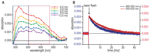
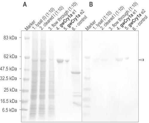
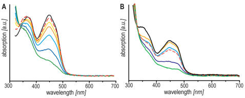
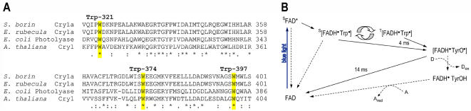
Cryptochromes (Cry) have been suggested to form the basis of light-dependent magnetic compass orientation in birds. However, to function as magnetic compass sensors, the cryptochromes of migratory birds must possess a number of key biophysical characteristics. Most importantly, absorption of blue light must produce radical pairs with lifetimes longer than about a microsecond. Cryptochrome 1a (gwCry1a) and the photolyase-homology-region of Cry1 (gwCry1-PHR) from the migratory garden warbler were recombinantly expressed and purified from a baculovirus/Sf9 cell expression system. Transient absorption measurements show that these flavoproteins are indeed excited by light in the blue spectral range leading to the formation of radicals with millisecond lifetimes. These biophysical characteristics suggest that gwCry1a is ideally suited as a primary light-mediated, radical-pair-based magnetic compass receptor.
Overview
The claims and statistics of this paper haven’t been checked yet.
Run the check — free, no account needed. It reads the open-access text and checks the sources it cites.
Integrity signals
Registry
Cites
Statistics
Figures
Verification
Claims the paper makes
The claims and statistics of this paper haven’t been checked yet.
Run the check — free, no account needed. It reads the open-access text and checks the sources it cites.
Claims other papers make about this one
Each is checked against this paper’s own text: does it say what the citing paper says it does?
In the citing paper
“It seems a logical next step to more complex living creatures, especially since four different types of cryptochrome have been confirmed in the eyes of migratory birds [103] and cryptochromes from the migratory garden warbler form radicals with millisecond lifetimes under the influence of the blue spectral range [104].”
From The future of quantum biology (2018) · cites this paper as [104]
Self-check
Percentages, sums and reported differences re-computed from the paper’s own numbers.
The self-check needs the paper’s full text.
Citations
Cites
Magnetic field effects in chemical kinetics and related phenomena
Steiner, U. E., & Ulrich, T. (1989) · Chemical Reviews · doi:10.1021/cr00091a003 · Cited by 1,660
HY4 gene of A. thaliana encodes a protein with characteristics of a blue-light photoreceptor
Ahmad, M., & Cashmore, A. R. (1993) · Nature · doi:10.1038/366162a0 · Cited by 1,279
A Model for Photoreceptor-Based Magnetoreception in Birds
Ritz, T., Adem, S., & Schulten, K. (2000) · Biophysical Journal · doi:10.1016/s0006-3495(00)76629-x · Cited by 1,176 · cited 4×
“This led to the suggestion that photochemical reactions in the birds’ eyes could produce spin-correlated radical pairs as the primary sensor in a magnetic field-dependent signalling cascade [7,8], based on the fact that the recombination reactions of spin-correlated radical pairs are known to be magnetic field-sensiti…”
BackgroundInfluentialCryptochromes: Blue Light Receptors for Plants and Animals
Cashmore, A. R., Jarillo, J. A., Wu, Y., & Liu, D. (1999) · Science · doi:10.1126/science.284.5415.760 · Cited by 1,027
Trends in the Development of Baculovirus Expression Vectors
Luckow, V. A., & Summers, M. D. (1988) · Nature Biotechnology · doi:10.1038/nbt0188-47 · Cited by 1,016
Magnetic Compass of European Robins
Wiltschko, W., & Wiltschko, R. (1972) · Science · doi:10.1126/science.176.4030.62 · Cited by 752
Structure and function of DNA photolyase
Sancar, A. (1994) · Biochemistry · doi:10.1021/bi00167a001 · Cited by 664
Resonance effects indicate a radical-pair mechanism for avian magnetic compass
Ritz, T., Thalau, P., Phillips, J. B., Wiltschko, R., & Wiltschko, W. (2004) · Nature · doi:10.1038/nature02534 · Cited by 617 · cited 1×
“The hypothesis that the avian magnetic compass is based on a radical pair mechanism is strongly supported by the observation that magnetic compass orientation of migratory birds can indeed be disturbed by weak radiofrequency magnetic fields with frequencies in the MHz range [17,18].”
BackgroundCrystal Structure of DNA Photolyase from Escherichia coli
Park, H.-W., Kim, S., Sancar, A., & Deisenhofer, J. (1995) · Science · doi:10.1126/science.7604260 · Cited by 584
Baculovirus Diversity and Molecular Biology
Blissard, G. W., & Rohrmann, G. F. (1990) · Annual Review of Entomology · doi:10.1146/annurev.en.35.010190.001015 · Cited by 535
Cited by
Sorted by citations
The cryptochromes: blue light photoreceptors in plants and animals
Chaves, I., Pokorny, R., Byrdin, M., Hoang, N., Ritz, T., Brettel, K., Essen, L.-O., van der Horst, G. T. J., Batschauer, A., & Ahmad, M. (2011) · Annual Review of Plant Biology · doi:10.1146/annurev-arplant-042110-103759 · Cited by 845
The Radical-Pair Mechanism of Magnetoreception
Hore, P. J., & Mouritsen, H. (2016) · Annual Review of Biophysics · doi:10.1146/annurev-biophys-032116-094545 · Cited by 813
Self-citationChemical magnetoreception in birds: The radical pair mechanism
Rodgers, C. T., & Hore, P. J. (2009) · Proceedings of the National Academy of Sciences · doi:10.1073/pnas.0711968106 · Cited by 542
Self-citationLong-distance navigation and magnetoreception in migratory animals
Mouritsen, H. (2018) · Nature · doi:10.1038/s41586-018-0176-1 · Cited by 547
Self-citationPlant photoreceptors: Multi-functional sensory proteins and their signaling networks
Paik, I., & Huq, E. (2019) · Seminars in Cell and Developmental Biology · doi:10.1016/j.semcdb.2019.03.007 · Cited by 379
Metadata
- Authors
- Liedvogel, M., Maeda, K., Henbest, K., Schleicher, E., Simon, T., Timmel, C. R., Hore, P. J., & Mouritsen, H.
- Affiliations
- Carl von Ossietzky Universität Oldenburg (DE)
- University of Oxford (GB)
- Freie Universität Berlin (DE)
- Corresponding author
- Liedvogel, Miriam
- Maeda, Kiminori
- Henbest, Kevin
- Schleicher, Erik
- Simon, Thomas
- Timmel, Christiane R.
- Hore, P. J.
- Mouritsen, Henrik
- ORCID
- Venue
- PLoS ONE
- Publisher
- Public Library of Science (PLoS)
- Published
- 2007-10-31
- Volume
- 2
- Issue
- 10
- Pages
- e1106
- ISSN
- 1932-6203 (ISSN-L)
- PMC
- PMC2040520
- OpenAlex
- W2163169468
- Cited by
- 183
- References
- 54
- Citations by year
- 2026: 3 · 2025: 7 · 2024: 7 · 2023: 6 · 2022: 7 · 2021: 11 · 2020: 9 · 2019: 3 · 2018: 12 · 2017: 12 · 2016: 14 · 2015: 11 · 2014: 11 · 2013: 8 · 2012: 11
- FWCI
- 7.68
- Type
- journal article
- Language
- en
- Open access
- gold
- doi.org
- Locations
- PLoS ONE (publishedVersion, cc-by)
- PubMed Central (publishedVersion, cc-by)
- PubMed (publishedVersion)
- MPG.PuRe (Max Planck Society) (submittedVersion, other-oa)
- DOAJ (DOAJ: Directory of Open Access Journals) (submittedVersion, cc-by-sa)
- Max Planck Digital Library (submittedVersion, other-oa)
- Figshare (submittedVersion, cc-by)
- Lund University Publications (Lund University) (submittedVersion)
- Refubium (Universitätsbibliothek der Freien Universität Berlin) (submittedVersion, cc-by)
- Funders
- Carl von Ossietzky Universität Oldenburg
- Royal Society
- EMF Biological Research Trust
- Deutsche Forschungsgemeinschaft (FOR 526, MO 1408/1-2, FOR 701)
- Volkswagen Foundation
- Engineering and Physical Sciences Research Council (GR/T05424/01)
- Topics
- Life Sciences › Biochemistry, Genetics and Molecular Biology › Biophysics › Electromagnetic Fields and Biological Effects
- Physical Sciences › Chemistry › Spectroscopy › Electron Spin Resonance Studies
- Life Sciences › Neuroscience › Endocrine and Autonomic Systems › Circadian rhythm and melatonin
- Keywords
- radical pairs, blue light absorption, flavoprotein, magnetic compass orientation, bird migration, garden warbler, transient absorption spectroscopy, recombinant protein expression
- MeSH terms
- Animals, Birds, Cell Line, Cloning, Molecular, Flavoproteins, Free Radicals, Insecta, Light, Magnetics, Models, Biological, Magnetic Resonance Spectroscopy, Recombinant Proteins, Ultraviolet Rays, Protein Structure, Tertiary, Animal Migration, Cryptochromes
About these checks
Registry, retraction, Cites and statistics results are computed, not judged, with one exception: a printed reference that no index lists is read once by a model before it counts as a citation error. Claims are extracted by a model and kept only when their quote appears verbatim in the text; each cited source is then checked, with a model’s help, for whether it says what the claim relies on. Those verdicts can be wrong — read the claim’s quote and the cited source before relying on one.
Statistics and arithmetic are recomputed, not judged.
Confidence bands are shown only for fields calibrated on our annotated test set. None is calibrated yet, so no band is shown.
What counts as a claim ›How we measure confidence ›How we check this ›
Are you an author, or do you see a problem with this page? Request a review
