

The DOI matches its Crossref record; 125 of the 126 references it cites were found in a registry or an index.
Opposite Effects of Early Maternal Deprivation on Neurogenesis in Male versus Female Rats
Open accessAbstract
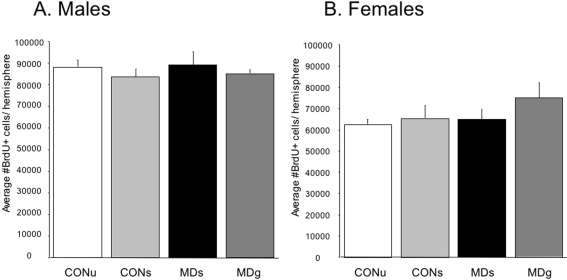
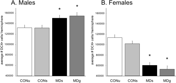
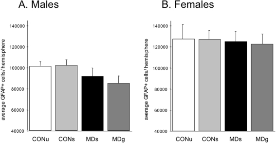
BACKGROUND: Major depression is more prevalent in women than in men. The underlying neurobiological mechanisms are not well understood, but recent data shows that hippocampal volume reductions in depressed women occur only when depression is preceded by an early life stressor. This underlines the potential importance of early life stress, at least in women, for the vulnerability to develop depression. Perinatal stress exposure in rodents affects critical periods of brain development that persistently alter structural, emotional and neuroendocrine parameters in adult offspring. Moreover, stress inhibits adult hippocampal neurogenesis, a form of structural plasticity that has been implicated a.o. in antidepressant action and is highly abundant early postnatally. We here tested the hypothesis that early life stress differentially affects hippocampal structural plasticity in female versus male offspring. PRINCIPAL FINDINGS: We show that 24 h of maternal deprivation (MD) at PND3 affects hippocampal structural plasticity at PND21 in a sex-dependent manner. Neurogenesis was significantly increased in male but decreased in female offspring after MD. Since no other structural changes were found in granule cell layer volume, newborn cell survival or proliferation rate, astrocyte number or gliogenesis, this indicates that MD elicits specific changes in subsets of differentiating cells and differentially affects immature neurons. The MD induced sex-specific effects on neurogenesis cannot be explained by differences in maternal care. CONCLUSIONS: Our data shows that early environment has a critical influence on establishing sex differences in neural plasticity and supports the concept that the setpoint for neurogenesis may be determined during perinatal life. It is tempting to speculate that a reduced level of neurogenesis, secondary to early stress exposure, may contribute to maladaptation of the HPA axis and possibly to the increased vulnerability of women to stress-related disorders.
Overview
The claims and statistics of this paper haven’t been checked yet.
Run the check — free, no account needed. It reads the open-access text and checks the sources it cites.
Integrity signals
Registry
Cites
Statistics
Figures
Verification
Claims the paper makes
The claims and statistics of this paper haven’t been checked yet.
Run the check — free, no account needed. It reads the open-access text and checks the sources it cites.
Claims other papers make about this one
Each is checked against this paper’s own text: does it say what the citing paper says it does?
In the citing paper
“Previous work showed maternal separation stress reduced adult neurogenesis in the rat hippocampus (37; 62–64), although other studies failed to show this (65).”
From Perineuronal nets, inhibitory interneurons and anxiety-related ventral hippocampal neuronal oscillations are altered by early life adversity (2019) · cites this paper as [65]
Self-check
Percentages, sums and reported differences re-computed from the paper’s own numbers.
The self-check needs the paper’s full text.
Citations
Cites
Developments of a water-maze procedure for studying spatial learning in the rat
Morris, R. (1984) · Journal of Neuroscience Methods · doi:10.1016/0165-0270(84)90007-4 · Cited by 7,180
Stress and the brain: from adaptation to disease
de Kloet, E. R., Joëls, M., & Holsboer, F. (2005) · Nature Reviews Neuroscience · doi:10.1038/nrn1683 · Cited by 4,621
Self-citationRequirement of hippocampal neurogenesis for the behavioral effects of antidepressants
Santarelli, L., Saxe, M., Gross, C., Surget, A., Battaglia, F., Dulawa, S., Weisstaub, N., Lee, J., Duman, R., Arancio, O., et al. (2003) · Science · doi:10.1126/science.1083328 · Cited by 4,321
Liu, D., Diorio, J., Tannenbaum, B., Caldji, C., Francis, D., Freedman, A., Sharma, S., Pearson, D., Plotsky, P. M., & Meaney, M. J. (1997) · Science · doi:10.1126/science.277.5332.1659 · Cited by 3,170
Heim, C., & Nemeroff, C. B. (2001) · Biological Psychiatry · doi:10.1016/s0006-3223(01)01157-x · Cited by 2,791
Kessler, R. (1993) · Journal of Affective Disorders · doi:10.1016/0165-0327(93)90026-g · Cited by 2,207
The Corticosteroid Receptor Hypothesis of Depression
Holsboer, F. (2000) · Neuropsychopharmacology · doi:10.1016/s0893-133x(00)00159-7 · Cited by 2,181
Heim, C. (2000) · JAMA · doi:10.1001/jama.284.5.592 · Cited by 1,968
Plotsky, P. M., & Meaney, M. J. (1993) · Molecular Brain Research · doi:10.1016/0169-328x(93)90189-v · Cited by 1,638 · cited 2×
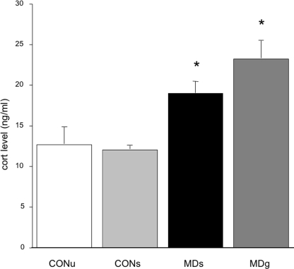
“Also, stress-induced ACTH [59,61] and corticosterone responses were increased in 24 h MD rats [53,60] while similar patterns are found in repeated separation paradigms [56,62–65].”
BackgroundSmaller hippocampal volume predicts pathologic vulnerability to psychological trauma
Gilbertson, M. W., Shenton, M. E., Ciszewski, A., Kasai, K., Lasko, N. B., Orr, S. P., & Pitman, R. K. (2002) · Nature Neuroscience · doi:10.1038/nn958 · Cited by 1,635
Cited by
Sorted by citations
Modulation of the Hypothalamic-Pituitary-Adrenal Axis by Early Life Stress Exposure
van Bodegom, M., Homberg, J. R., & Henckens, M. J. A. G. (2017) · Frontiers in Cellular Neuroscience · doi:10.3389/fncel.2017.00087 · Cited by 606
Lucassen, P. J., Meerlo, P., Naylor, A. S., van Dam, A. M., Dayer, A. G., Fuchs, E., Oomen, C. A., & Czéh, B. (2010) · European Neuropsychopharmacology · doi:10.1016/j.euroneuro.2009.08.003 · Cited by 449
Self-citationJoëls, M., Sarabdjitsingh, R. A., & Karst, H. (2012) · Pharmacological Reviews · doi:10.1124/pr.112.005892 · Cited by 445
Self-citationWalker, C., Bath, K. G., Joëls, M., Kőrösi, A., Larauche, M. H., Lucassen, P. J., Morris, M. J., Raineki, C., Roth, T. L., Sullivan, R. M., et al. (2017) · Stress · doi:10.1080/10253890.2017.1343296 · Cited by 432
Self-citationOomen, C. A., Soeters, H., Audureau, N., Vermunt, L., van Hasselt, F. N., Manders, E. M. M., Joëls, M., Lucassen, P. J., & Krugers, H. J. (2010) · Journal of Neuroscience · doi:10.1523/jneurosci.0247-10.2010 · Cited by 367
Self-citation
Metadata
- Authors
- Oomen, C. A., Girardi, C. E. N., Cahyadi, R., Verbeek, E. C., Krugers, H., Joëls, M., & Lucassen, P. J.
- Affiliations
- University of Amsterdam (NL)
- Corresponding author
- Lucassen, Paul J.
- Venue
- PLoS ONE
- Publisher
- Public Library of Science (PLoS)
- Published
- 2009-01-29
- Volume
- 4
- Issue
- 1
- Pages
- e3675
- ISSN
- 1932-6203 (ISSN-L)
- PMC
- PMC2629844
- OpenAlex
- W2105673253
- Cited by
- 190
- References
- 126
- Citations by year
- 2026: 5 · 2025: 3 · 2024: 3 · 2023: 6 · 2022: 6 · 2021: 6 · 2020: 12 · 2019: 13 · 2018: 14 · 2017: 17 · 2016: 15 · 2015: 12 · 2014: 14 · 2013: 10 · 2012: 9
- FWCI
- 5.54
- Type
- journal article
- Language
- en
- Open access
- gold
- doi.org
- Locations
- PLoS ONE (publishedVersion, cc-by)
- PubMed Central (publishedVersion, cc-by)
- PubMed (publishedVersion)
- UvA-DARE (University of Amsterdam) (publishedVersion, cc-by)
- UvA-DARE (University of Amsterdam) (submittedVersion, cc-by)
- DOAJ (DOAJ: Directory of Open Access Journals) (submittedVersion, cc-by-sa)
- Data Archiving and Networked Services (DANS) (submittedVersion, other-oa)
- Topics
- Life Sciences › Neuroscience › Developmental Neuroscience › Neurogenesis and neuroplasticity mechanisms
- Life Sciences › Neuroscience › Behavioral Neuroscience › Stress Responses and Cortisol
- Health Sciences › Medicine › Public Health, Environmental and Occupational Health › Maternal Mental Health During Pregnancy and Postpartum
- Keywords
- maternal deprivation, early life stress, hippocampal neurogenesis, sex differences, structural plasticity, perinatal stress, hypothalamic-pituitary-adrenal axis, astrocytes, gliogenesis, maternal care
- MeSH terms
- Analysis of Variance, Animals, Animals, Newborn, Astrocytes, Behavior, Animal, Cell Differentiation, Cell Survival, Corticosterone, Female, Male, Maternal Deprivation, Neuroglia, Neuronal Plasticity, Sex Factors, Weight Loss, Rats, Wistar, Dentate Gyrus, Cell Proliferation, Rats, Neurogenesis
About these checks
Registry, retraction, Cites and statistics results are computed, not judged, with one exception: a printed reference that no index lists is read once by a model before it counts as a citation error. Claims are extracted by a model and kept only when their quote appears verbatim in the text; each cited source is then checked, with a model’s help, for whether it says what the claim relies on. Those verdicts can be wrong — read the claim’s quote and the cited source before relying on one.
Statistics and arithmetic are recomputed, not judged.
Confidence bands are shown only for fields calibrated on our annotated test set. None is calibrated yet, so no band is shown.
What counts as a claim ›How we measure confidence ›How we check this ›
Are you an author, or do you see a problem with this page? Request a review
