

The DOI matches its Crossref record; 49 of the 49 references it cites were found in a registry or an index.
p38γ Mitogen-Activated Protein Kinase Is a Key Regulator in Skeletal Muscle Metabolic Adaptation in Mice
Open accessAbstract
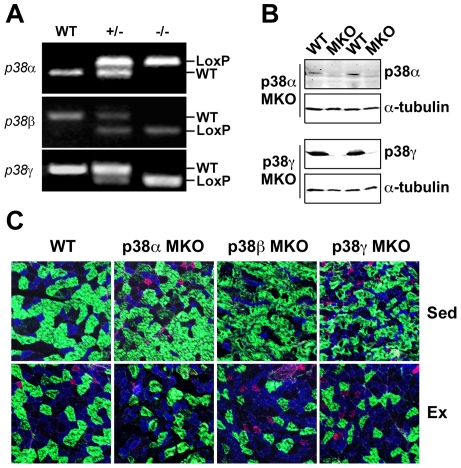
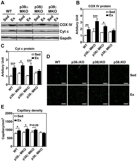
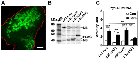
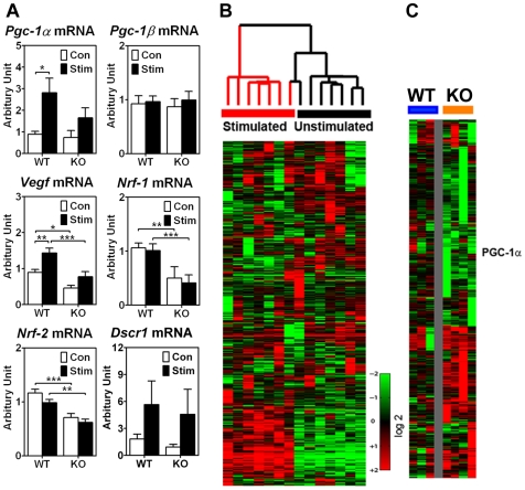
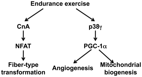
Regular endurance exercise induces skeletal muscle contractile and metabolic adaptations, conferring salutary health benefits, such as protection against the metabolic syndrome. The plasticity of skeletal muscle has been extensively investigated, but how the adaptive processes are precisely controlled is largely unknown. Using muscle-specific gene deletion in mice, we now show that p38gamma mitogen-activated protein kinase (MAPK), but not p38alpha and p38beta, is required for endurance exercise-induced mitochondrial biogenesis and angiogenesis, whereas none of the p38 isoforms are required for IIb-to-IIa fiber-type transformation. These phenotypic findings were further supported by microarray and real-time PCR analyses revealing contractile activity-dependent p38gamma target genes, including peroxisome proliferator-activated receptor gamma co-activator-1alpha (Pgc-1alpha) and vascular endothelial growth factor (Vegf), in skeletal muscle following motor nerve stimulation. Gene transfer-mediated overexpression of a dominant negative form of p38gamma, but not that of p38alpha or p38beta, blocked motor nerve stimulation-induced Pgc-1alpha transcription. These findings provide direct evidence for an obligated role of p38gamma MAPK-PGC-1alpha regulatory axis in endurance exercise-induced metabolic adaptation, but not contractile adaptation, in skeletal muscle.
Overview
The claims and statistics of this paper haven’t been checked yet.
Run the check — free, no account needed. It reads the open-access text and checks the sources it cites.
Integrity signals
Registry
Cites
Statistics
Figures
Verification
Claims the paper makes
The claims and statistics of this paper haven’t been checked yet.
Run the check — free, no account needed. It reads the open-access text and checks the sources it cites.
Claims other papers make about this one
Each is checked against this paper’s own text: does it say what the citing paper says it does?
The source backs part of the claim, not all of it — or backs it for a different population, condition or comparison.
In the citing paper
“The decrease in glycogen also activates the 38 kDa mitogen-activated protein kinase (p38), which, like AMPK, can increase the transcription and activity of PGC-1α [35–37].”
From Using Molecular Biology to Maximize Concurrent Training (2014) · cites this paper as [36]
Experimental — accuracy not yet measured
Self-check
Percentages, sums and reported differences re-computed from the paper’s own numbers.
The self-check needs the paper’s full text.
Citations
Cites
Calcium's Role in Mechanotransduction during Muscle Development
Damm, T. B., & Egli, M. (2014) · Cellular Physiology and Biochemistry · doi:10.1159/000356667 · Cited by 14,552
Gene Expression Omnibus: NCBI gene expression and hybridization array data repository
Edgar, R. (2002) · Nucleic Acids Research · doi:10.1093/nar/30.1.207 · Cited by 13,288
AMPK regulates energy expenditure by modulating NAD+ metabolism and SIRT1 activity
Cantó, C., Gerhart-Hines, Z., Feige, J. N., Lagouge, M., Noriega, L., Milne, J. C., Elliott, P. J., Puigserver, P., & Auwerx, J. (2009) · Nature · doi:10.1038/nature07813 · Cited by 3,300
Transcriptional co-activator PGC-1α drives the formation of slow-twitch muscle fibres
Lin, J., Wu, H., Tarr, P. T., Zhang, C.-Y., Wu, Z., Boss, O., Michael, L. F., Puigserver, P., Isotani, E., Olson, E. N., et al. (2002) · Nature · doi:10.1038/nature00904 · Cited by 2,546
Muscle deficiency and neonatal death in mice with a targeted mutation in the myogenin gene
Hasty, P., Bradley, A., Morris, J. H., Edmondson, D. G., Venuti, J. M., Olson, E. N., & Klein, W. H. (1993) · Nature · doi:10.1038/364501a0 · Cited by 1,364
Regulation of Muscle Fiber Type and Running Endurance by PPARδ
Wang, Y.-X., Zhang, C.-L., Yu, R. T., Cho, H. K., Nelson, M. C., Bayuga-Ocampo, C. R., Ham, J., Kang, H., & Evans, R. M. (2004) · PLoS Biology · doi:10.1371/journal.pbio.0020294 · Cited by 1,089
HIF-independent regulation of VEGF and angiogenesis by the transcriptional coactivator PGC-1α
Arany, Z., Foo, S.-Y., Ma, Y., Ruas, J. L., Bommi-Reddy, A., Girnun, G., Cooper, M., Laznik, D., Chinsomboon, J., Rangwala, S. M., et al. (2008) · Nature · doi:10.1038/nature06613 · Cited by 1,054
Adaptations of skeletal muscle to exercise: rapid increase in the transcriptional coactivator PGC-1
Baar, K., Wende, A. R., Jones, T. E., Marison, M., Nolte, L. A., Chen, M., Kelly, D. P., & Holloszy, J. O. (2002) · The FASEB Journal · doi:10.1096/fj.02-0367com · Cited by 1,020
A calcineurin-dependent transcriptional pathway controls skeletal muscle fiber typePreprint
Chin, E. R., Olson, E. N., Richardson, J. A., Yang, Q., Humphries, C. G., Shelton, J. M., Wu, H., Zhu, W., Bassel‐Duby, R., & Williams, R. S. (1998) · Genes & Development · doi:10.1101/gad.12.16.2499 · Cited by 1,015
Exercise induces transient transcriptional activation of the PGC‐1α gene in human skeletal muscle
Pilegaard, H., Saltin, B., & Neufer, P. D. (2003) · The Journal of Physiology · doi:10.1113/jphysiol.2002.034850 · Cited by 885
Handschin, C., Rhee, J., Lin, J., Tarr, P. T., & Spiegelman, B. M. (2003) · Proceedings of the National Academy of Sciences · doi:10.1073/pnas.1232352100 · Cited by 729
Akimoto, T., Pohnert, S. C., Li, P., Zhang, M., Gumbs, C., Rosenberg, P. B., Williams, R. S., & Yan, Z. (2005) · Journal of Biological Chemistry · doi:10.1074/jbc.m408862200 · Cited by 714
Self-citationGleyzer, N., Vercauteren, K., & Scarpulla, R. C. (2005) · Molecular and Cellular Biology · doi:10.1128/mcb.25.4.1354-1366.2005 · Cited by 711
Regulation of mitochondrial biogenesis in skeletal muscle by CaMK
Wu, H., Kanatous, S. B., Thurmond, F. A., Gallardo, T., Isotani, E., Bassel-Duby, R., & Williams, R. S. (2002) · Science · doi:10.1126/science.1071163 · Cited by 684
Handschin, C., Chin, S., Li, P., Liu, F., Maratos-Flier, E., LeBrasseur, N. K., Yan, Z., & Spiegelman, B. M. (2007) · Journal of Biological Chemistry · doi:10.1074/jbc.m704817200 · Cited by 611
Self-citationMEF2 responds to multiple calcium‐regulated signals in the control of skeletal muscle fiber type
Wu, H., Naya, F. J., McKinsey, T. A., Mercer, B., Shelton, J. M., Chin, E. R., Simard, A. R., Michel, R. N., Bassel‐Duby, R., Olson, E. N., et al. (2000) · The EMBO Journal · doi:10.1093/emboj/19.9.1963 · Cited by 468
Stimulation of Slow Skeletal Muscle Fiber Gene Expression by Calcineurin in Vivo
Naya, F. J., Mercer, B. M., Shelton, J. M., Richardson, J. A., Williams, R. S., & Olson, E. N. (2000) · Journal of Biological Chemistry · doi:10.1074/jbc.275.7.4545 · Cited by 420
Rothermel, B. A., Vega, R. B., Yang, J., Wu, H., Bassel‐Duby, R., & Williams, R. S. (2000) · Journal of Biological Chemistry · doi:10.1074/jbc.275.12.8719 · Cited by 414
Influence of exercise intensity and duration on biochemical adaptations in skeletal muscle
Dudley, G. A., Abraham, W. M., & Terjung, R. L. (1982) · Journal of Applied Physiology · doi:10.1152/jappl.1982.53.4.844 · Cited by 413
Cardiac and skeletal muscle adaptations to voluntary wheel running in the mouse
Allen, D. L., Harrison, B. C., Maass, A. H., Bell, M., Byrnes, W. C., & Leinwand, L. A. (2001) · Journal of Applied Physiology · doi:10.1152/jappl.2001.90.5.1900 · Cited by 377
Intracellular signaling specificity in skeletal muscle in response to different modes of exercise
Nader, G. A., & Esser, K. A. (2001) · Journal of Applied Physiology · doi:10.1152/jappl.2001.90.5.1936 · Cited by 372
Arany, Z., Lebrasseur, N., Morris, C., Smith, E., Yang, W., Ma, Y., Chin, S., & Spiegelman, B. M. (2007) · Cell Metabolism · doi:10.1016/j.cmet.2006.12.003 · Cited by 370
Activation of MEF2 by muscle activity is mediated through a calcineurin‐dependent pathway
Wu, H., Rothermel, B., Kanatous, S., Rosenberg, P. B., Naya, F. J., Shelton, J. M., Hutcheson, K. A., DiMaio, J. M., Olson, E. N., Bassel‐Duby, R., et al. (2001) · The EMBO Journal · doi:10.1093/emboj/20.22.6414 · Cited by 366
Effects of low-intensity prolonged exercise on PGC-1 mRNA expression in rat epitrochlearis muscle
Terada, S., Goto, M., Kato, M., Kawanaka, K., Shimokawa, T., & Tabata, I. (2002) · Biochemical and Biophysical Research Communications · doi:10.1016/s0006-291x(02)00881-1 · Cited by 354
Early signaling responses to divergent exercise stimuli in skeletal muscle from well-trained humans
Coffey, V. G., Zhong, Z., Shield, A., Canny, B. J., Chibalin, A. V., Zierath, J. R., & Hawley, J. A. (2006) · The FASEB Journal · doi:10.1096/fj.05-4809fje · Cited by 336
Irrcher, I., Adhihetty, P. J., Sheehan, T. E., Joseph, A.-M., & Hood, D. A. (2003) · American Journal of Physiology-Cell Physiology · doi:10.1152/ajpcell.00409.2002 · Cited by 315
Li, S., Czubryt, M. P., McAnally, J., Bassel‐Duby, R., Richardson, J. A., Wiebel, F. F., Nordheim, A., & Olson, E. N. (2005) · Proceedings of the National Academy of Sciences · doi:10.1073/pnas.0409103102 · Cited by 295
Myocyte-enriched calcineurin-interacting protein, MCIP1, inhibits cardiac hypertrophy in vivo
Rothermel, B. A., McKinsey, T. A., Vega, R. B., Nicol, R. L., Mammen, P. P. A., Yang, J., Antos, C. L., Shelton, J. M., Bassel‐Duby, R., Olson, E. N., et al. (2001) · Proceedings of the National Academy of Sciences · doi:10.1073/pnas.041614798 · Cited by 294
Wu, Z., Huang, X., Feng, Y., Handschin, C., Feng, Y., Gullicksen, P. S., Bare, O., Labow, M., Spiegelman, B., & Stevenson, S. C. (2006) · Proceedings of the National Academy of Sciences · doi:10.1073/pnas.0606714103 · Cited by 291
Extracellular matrix adaptation of tendon and skeletal muscle to exercise
Kjær, M., Magnusson, S. P., Krogsgaard, M. R., Bojsen‐Møller, J., Olesen, J. L., Heinemeier, K. M., Hansen, M., Haraldsson, B. T., Koskinen, S., Esmarck, B., et al. (2006) · Journal of Anatomy · doi:10.1111/j.1469-7580.2006.00549.x · Cited by 249
Divergent effects of exercise on metabolic and mitogenic signaling pathways in human skeletal muscle
Widegren, U., Jiang, X. J., Krook, A., Chibalin, A., Björnholm, M., Tally, M., Roth, R. A., Henriksson, J., Wallberg‐Henriksson, H., & Zierath, J. R. (1998) · The FASEB Journal · doi:10.1096/fasebj.12.13.1379 · Cited by 248
NFAT is a nerve activity sensor in skeletal muscle and controls activity-dependent myosin switching
McCullagh, K. J. A., Calabria, E., Pallafacchina, G., Ciciliot, S., Serrano, A. L., Argentini, C., Kalhovde, J. M., Lømo, T., & Schiaffino, S. (2004) · Proceedings of the National Academy of Sciences · doi:10.1073/pnas.0308035101 · Cited by 225
Voluntary running induces fiber type-specific angiogenesis in mouse skeletal muscle
Waters, R. E., Rotevatn, S., Li, P., Annex, B. H., & Yan, Z. (2004) · American Journal of Physiology-Cell Physiology · doi:10.1152/ajpcell.00247.2004 · Cited by 214
Self-citationExercise and Myocyte Enhancer Factor 2 Regulation in Human Skeletal Muscle
McGee, S. L., & Hargreaves, M. (2004) · Diabetes · doi:10.2337/diabetes.53.5.1208 · Cited by 205
Altered Skeletal Muscle Phenotypes in Calcineurin Aα and Aβ Gene-Targeted Mice
Parsons, S. A., Wilkins, B. J., Bueno, O. F., & Molkentin, J. D. (2003) · Molecular and Cellular Biology · doi:10.1128/mcb.23.12.4331-4343.2003 · Cited by 162
PGC-1α mediates exercise-induced skeletal muscle VEGF expression in mice
Leick, L., Hellsten, Y., Fentz, J., Lyngby, S. S., Wojtaszewski, J. F. P., Hidalgo, J., & Pilegaard, H. (2009) · American Journal of Physiology-Endocrinology and Metabolism · doi:10.1152/ajpendo.00076.2009 · Cited by 126
Akimoto, T., Ribar, T. J., Williams, R. S., & Yan, Z. (2004) · American Journal of Physiology-Cell Physiology · doi:10.1152/ajpcell.00248.2004 · Cited by 124
Self-citationYu, M., Blomstrand, E., Chibalin, A., Krook, A., & Zierath, J. R. (2001) · The Journal of Physiology · doi:10.1111/j.1469-7793.2001.00273.x · Cited by 121
Akimoto, T., Sorg, B. S., & Yan, Z. (2004) · American Journal of Physiology-Cell Physiology · doi:10.1152/ajpcell.00425.2003 · Cited by 117
Self-citationMatching of Calcineurin Activity to Upstream Effectors Is Critical for Skeletal Muscle Fiber Growth
Dunn, S. E., Chin, E. R., & Michel, R. N. (2000) · The Journal of Cell Biology · doi:10.1083/jcb.151.3.663 · Cited by 114
Boppart, M. D., Asp, S., Wojtaszewski, J. F. P., Fielding, R. A., Mohr, T. T., & Goodyear, L. J. (2000) · The Journal of Physiology · doi:10.1111/j.1469-7793.2000.00663.x · Cited by 105
Skeletal muscle cytochrome c and myoglobin, endurance, and frequency of training
Hickson, R. C. (1981) · Journal of Applied Physiology · doi:10.1152/jappl.1981.51.3.746 · Cited by 93
Calcineurin Is Necessary for the Maintenance but Not Embryonic Development of Slow Muscle Fibers
Oh, M., Rybkin, I. I., Copeland, V., Czubryt, M. P., Shelton, J. M., van Rooij, E., Richardson, J. A., Hill, J. A., Windt, L. J. D., Bassel‐Duby, R., et al. (2005) · Molecular and Cellular Biology · doi:10.1128/mcb.25.15.6629-6638.2005 · Cited by 92
Gute, D. C., Laughlin, M. H., & Amann, J. F. (1994) · Microcirculation · doi:10.3109/10739689409148273 · Cited by 90
Boppart, M. D., Hirshman, M. F., Sakamoto, K., Fielding, R. A., & Goodyear, L. J. (2001) · American Journal of Physiology-Cell Physiology · doi:10.1152/ajpcell.2001.280.2.c352 · Cited by 90
Mitochondrial cytochrome c oxidase subunit IV is phosphorylated by an endogenous kinase
Steenaart, N. A. E., & Shore, G. C. (1997) · FEBS Letters · doi:10.1016/s0014-5793(97)01145-9 · Cited by 82
Akimoto, T., Li, P., & Yan, Z. (2008) · American Journal of Physiology-Cell Physiology · doi:10.1152/ajpcell.00104.2008 · Cited by 61
Self-citationChoi, S., Liu, X., Li, P., Akimoto, T., Lee, S. Y., Zhang, M., & Yan, Z. (2005) · Journal of Applied Physiology · doi:10.1152/japplphysiol.00545.2005 · Cited by 39
Self-citationDudley, G. A., Abraham, W. M., & Terjung, R. L. (1981) · Medicine & Science in Sports & Exercise · doi:10.1249/00005768-198101320-00349 · Cited by 5
Cited by
Sorted by citations
Exercise Metabolism and the Molecular Regulation of Skeletal Muscle Adaptation
Egan, B., & Zierath, J. R. (2013) · Cell Metabolism · doi:10.1016/j.cmet.2012.12.012 · Cited by 2,084
Radak, Z. O., Zhao, Z., Koltai, E., Ohno, H., & Atalay, M. (2012) · Antioxidants and Redox Signaling · doi:10.1089/ars.2011.4498 · Cited by 643
Maintenance of Skeletal Muscle Mitochondria in Health, Exercise, and Aging
Hood, D. A., Memme, J. M., Oliveira, A. N., & Triolo, M. (2019) · Annual Review of Physiology · doi:10.1146/annurev-physiol-020518-114310 · Cited by 567
AMPK in skeletal muscle function and metabolism
Kjøbsted, R., Hingst, J. R., Fentz, J., Foretz, M., Sanz, M.-N., Pehmøller, C. K., Shum, M., Marette, A., Mounier, R., Treebak, J. T., et al. (2017) · The FASEB Journal · doi:10.1096/fj.201700442r · Cited by 517
Lira, V. A., Okutsu, M., Zhang, M., Greene, N. P., Laker, R. C., Breen, D. S., Hoehn, K. L., & Yan, Z. (2013) · The FASEB Journal · doi:10.1096/fj.13-228486 · Cited by 432
Self-citation
Metadata
- Authors
- Pogozelski, A. R., Geng, T., Li, P., Yin, X., Lira, V. A., Zhang, M., Chi, J.-T., & Yan, Z.
- Affiliations
- Duke Medical Center (US)
- University of Virginia (US)
- Corresponding author
- Yan, Zhen
- Venue
- PLoS ONE
- Publisher
- Public Library of Science (PLoS)
- Published
- 2009-11-20
- Volume
- 4
- Issue
- 11
- Pages
- e7934
- ISSN
- 1932-6203 (ISSN-L)
- PMC
- PMC2775956
- OpenAlex
- W2060618287
- Cited by
- 164
- References
- 49
- Citations by year
- 2026: 6 · 2025: 3 · 2024: 6 · 2023: 7 · 2022: 4 · 2021: 4 · 2020: 12 · 2019: 9 · 2018: 6 · 2017: 7 · 2016: 10 · 2015: 16 · 2014: 17 · 2013: 18 · 2012: 15
- FWCI
- 5.24
- Type
- journal article
- Language
- en
- Open access
- gold
- doi.org
- Locations
- PLoS ONE (publishedVersion, cc-by)
- PubMed Central (publishedVersion, cc-by)
- PubMed (publishedVersion)
- DOAJ (DOAJ: Directory of Open Access Journals) (submittedVersion, cc-by-sa)
- Figshare (submittedVersion, cc-by)
- Funders
- Howard Hughes Medical Institute
- Boehringer Ingelheim
- National Institutes of Health
- National Institute of Arthritis and Musculoskeletal and Skin Diseases (R01AR050429, AR 050429)
- Topics
- Life Sciences › Biochemistry, Genetics and Molecular Biology › Molecular Biology › Muscle Physiology and Disorders
- Health Sciences › Medicine › Physiology (medical) › Adipose Tissue and Metabolism
- Health Sciences › Medicine › Physiology (medical) › Cardiovascular and exercise physiology
- Keywords
- p38γ MAPK, endurance exercise, mitochondrial biogenesis, angiogenesis, PGC-1α, vascular endothelial growth factor, muscle fiber type shift, motor nerve stimulation
- MeSH terms
- Peroxisome Proliferator-Activated Receptor Gamma Coactivator 1-alpha, Animals, Genes, Dominant, Male, Mice, Inbred C57BL, Muscle Contraction, Neovascularization, Pathologic, Physical Conditioning, Animal, Transcription Factors, Trans-Activators, Gene Expression Regulation, Enzymologic, Muscle, Skeletal, Protein Isoforms, Oligonucleotide Array Sequence Analysis, MAP Kinase Signaling System, Vascular Endothelial Growth Factor A, p38 Mitogen-Activated Protein Kinases, Mice
About these checks
Registry, retraction, Cites and statistics results are computed, not judged, with one exception: a printed reference that no index lists is read once by a model before it counts as a citation error. Claims are extracted by a model and kept only when their quote appears verbatim in the text; each cited source is then checked, with a model’s help, for whether it says what the claim relies on. Those verdicts can be wrong — read the claim’s quote and the cited source before relying on one.
Statistics and arithmetic are recomputed, not judged.
Confidence bands are shown only for fields calibrated on our annotated test set. None is calibrated yet, so no band is shown.
What counts as a claim ›How we measure confidence ›How we check this ›
Are you an author, or do you see a problem with this page? Request a review
