

The DOI matches its Crossref record; 5 of the 5 references it cites were found in a registry or an index.
The Human Proteins MBD5 and MBD6 Associate with Heterochromatin but They Do Not Bind Methylated DNA
Overview
The claims and statistics of this paper haven’t been checked yet.
Run the check — free, no account needed. It reads the open-access text and checks the sources it cites.
Integrity signals
Registry
Cites
Statistics
Figures
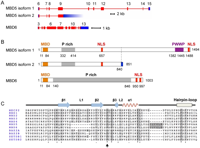
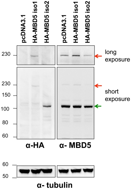
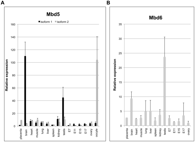
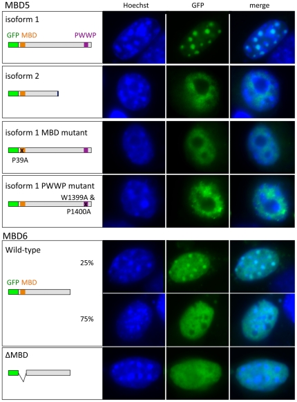
Verification
Claims the paper makes
The claims and statistics of this paper haven’t been checked yet.
Run the check — free, no account needed. It reads the open-access text and checks the sources it cites.
Claims other papers make about this one
Each is checked against this paper’s own text: does it say what the citing paper says it does?
In the citing paper
“MBD5 and MBD6 both possess a conserved but structurally distinct methyl-binding domain (MBD) but do not bind to DNA38 (Extended Data Fig. 6a).”
From RNA m5C oxidation by TET2 regulates chromatin state and leukaemogenesis (2024) · cites this paper as [38]
In the citing paper
“Previous studies have identified MBD5 and MBD6 as partner proteins of PR-DUB, and their localization to heterochromatin appeared to be independent of DNA 5mC38,39.”
From RNA m5C oxidation by TET2 regulates chromatin state and leukaemogenesis (2024) · cites this paper as [38]
Self-check
Percentages, sums and reported differences re-computed from the paper’s own numbers.
The self-check needs the paper’s full text.
Citations
Cites
DNA methylation landscapes: Provocative insights from epigenomics
Suzuki, M. M., & Bird, A. (2008) · Nature Reviews Genetics · doi:10.1038/nrg2341 · Cited by 3,039
Genomic DNA methylation: the mark and its mediators.
Klose, R. J., & Bird, A. P. (2006) · Trends in Biochemical Sciences · doi:10.1016/j.tibs.2005.12.008 · Cited by 2,501
Stability and flexibility of epigenetic gene regulation in mammalian development
Reik, W. (2007) · Nature · doi:10.1038/nature05918 · Cited by 1,963 · cited 1×
“The DNA methyltransferases DNMT1, DNMT3a, and DNMT3b are essential for mouse viability [4].”
Deficiency of methyl-CpG binding protein-2 in CNS neurons results in a Rett-like phenotype in mice
Chen, R. Z., Akbarian, S., Tudor, M., & Jaenisch, R. (2001) · Nature Genetics · doi:10.1038/85906 · Cited by 1,275
Lim, J., Hao, T., Shaw, C., Patel, A. J., Szabó, G., Rual, J.-F., Fisk, C. J., Li, N., Smolyar, A., Hill, D. E., et al. (2006) · Cell · doi:10.1016/j.cell.2006.03.032 · Cited by 789
Research works printed but not linked: 40
- M Weber, 2007, Genomic patterns of DNA methylation: targets and function of an epigenetic mark., Curr Opin Cell Biol, 19, 273, 10.1016/j.ceb.2007.04.011 · DOI
- N Sasai, 2009, Many paths to one goal? The proteins that recognize methylated DNA in eukaryotes., Int J Dev Biol, 53, 323, 10.1387/ijdb.082652ns · DOI
- X Zhao, 2003, Mice lacking methyl-CpG binding protein 1 have deficits in adult neurogenesis and hippocampal function., Proc Natl Acad Sci U S A, 100, 6777, 10.1073/pnas.1131928100 · DOI
- B Hendrich, 2001, Closely related proteins MBD2 and MBD3 play distinctive but interacting roles in mouse development., Genes Dev, 15, 710, 10.1101/gad.194101 · DOI
- CB Millar, 2002, Enhanced CpG mutability and tumorigenesis in MBD4-deficient mice., Science, 297, 403, 10.1126/science.1073354 · DOI
- J Guy, 2001, A mouse Mecp2-null mutation causes neurological symptoms that mimic Rett syndrome., Nat Genet, 27, 322, 10.1038/85899 · DOI · Paper page
- A Prokhortchouk, 2006, Kaiso-deficient mice show resistance to intestinal cancer., Mol Cell Biol, 26, 199, 10.1128/MCB.26.1.199-208.2006 · DOI
- I Martin Caballero, 2009, The methyl-CpG binding proteins Mecp2, Mbd2 and Kaiso are dispensable for mouse embryogenesis, but play a redundant function in neural differentiation., PLoS One, 4, e4315, 10.1371/journal.pone.0004315 · DOI
- J Sharif, 2007, The SRA protein Np95 mediates epigenetic inheritance by recruiting Dnmt1 to methylated DNA., Nature, 450, 908, 10.1038/nature06397 · DOI
- RA Rollins, 2006, Large-scale structure of genomic methylation patterns., Genome Res, 16, 157, 10.1101/gr.4362006 · DOI
- B Hendrich, 2003, The methyl-CpG binding domain and the evolving role of DNA methylation in animals., Trends Genet, 19, 269, 10.1016/S0168-9525(03)00080-5 · DOI
- TC Roloff, 2003, Comparative study of methyl-CpG-binding domain proteins., BMC Genomics, 4, 1, 10.1186/1471-2164-4-1 · DOI
- S Jaillard, 2009, 2q23.1 microdeletion identified by array comparative genomic hybridisation: an emerging phenotype with Angelman-like features?, J Med Genet, 46, 847, 10.1136/jmg.2008.058156 · DOI
- J Wagenstaller, 2007, Copy-number variations measured by single-nucleotide-polymorphism oligonucleotide arrays in patients with mental retardation., Am J Hum Genet, 81, 768, 10.1086/521274 · DOI
- SR Williams, 2010, Haploinsufficiency of MBD5 associated with a syndrome involving microcephaly, intellectual disabilities, severe speech impairment, and seizures., Eur J Hum Genet, 18, 436, 10.1038/ejhg.2009.199 · DOI
- S Grosso, 2008, Epilepsy and electroencephalographic anomalies in chromosome 2 aberrations. A review., Epilepsy Res, 79, 63, 10.1016/j.eplepsyres.2007.12.011 · DOI
- HY Zoghbi, 2009, Pathogenic mechanisms of a polyglutamine-mediated neurodegenerative disease, spinocerebellar ataxia type 1., J Biol Chem, 284, 7425, 10.1074/jbc.R800041200 · DOI
- AB Bowman, 2007, Duplication of Atxn1l suppresses SCA1 neuropathology by decreasing incorporation of polyglutamine-expanded ataxin-1 into native complexes., Nat Genet, 39, 373, 10.1038/ng1977 · DOI · Paper page
- M Brameier, 2007, NucPred–predicting nuclear localization of proteins., Bioinformatics, 23, 1159, 10.1093/bioinformatics/btm066 · DOI
- I Ohki, 2001, Solution structure of the methyl-CpG binding domain of human MBD1 in complex with methylated DNA., Cell, 105, 487, 10.1016/S0092-8674(01)00324-5 · DOI
- KL Ho, 2008, MeCP2 binding to DNA depends upon hydration at methyl-CpG., Mol Cell, 29, 525, 10.1016/j.molcel.2007.12.028 · DOI
- B Hendrich, 1998, Identification and characterization of a family of mammalian methyl-CpG binding proteins., Mol Cell Biol, 18, 6538, 10.1128/MCB.18.11.6538 · DOI
- HF Jorgensen, 2004, Mbd1 is recruited to both methylated and nonmethylated CpGs via distinct DNA binding domains., Mol Cell Biol, 24, 3387, 10.1128/MCB.24.8.3387-3395.2004 · DOI
- GJ Filion, 2006, A family of human zinc finger proteins that bind methylated DNA and repress transcription., Mol Cell Biol, 26, 169, 10.1128/MCB.26.1.169-181.2006 · DOI
- L Lande-Diner, 2007, Role of DNA methylation in stable gene repression., J Biol Chem, 282, 12194, 10.1074/jbc.M607838200 · DOI
- N Sasai, 2010, Sequence-specific recognition of methylated DNA by human zinc-finger proteins., Nucleic Acids Res
- RJ Klose, 2005, DNA Binding Selectivity of MeCP2 Due to a Requirement for A/T Sequences Adjacent to Methyl-CpG., Mol Cell, 19, 667, 10.1016/j.molcel.2005.07.021 · DOI
- PA Wade, 1999, Mi-2 complex couples DNA methylation to chromatin remodelling and histone deacetylation., Nat Genet, 23, 62, 10.1038/12664 · DOI
- C Mayer, 2006, Intergenic transcripts regulate the epigenetic state of rRNA genes., Mol Cell, 22, 351, 10.1016/j.molcel.2006.03.028 · DOI
- C Maison, 2002, Higher-order structure in pericentric heterochromatin involves a distinct pattern of histone modification and an RNA component., Nat Genet, 30, 329, 10.1038/ng843 · DOI
- H Bouzinba-Segard, 2006, Accumulation of small murine minor satellite transcripts leads to impaired centromeric architecture and function., Proc Natl Acad Sci U S A, 103, 8709, 10.1073/pnas.0508006103 · DOI
- R Margueron, 2010, Chromatin structure and the inheritance of epigenetic information., Nat Rev Genet, 11, 285, 10.1038/nrg2752 · DOI
- Y Wang, 2009, Regulation of Set9-mediated H4K20 methylation by a PWWP domain protein., Mol Cell, 33, 428, 10.1016/j.molcel.2009.02.002 · DOI
- A Vezzoli, 2010, Molecular basis of histone H3K36me3 recognition by the PWWP domain of Brpf1., Nat Struct Mol Biol, 17, 617, 10.1038/nsmb.1797 · DOI
- DT Carrell, The human sperm epigenome and its potential role in embryonic development., Mol Hum Reprod, 16, 37, 10.1093/molehr/gap090 · DOI
- J Gaucher, 2010, From meiosis to postmeiotic events: the secrets of histone disappearance., FEBS J, 277, 599, 10.1111/j.1742-4658.2009.07504.x · DOI
- B Payer, 2003, Stella is a maternal effect gene required for normal early development in mice., Curr Biol, 13, 2110, 10.1016/j.cub.2003.11.026 · DOI
- T Nakamura, 2007, PGC7/Stella protects against DNA demethylation in early embryogenesis., Nat Cell Biol, 9, 64, 10.1038/ncb1519 · DOI
- A Metchat, 2009, Mammalian heat shock factor 1 is essential for oocyte meiosis and directly regulates Hsp90alpha expression., J Biol Chem, 284, 9521, 10.1074/jbc.M808819200 · DOI
- KJ Livak, 2001, Analysis of relative gene expression data using real-time quantitative PCR and the 2(-Delta Delta C(T)) Method., Methods, 25, 402, 10.1006/meth.2001.1262 · DOI · Paper page
Cited by
Talkowski, M. E., Rosenfeld, J. A., Blumenthal, I., Pillalamarri, V., Chiang, C., Heilbut, A., Ernst, C., Hanscom, C., Rossin, E., Lindgren, A. M., et al. (2012) · Cell · doi:10.1016/j.cell.2012.03.028 · Cited by 577
Methyl-cpG-binding domain proteins: readers of the epigenome
Du, Q., Luu, P.-L., Stirzaker, C., & Clark, S. J. (2015) · Epigenomics · doi:10.2217/epi.15.39 · Cited by 544
Talkowski, M. E., Mullegama, S. V., Rosenfeld, J. A., van Bon, B. W. M., Shen, Y., Repnikova, E. A., Gastier-Foster, J., Thrush, D. L., Kathiresan, S., Ruderfer, D. M., et al. (2011) · The American Journal of Human Genetics · doi:10.1016/j.ajhg.2011.09.011 · Cited by 249
RNA m5C oxidation by TET2 regulates chromatin state and leukaemogenesis
Zou, Z., Dou, X., Li, Y., Zhang, Z., Wang, J., Gao, B., Xiao, Y., Wang, Y., Zhao, L., Sun, C., et al. (2024) · Nature · doi:10.1038/s41586-024-07969-x · Cited by 103
Hogart, A., Lichtenberg, J., Ajay, S. S., Anderson, S., NIH Intramural Sequencing Center, Margulies, E. H., & Bodine, D. M. (2012) · Genome Research · doi:10.1101/gr.132878.111 · Cited by 101
Metadata
- Authors
- Laget, S., Joulie, M., Le Masson, F., Sasai, N., Christians, E., Pradhan, S., Roberts, R. J., & Defossez, P.-A.
- Venue
- PLoS ONE
- Publisher
- Public Library of Science (PLoS)
- Published
- 2010-08-06
- Volume
- 5
- Issue
- 8
- Pages
- e11982
- ISSN
- 1932-6203
- PMC
- PMC2917364
- Cited by
- 110
- References
- 5
- Language
- en
About these checks
Registry, retraction, Cites and statistics results are computed, not judged, with one exception: a printed reference that no index lists is read once by a model before it counts as a citation error. Claims are extracted by a model and kept only when their quote appears verbatim in the text; each cited source is then checked, with a model’s help, for whether it says what the claim relies on. Those verdicts can be wrong — read the claim’s quote and the cited source before relying on one.
Statistics and arithmetic are recomputed, not judged.
Confidence bands are shown only for fields calibrated on our annotated test set. None is calibrated yet, so no band is shown.
What counts as a claim ›How we measure confidence ›How we check this ›
Are you an author, or do you see a problem with this page? Request a review
How to cite
Cite CoChat for the checks on this page, and the paper for its own findings.
- This page
- CoChat. (2026). Citation check: The Human Proteins MBD5 and MBD6 Associate with Heterochromatin but They Do Not Bind Methylated DNA. https://app.cochat.ai/papers/10.1371/journal.pone.0011982
- The paper
- Laget, S., Joulie, M., Le Masson, F., Sasai, N., Christians, E., Pradhan, S., Roberts, R. J., & Defossez, P.-A. (2010). The Human Proteins MBD5 and MBD6 Associate with Heterochromatin but They Do Not Bind Methylated DNA. PLoS ONE. https://doi.org/10.1371/journal.pone.0011982
