

The DOI matches its Crossref record; 31 of the 31 references it cites were found in a registry or an index.
Bacteria Penetrate the Inner Mucus Layer before Inflammation in the Dextran Sulfate Colitis Model
Open accessAbstract
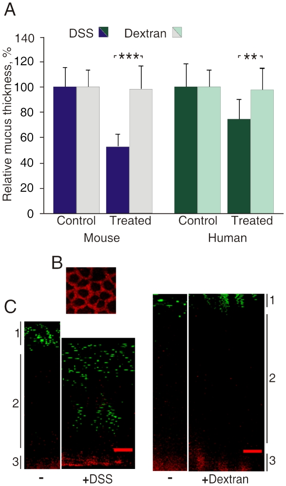
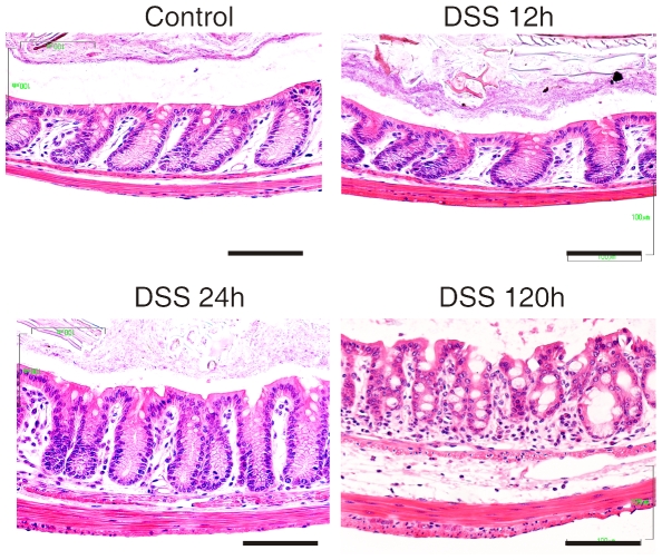
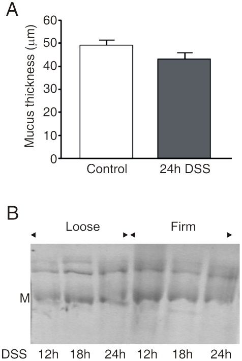
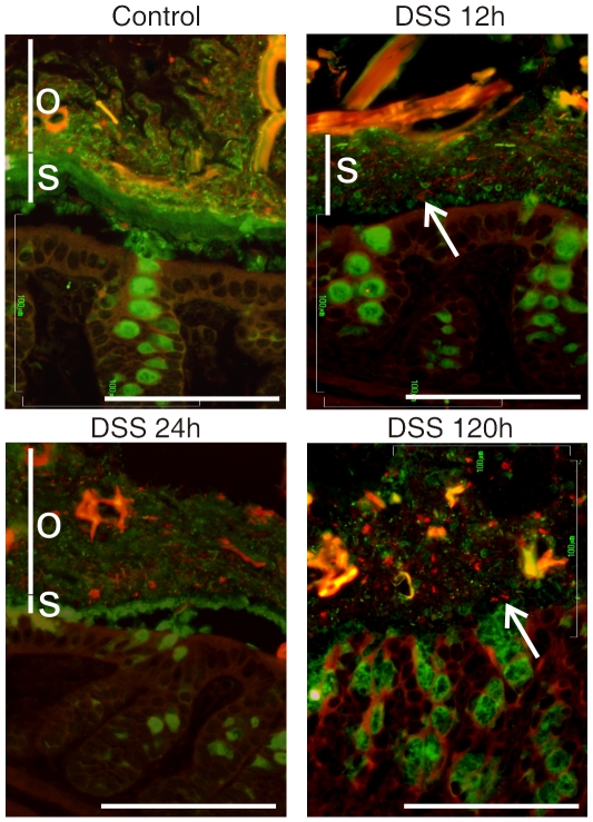
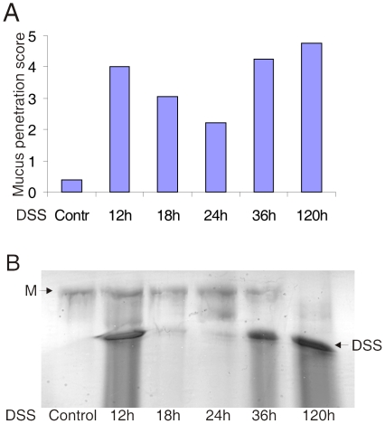
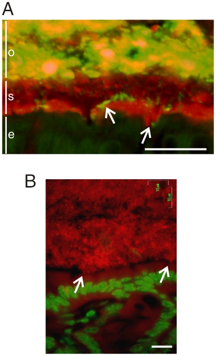
BACKGROUND: Protection of the large intestine with its enormous amount of commensal bacteria is a challenge that became easier to understand when we recently could describe that colon has an inner attached mucus layer devoid of bacteria (Johansson et al. (2008) Proc. Natl. Acad. Sci. USA 105, 15064-15069). The bacteria are thus kept at a distance from the epithelial cells and lack of this layer, as in Muc2-null mice, allow bacteria to contact the epithelium. This causes colitis and later on colon cancer, similar to the human disease Ulcerative Colitis, a disease that still lacks a pathogenetic explanation. Dextran Sulfate (DSS) in the drinking water is the most widely used animal model for experimental colitis. In this model, the inflammation is observed after 3-5 days, but early events explaining why DSS causes this has not been described. PRINCIPAL FINDINGS: When mucus formed on top of colon explant cultures were exposed to 3% DSS, the thickness of the inner mucus layer decreased and became permeable to 2 microm fluorescent beads after 15 min. Both DSS and Dextran readily penetrated the mucus, but Dextran had no effect on thickness or permeability. When DSS was given in the drinking water to mice and the colon was stained for bacteria and the Muc2 mucin, bacteria were shown to penetrate the inner mucus layer and reach the epithelial cells already within 12 hours, long before any infiltration of inflammatory cells. CONCLUSION: DSS thus causes quick alterations in the inner colon mucus layer that makes it permeable to bacteria. The bacteria that reach the epithelial cells probably trigger an inflammatory reaction. These observations suggest that altered properties or lack of the inner colon mucus layer may be an initial event in the development of colitis.
Overview
The claims and statistics of this paper haven’t been checked yet.
Run the check — free, no account needed. It reads the open-access text and checks the sources it cites.
Integrity signals
Registry
Cites
Statistics
Figures
Verification
Claims the paper makes
The claims and statistics of this paper haven’t been checked yet.
Run the check — free, no account needed. It reads the open-access text and checks the sources it cites.
Claims other papers make about this one
Each is checked against this paper’s own text: does it say what the citing paper says it does?
In the citing paper
“Failure of this protective barrier to separate bacteria from the epithelium is observed in a number of murine colitis models 15, 17.”
From The composition of the gut microbiota shapes the colon mucus barrier (2015) · cites this paper as [17]
Self-check
Percentages, sums and reported differences re-computed from the paper’s own numbers.
The self-check needs the paper’s full text.
Citations
Cites
Recognition of Commensal Microflora by Toll-Like Receptors Is Required for Intestinal Homeostasis
Rakoff-Nahoum, S., Paglino, J., Eslami-Varzaneh, F., Edberg, S., & Medzhitov, R. (2004) · Cell · doi:10.1016/j.cell.2004.07.002 · Cited by 4,251
Intestinal mucosal barrier function in health and disease
Turner, J. R. (2009) · Nature reviews. Immunology · doi:10.1038/nri2653 · Cited by 3,952
Okayasu, I., Hatakeyama, S., Yamada, M., Ohkusa, T., Inagaki, Y., & NAKAYA, R. (1990) · Gastroenterology · doi:10.1016/0016-5085(90)90290-h · Cited by 2,408
The inner of the two Muc2 mucin-dependent mucus layers in colon is devoid of bacteria
Johansson, M. E. V., Phillipson, M., Petersson, J., Velcich, A., Holm, L., & Hansson, G. C. (2008) · Proceedings of the National Academy of Sciences · doi:10.1073/pnas.0803124105 · Cited by 2,200
Self-citationClinicopathologic study of dextran sulfate sodium experimental acute colitis
Shichijo, K., Matuu, M., Ikeda, Y., Naito, S., Ito, M., & Sekine, I. (1998) · The Japanese Journal of Pharmacology · doi:10.1016/s0021-5198(19)41298-5 · Cited by 1,945
Van der Sluis, M., De Koning, B. A. E., De Bruijn, A. C. J. M., Velcich, A., Meijerink, J. P. P., Van Goudoever, J. B., Büller, H. A., Dekker, J., Van Seuningen, I., Renes, I. B., et al. (2006) · Gastroenterology · doi:10.1053/j.gastro.2006.04.020 · Cited by 1,684
The adherent gastrointestinal mucus gel layer: thickness and physical state in vivo
Atuma, C., Strugala, V., Allen, A., & Holm, L. (2001) · American Journal of Physiology-Gastrointestinal and Liver Physiology · doi:10.1152/ajpgi.2001.280.5.g922 · Cited by 1,083
Self-citationColorectal Cancer in Mice Genetically Deficient in the Mucin Muc2
Velcich, A., Yang, W., Heyer, J., Fragale, A., Nicholas, C., Viani, S., Kucherlapati, R., Lipkin, M., Yang, K., & Augenlicht, L. (2002) · Science · doi:10.1126/science.1069094 · Cited by 936
Heazlewood, C. K., Cook, M. C., Eri, R., Price, G. R., Tauro, S. B., Taupin, D., Thornton, D. J., Png, C. W., Crockford, T. L., Cornall, R. J., et al. (2008) · PLoS Medicine · doi:10.1371/journal.pmed.0050054 · Cited by 701
Inflammatory Bowel Disease and Adenomas in Mice Expressing a Dominant Negative N-Cadherin
Hermiston, M. L., & Gordon, J. I. (1995) · Science · doi:10.1126/science.270.5239.1203 · Cited by 669
Cited by
Sorted by citations
Items 101–200 of 363
Krajina, B. A., Tropini, C., Zhu, A., DiGiacomo, P., Sonnenburg, J. L., Heilshorn, S. C., & Spakowitz, A. J. (2017) · ACS Central Science · doi:10.1021/acscentsci.7b00449 · Cited by 89
Xiao, F., Yu, Q., Li, J., Johansson, M., Singh, A. K., Xia, W., Riederer, B., Engelhardt, R., Montrose, M. H., Soleimani, M., et al. (2013) · Acta Physiologica · doi:10.1111/apha.12220 · Cited by 88
Self-citationMicrobiota-Sourced Purines Support Wound Healing and Mucous Barrier Function
Lee, J. S., Wang, R. X., Goldberg, M. S., Clifford, G. P., Kao, D. J., & Colgan, S. P. (2020) · iScience · doi:10.1016/j.isci.2020.101226 · Cited by 86
A Biomimetic Human Gut‐on‐a‐Chip for Modeling Drug Metabolism in Intestine
Guo, Y., Li, Z., Su, W., Wang, L., Zhu, Y., & Qin, J. (2018) · Artificial Organs · doi:10.1111/aor.13163 · Cited by 85
Catalán, N., Villasante, A., Wacyk, J. M., Ramírez, C., & Romero, J. (2017) · Probiotics and Antimicrobial Proteins · doi:10.1007/s12602-017-9366-7 · Cited by 89
Kissoon‐Singh, V., Moreau, F., Trusevych, E., & Chadee, K. (2013) · American Journal Of Pathology · doi:10.1016/j.ajpath.2012.11.035 · Cited by 79
Tang, X., Ma, W.-P., Zhan, W., Wang, X., Dong, H., Zhao, H., Yang, L., Ji, C., Han, Q., Ji, C., et al. (2018) · Bioscience Reports · doi:10.1042/bsr20171241 · Cited by 78
Zheng, B., van Bergenhenegouwen, J., Overbeek, S. A., van de Kant, H. J. G., Garssen, J., Folkerts, G., Vos, P., Morgan, M. E., & Kraneveld, A. D. (2014) · PLoS ONE · doi:10.1371/journal.pone.0095441 · Cited by 78
Murine Models of Inflammatory Bowel Disease (IBD)
DeVoss, J., & Diehl, L. J. (2013) · Toxicologic Pathology · doi:10.1177/0192623313509729 · Cited by 78
Distinct cellular roles for PDCD10 define a gut-brain axis in cerebral cavernous malformation
Tang, A. T., Sullivan, K. R., Hong, C. C., Goddard, L. M., Mahadevan, A., Ren, A. A., Pardo, H., Peiper, A. M., Griffin, E., Tanes, C., et al. (2019) · Science Translational Medicine · doi:10.1126/scitranslmed.aaw3521 · Cited by 78
Paassen, N. B., Loonen, L. M. P., Witte‐Bouma, J., Male, A. K.-V., de Bruijn, A. C. J. M., van der Sluis, M., Lü, P., van Goudoever, J. B., Wells, J. M., Dekker, J. P., et al. (2012) · PLoS ONE · doi:10.1371/journal.pone.0038798 · Cited by 77
Kuttappan, V. A., Berghman, L., Vicuña, E. A., Latorre, J. D., Menconi, A., Wolchok, J. D., Wolfenden, A. D., Faulkner, O. B., Tellez, G. I., Hargis, B. M., et al. (2015) · Poultry Science · doi:10.3382/ps/pev114 · Cited by 76
Chen, X., Zuo, Q., Hai, Y., & Sun, X. (2010) · Medical Hypotheses · doi:10.1016/j.mehy.2010.09.026 · Cited by 75
Allaire, J. M., Morampudi, V., Crowley, S. M., Stahl, M., Yu, H., Bhullar, K., Knodler, L. A., Bressler, B. L., Jacobson, K., & Vallance, B. A. (2018) · American Journal of Physiology-Gastrointestinal and Liver Physiology · doi:10.1152/ajpgi.00181.2017 · Cited by 79
Guo, Y., Zhou, G., He, C., Yang, W., He, Z., & Liu, Z. (2015) · Mediators of Inflammation · doi:10.1155/2015/843089 · Cited by 74
Jädert, C., Phillipson, M., Holm, L., Lundberg, J. O., & Borniquel, S. (2014) · Redox Biology · doi:10.1016/j.redox.2013.12.012 · Cited by 73
Self-citationDuncan, K., Carey-Ewend, K., & Vaishnava, S. (2021) · Gut Microbes · doi:10.1080/19490976.2021.1874815 · Cited by 79
Kawashima, H. (2012) · Biological and Pharmaceutical Bulletin · doi:10.1248/bpb.b12-00412 · Cited by 73
Liu, J., Wang, Z., Mai, P., Hao, Y., Wang, Z., & Wang, J. (2022) · International Journal of Biological Macromolecules · doi:10.1016/j.ijbiomac.2022.06.194 · Cited by 74
The natural compound celastrol inhibits necroptosis and alleviates ulcerative colitis in mice
Jia, Z., Xu, C., Shen, J., Xia, T., Yang, J., & He, Y. (2015) · International Immunopharmacology · doi:10.1016/j.intimp.2015.09.029 · Cited by 71
Dietary red raspberries attenuate dextran sulfate sodium-induced acute colitis
Bibi, S., Kang, Y., Du, ., & Zhu, M. J. (2017) · The Journal of Nutritional Biochemistry · doi:10.1016/j.jnutbio.2017.08.017 · Cited by 71
Polymers in the gut compress the colonic mucus hydrogel
Datta, S. S., Steinberg, A. P., & Ismagilov, R. F. (2016) · Proceedings of the National Academy of Sciences · doi:10.1073/pnas.1602789113 · Cited by 66
Prado-Rebolledo, O. F., Delgado-Machuca, J. D. J., de Macedo, R. R. B., García-Márquez, L. J., Morales-Barrera, J. E., Latorre, J. D., Hernández-Velasco, X., & Téllez‐Isaías, G. (2016) · Avian Pathology · doi:10.1080/03079457.2016.1222808 · Cited by 67
Role of the mucin-like glycoprotein FCGBP in mucosal immunity and cancer
Liu, Q., Niu, X., Li, Y., Zhang, J.-R., Zhu, S., Yang, Q.-Y., Zhang, W., & Li, G. (2022) · Frontiers in Immunology · doi:10.3389/fimmu.2022.863317 · Cited by 66
Smelt, M. J., de Haan, B. J., Bron, P. A., van Swam, I. I., Meijerink, M., Wells, J. M., Kleerebezem, M., Faas, M. M., & de Vos, P. (2013) · PLoS ONE · doi:10.1371/journal.pone.0063099 · Cited by 66
Fungal lysozyme leverages the gut microbiota to curb DSS-induced colitis
Larsen, I., Jensen, B. A. H., Bonazzi, E., Choi, B. S.-Y., Kristensen, N. N., Schmidt, E. G. W., Sünderhauf, A., Morin, L., Olsen, P., Hansen, L. B. S., et al. (2021) · Gut Microbes · doi:10.1080/19490976.2021.1988836 · Cited by 66
The Role of CXCR3 in DSS-Induced Colitis
Chami, B., Yeung, A. W. S., van Vreden, C., King, N. J. C., & Bao, S. (2014) · PLoS ONE · doi:10.1371/journal.pone.0101622 · Cited by 63
Sovran, B., Loonen, L. M. P., Lü, P., Hugenholtz, F., Belzer, C., Stolte, E. H., Boekschoten, M. V., van Baarlen, P., Kleerebezem, M., de Vos, P., et al. (2015) · Inflammatory Bowel Diseases · doi:10.1097/mib.0000000000000319 · Cited by 62
CD4 + T Cells Drive Goblet Cell Depletion during Citrobacter rodentium Infection
Chan, J. M., Bhinder, G., Sham, H. P., Ryz, N. R., Huang, T., Bergstrom, K., & Vallance, B. A. (2013) · Infection and Immunity · doi:10.1128/iai.00655-13 · Cited by 59
Yang, X., Mao, Z., Huang, Y., Yan, H., Yan, Q., Hong, J., Fan, J., & Yao, J. Q. (2021) · Redox Biology · doi:10.1016/j.redox.2021.101881 · Cited by 60
Hoffmann, W. (2021) · International Journal of Molecular Sciences · doi:10.3390/ijms22094909 · Cited by 65
Hudcovic, T., Kolinska, J., Klepetar, J., Stepankova, R., Řezanka, T., Šrůtková, D., Schwarzer, M., Erban, V., Du, Z., Wells, J. M., et al. (2011) · Clinical & Experimental Immunology · doi:10.1111/j.1365-2249.2011.04498.x · Cited by 60
Wheat Consumption Aggravates Colitis in Mice via Amylase Trypsin Inhibitor–mediated Dysbiosis
Pickert, G., Wirtz, S. J., Matzner, J., Ashfaq–Khan, M., Heck, R., Rosigkeit, S., Thies, D., Surabattula, R., Ehmann, D., Wehkamp, J., et al. (2020) · Gastroenterology · doi:10.1053/j.gastro.2020.03.064 · Cited by 60
PPARγ: The Central Mucus Barrier Coordinator in Ulcerative Colitis
Fang, J. C., Wang, H., Xue, Z., Cheng, Y., & Zhang, X. (2021) · Inflammatory Bowel Diseases · doi:10.1093/ibd/izaa273 · Cited by 56
Mucus layer modeling of human colonoids during infection with enteroaggragative E. coli
Liu, L., Saitz, W., Smith, R., Gonyar, L. A., In, J., Kovbasnjuk, O. N., Zachos, N. C., Donowitz, M., Nataro, J. P., & Ruı́z-Pérez, F. (2020) · Scientific Reports · doi:10.1038/s41598-020-67104-4 · Cited by 58
Gp91phox contributes to the development of experimental inflammatory bowel disease
Bao, S., Carr, E. D., Xu, Y., & Hunt, N. H. (2011) · Immunology and Cell Biology · doi:10.1038/icb.2011.4 · Cited by 58
Gleinser, M., Grimm, V., Zhurina, D. S., Yuan, J., & Riedel, C. U. (2012) · Microbial Cell Factories · doi:10.1186/1475-2859-11-80 · Cited by 61
Methods of Inducing Inflammatory Bowel Disease in Mice
Bang, B., & Lichtenberger, L. M. (2016) · Current Protocols in Pharmacology · doi:10.1002/0471141755.ph0558s72 · Cited by 55
iNOS-Dependent Increase in Colonic Mucus Thickness in DSS-Colitic Rats
Schreiber, O., Petersson, J. H., Waldén, T. B., Ahl, D., Sandler, S., Phillipson, M., & Holm, L. P. (2013) · PLoS ONE · doi:10.1371/journal.pone.0071843 · Cited by 54
Self-citationMucin-Type O-Glycans: Barrier, Microbiota, and Immune Anchors in Inflammatory Bowel Disease
Zhang, Y., Wang, L., Ocansey, D. K. W., Wang, B., Wang, L., & Xu, Z. (2021) · Journal of Inflammation Research · doi:10.2147/jir.s327609 · Cited by 58
Sharpen, J. D. A., Dolan, B., Nyström, E. E. L., Birchenough, G. M. H., Arike, L., Martínez‐Abad, B., Johansson, M., Hansson, G. C., & Recktenwald, C. V. (2022) · Nature Communications · doi:10.1038/s41467-021-27743-1 · Cited by 53
Self-citationExacerbated intestinal inflammation in P2Y6 deficient mice is associated with Th17 activation
Salem, M., Azreq, M.-A. E., Pelletier, J., Robaye, B., Aoudjit, F., & Sévigny, J. (2019) · Biochimica et Biophysica Acta (BBA) - Molecular Basis of Disease · doi:10.1016/j.bbadis.2019.06.019 · Cited by 52
SMITH, P. F., Siddharth, J., Pearson, R., Holway, N., Shaxted, M., Butler, M., Clark, N., Jamontt, J., Watson, R. P., Sanmugalingam, D., et al. (2012) · PLoS ONE · doi:10.1371/journal.pone.0030273 · Cited by 52
Kim, W.-K., Jang, Y. J., Seo, B., Han, D. H., Park, S., & Ko, G. (2018) · Journal of Functional Foods · doi:10.1016/j.jff.2018.11.035 · Cited by 52
TNFR1-induced lethal inflammation is mediated by goblet and Paneth cell dysfunction
Hauwermeiren, F. V., Vandenbroucke, R. E., Grine, L., Lodens, S., Wonterghem, E. V., Rycke, R. M. D., Geest, N. D., Hassan, B. A., & Libert, C. (2014) · Mucosal Immunology · doi:10.1038/mi.2014.112 · Cited by 52
Evaluation of Gastrointestinal Leakage in Multiple Enteric Inflammation Models in Chickens
Kuttappan, V. A., Vicuña, E. A., Latorre, J. D., Wolfenden, A. D., Tellez, G., Hargis, B. M., & Bielke, L. R. (2015) · Frontiers in Veterinary Science · doi:10.3389/fvets.2015.00066 · Cited by 51
Host-Recognition of Pathogens and Commensals in the Mammalian Intestine
Rossi, O., van Baarlen, P., & Wells, J. M. (2011) · Current topics in microbiology and immunology · doi:10.1007/82_2011_191 · Cited by 50
Smelt, M. J., de Haan, B. J., Bron, P. A., van Swam, I. I., Meijerink, M., Wells, J. M., Faas, M. M., & de Vos, P. (2013) · PLoS ONE · doi:10.1371/journal.pone.0068952 · Cited by 50
Ferenczi, S., Szegi, K., Winkler, Z., Barna, T., & Kovács, K. (2016) · Scientific Reports · doi:10.1038/srep34132 · Cited by 50
Devaraj, N. K., Suppiah, S., Veettil, S. K., Ching, S. M., Lee, K. W., Menon, R. K., Soo, M. J., Deuraseh, I., Hoo, F. K., & Sivaratnam, D. (2019) · Nutrients · doi:10.3390/nu11122886 · Cited by 50
A theoretical model of temperate phages as mediators of gut microbiome dysbiosis
Lin, D. M., & Lin, H. C. (2019) · F1000Research · doi:10.12688/f1000research.18480.1 · Cited by 49
Tajasuwan, L., Kettawan, A., Rungruang, T., Wunjuntuk, K., & Prombutara, P. (2023) · Nutrients · doi:10.3390/nu15061528 · Cited by 49
Mucus and adiponectin deficiency: role in chronic inflammation-induced colon cancer
Saxena, A., Baliga, M. S., Ponemone, V., Kaur, K., Larsen, B., Fletcher, E., Greene, J., & Fayad, R. (2013) · International Journal of Colorectal Disease · doi:10.1007/s00384-013-1664-2 · Cited by 46
Muc5ac Expression Protects the Colonic Barrier in Experimental Colitis
Olli, K. E., Rapp, C., O’Connell, L., Collins, C. B., McNamee, E. N., Jensen, O., Jedlicka, P., Allison, K. C., Goldberg, M. S., Gerich, M. E., et al. (2020) · Inflammatory Bowel Diseases · doi:10.1093/ibd/izaa064 · Cited by 45
Reichmann, F., Painsipp, E., & Holzer, P. D. (2013) · PLoS ONE · doi:10.1371/journal.pone.0054811 · Cited by 45
Prebiotics and synbiotics in ulcerative colitis
Laurell, A., & Sjöberg, K. (2016) · Scandinavian Journal of Gastroenterology · doi:10.1080/00365521.2016.1263680 · Cited by 44
Self-citationEriksson, M., Johannssen, T., von Smolinski, D., Gruber, A. D., Seeberger, P. H., & Lepenies, B. (2013) · Frontiers in Immunology · doi:10.3389/fimmu.2013.00196 · Cited by 44
Morgan, M. E., Zheng, B., Koelink, P. J., van de Kant, H. J. G., Haazen, L. C. J. M., van Roest, M., Garssen, J., Folkerts, G., & Kraneveld, A. D. (2013) · PLoS ONE · doi:10.1371/journal.pone.0069936 · Cited by 43
Host NLRP6 exacerbates graft-versus-host disease independent of gut microbial composition
Toubai, T., Fujiwara, H., Rossi, C., Riwes, M. M., Tamaki, H., Zajac, C. K., Liu, C., Mathew, A. V., Byun, J., Oravecz-Wilson, K. I., et al. (2019) · Nature Microbiology · doi:10.1038/s41564-019-0373-1 · Cited by 45
Hung, Y., Ko, W., Chou, P.-H., Chen, Y., Lin, H.-J., Liu, Y.-H., Tsai, H.-W., Lee, J., & Tsai, P.-J. (2015) · The Journal of Infectious Diseases · doi:10.1093/infdis/jiv184 · Cited by 42
In vivo models of mucin biosynthesis and function
Syed, Z. A., Zhang, L., & Hagen, K. G. T. (2022) · Advanced Drug Delivery Reviews · doi:10.1016/j.addr.2022.114182 · Cited by 46
Huang, X., Hu, J., Zhang, H., Li, J., Zhu, X., Liu, Y., Liang, Y., & Mei, Y. (2023) · Microbiology Spectrum · doi:10.1128/spectrum.04370-22 · Cited by 44
Saijo, H., Tatsumi, N., Arihiro, S., Kato, T., Okabe, M., Tajiri, H., & Hashimoto, H. (2015) · Laboratory Investigation · doi:10.1038/labinvest.2015.60 · Cited by 41
Scaldaferri, F., Lopetuso, L. R., Petito, V., Cufino, V., Bilotta, M., Arena, V., Stigliano, E., Maulucci, G., Papi, M., Caristo, M. E., et al. (2014) · United European Gastroenterology Journal · doi:10.1177/2050640614520867 · Cited by 41
Vong, L. B., Yoshitomi, T., Morikawa, K., Saito, S., Matsui, H., & Nagasaki, Y. (2013) · Journal of Gastroenterology · doi:10.1007/s00535-013-0836-8 · Cited by 39
The longitudinal and cross-sectional heterogeneity of the intestinal microbiota
Miller, B. M., Liou, M. J., Lee, J., & Bäumler, A. J. (2021) · Current Opinion in Microbiology · doi:10.1016/j.mib.2021.08.004 · Cited by 44
Modeling mucus physiology and pathophysiology in human organs-on-chips
Izadifar, Z., Sontheimer-Phelps, A., Lubamba, B., Bai, H., Fadel, C. W., Stejskalová, A., Özkan, A., Dasgupta, Q., Bein, A., Junaid, A., et al. (2022) · Advanced Drug Delivery Reviews · doi:10.1016/j.addr.2022.114542 · Cited by 39
Matute, J. D., Duan, J., Flak, M. B., Griebel, P., Tascón-Arcila, J., Doms, S., Hanley, T., Antanaviciute, A., Gundrum, J., Welch, J. L. M., et al. (2022) · The Journal of Experimental Medicine · doi:10.1084/jem.20211938 · Cited by 37
Immune Regulation of Goblet Cell and Mucus Functions in Health and Disease
Gustafsson, J. K., & Hansson, G. C. (2025) · Annual Review of Immunology · doi:10.1146/annurev-immunol-101721-065224 · Cited by 43
Self-citationDoes MHC heterozygosity influence microbiota form and function?
Khan, M. A. W., Stephens, W. Z., Mohammed, A. D., Round, J. L., & Kubinak, J. L. (2019) · PLoS ONE · doi:10.1371/journal.pone.0215946 · Cited by 37
Williams, B. B., Tebbutt, N. C., Büchert, M., Putoczki, T. L., Doggett, K., Bao, S., Johnstone, C. N., Masson, F., Hollande, F., Burgess, A. W., et al. (2015) · Disease Models & Mechanisms · doi:10.1242/dmm.019935 · Cited by 35
Shen, S., Kumar, K. P., Stanley, D., Moore, R. J., Van, T. T. H., Wen, S., Hickey, M. J., & Wong, C. H. Y. (2018) · Frontiers in Immunology · doi:10.3389/fimmu.2018.00999 · Cited by 35
Sun, B., Yuan, J., Wang, S., Lin, J., Wanjun, Z., Shao, J., Wang, R., Shi, B., & Hu, H. (2018) · Frontiers in Pharmacology · doi:10.3389/fphar.2018.01235 · Cited by 34
Neonatal hyperoxia disrupts the intestinal barrier and impairs intestinal function in rats
Chou, H., & Chen, C. (2017) · Experimental and Molecular Pathology · doi:10.1016/j.yexmp.2017.05.006 · Cited by 34
Kuprys, P. V., Cannon, A. R., Shieh, J., Iftekhar, N., Park, S. K., Eberhardt, J. M., Ding, X., & Choudhry, M. A. (2020) · Gut Microbes · doi:10.1080/19490976.2020.1838236 · Cited by 34
The Habitat Filters of Microbiota-Nourishing Immunity
Miller, B. M., & Bäumler, A. J. (2021) · Annual Review of Immunology · doi:10.1146/annurev-immunol-101819-024945 · Cited by 35
Different Forms of TFF3 in the Human Saliva: Heterodimerization with IgG Fc Binding Protein (FCGBP)
Houben, T., Harder, S., Schlüter, H., Kalbacher, H., & Hoffmann, W. (2019) · International Journal of Molecular Sciences · doi:10.3390/ijms20205000 · Cited by 32
Liberti, A., Natarajan, O., Atkinson, C. G. F., Sordino, P., & Dishaw, L. J. (2021) · Frontiers in Immunology · doi:10.3389/fimmu.2021.642687 · Cited by 32
Yokota, H., Tsuzuki, A., Shimada, Y., Imai, A., Utsumi, D., Tsukahara, T., Matsumoto, M., Amagase, K., Iwata, K., Nakamura, A., et al. (2016) · Journal of Pharmacology and Experimental Therapeutics · doi:10.1124/jpet.116.235580 · Cited by 33
Kozlowski, C., Jeet, S., Beyer, J. C., Guerrero, S., Lesch, J., Wang, X., DeVoss, J., & Diehl, L. J. (2013) · Disease Models & Mechanisms · doi:10.1242/dmm.011759 · Cited by 31
Fluoride-induced alteration in the diversity and composition of bacterial microbiota in mice colon
Fu, R., Niu, R., Li, R., Yue, B., Zhang, X., Cao, Q., Wang, J., & Sun, Z. (2020) · Biological Trace Element Research · doi:10.1007/s12011-019-01942-w · Cited by 27
Liang, X., Xie, J., Liu, H., Zhao, R., Zhang, W., Wang, H., Pan, H., Zhou, Y., & Han, W. (2022) · Cellular and Molecular Gastroenterology and Hepatology · doi:10.1016/j.jcmgh.2022.03.007 · Cited by 31
Loss of primary cilia promotes inflammation and carcinogenesis
Paul, C., Tang, R., Longobardi, C., Lattanzio, R., Eguether, T., Turali, H., Bremond, J., Maurizy, C., Gabola, M., Poupeau, S., et al. (2022) · EMBO Reports · doi:10.15252/embr.202255687 · Cited by 36
Molecular Alterations in the Stomach of Tff1-Deficient Mice: Early Steps in Antral Carcinogenesis
Znalesniak, E. B., Salm, F., & Hoffmann, W. (2020) · International Journal of Molecular Sciences · doi:10.3390/ijms21020644 · Cited by 30
Proteomic Study of the Mucin Granulae in an Intestinal Goblet Cell Model
Rodríguez-Piñeiro, A. M., van der Post, S., Johansson, M., Thomsson, K. A., Nesvizhskii, A. I., & Hansson, G. C. (2012) · Journal of Proteome Research · doi:10.1021/pr2010988 · Cited by 32
Self-citationMicrobes with higher metabolic independence are enriched in human gut microbiomes under stressPreprint
Veseli, I., Chen, Y. T., Schechter, M. S., Vanni, C., Fogarty, E. C., Watson, A. R., Jabrì, B., Blekhman, R., Willis, A. D., Yu, M., et al. (2023) · eLife · doi:10.7554/elife.89862 · Cited by 33
Dupont, A., Kaconis, Y., Yang, I., Albers, T., Woltemate, S., Heinbockel, L., Andersson, M., Suerbaum, S., Brandenburg, K., & Hornef, M. W. (2014) · Gut · doi:10.1136/gutjnl-2014-307150 · Cited by 31
The SOS Response Mediates Sustained Colonization of the Mammalian Gut
Samuels, A. N., Roggiani, M., Zhu, J., Goulian, M., & Kohli, R. M. (2018) · Infection and Immunity · doi:10.1128/iai.00711-18 · Cited by 29
Yang, J. C., Jacobs, J. P., Hwang, M., Sabui, S., Liang, F., Said, H. M., & Skupsky, J. (2023) · Nutrients · doi:10.3390/nu15020264 · Cited by 31
Kleiveland, C. R., Hult, L. T. O., Spetalen, S., Kaldhusdal, M., Christoffersen, T. E., Bengtsson, O., Romarheim, O. H., Jacobsen, M., & Lea, T. (2012) · Applied and Environmental Microbiology · doi:10.1128/aem.02464-12 · Cited by 29
Brasseit, J., Althaus-Steiner, E., Faderl, M., Dickgreber, N., Saurer, L., Genitsch, V., Dolowschiak, T., Li, H., Finke, D., Hardt, W., et al. (2015) · Mucosal Immunology · doi:10.1038/mi.2015.93 · Cited by 28
The role of mucoprotectants in the management of gastrointestinal disorders
Eutamène, H., Beaufrand, C., Harkat, C., & Théodorou, V. (2017) · Expert Review of Gastroenterology & Hepatology · doi:10.1080/17474124.2018.1378573 · Cited by 29
Velde, A. A. T., Brüll, F., Heinsbroek, S. E. M., Meijer, S. L., Lütjohann, D., Vreugdenhil, A. C. E., & Plat, J. (2015) · Nutrients · doi:10.3390/nu7105412 · Cited by 28
A glance at … dietary emulsifiers, the human intestinal mucus and microbiome, and dietary fiber
Glade, M. J., & Meguid, M. M. (2015) · Nutrition · doi:10.1016/j.nut.2015.12.036 · Cited by 28
Cheong, K., Biney, E., Wang, M., Zhong, H., Zhong, S., & Sathuvan, M. (2026) · npj Science of Food · doi:10.1038/s41538-026-00750-6 · Cited by 27
Reischl, S., Lee, J. H., Miltschitzky, E., Vieregge, V., Walter, R. L., Twardy, V., Kasajima, A., Frieß, H., Kamaly, N., & Neumann, P.-A. (2021) · Inflammatory Bowel Diseases · doi:10.1093/ibd/izab008 · Cited by 29
Guo, X.-K., Wang, J., van Hensbergen, V. P., Liu, J., Xu, H., & Hu, X. (2023) · Cell Reports · doi:10.1016/j.celrep.2023.112754 · Cited by 28
Luminal Microbes Promote Monocyte–Stem Cell Interactions Across a Healthy Colonic Epithelium
Skoczek, D. A., Walczysko, P., Horn, N., Parris, A., Clare, S., Williams, M. R., & Sobolewski, A. (2014) · The Journal of Immunology · doi:10.4049/jimmunol.1301497 · Cited by 26
Yang, Y., Latorre, J. D., Khatri, B., Kwon, Y. M., Kong, B. ‐., Teague, K. D., Graham, D. B., Wolfenden, A. D., Mahaffey, B. D., Baxter, M. F. A., et al. (2017) · Poultry Science · doi:10.3382/ps/pex311 · Cited by 26
Monk, J. M., Wu, W., Hutchinson, A. L., Pauls, P., Robinson, L. E., & Power, K. A. (2018) · The Journal of Nutritional Biochemistry · doi:10.1016/j.jnutbio.2018.02.013 · Cited by 28
Metadata
- Authors
- Johansson, M. E. V., Gustafsson, J. K., Sjöberg, K. E., Petersson, J., Holm, L., Sjövall, H., & Hansson, G. C.
- Affiliations
- University of Gothenburg (SE)
- Uppsala University (SE)
- Corresponding author
- Hansson, Gunnar C.
- Venue
- PLoS ONE
- Publisher
- Public Library of Science (PLoS)
- Published
- 2010-08-18
- Volume
- 5
- Issue
- 8
- Pages
- e12238
- ISSN
- 1932-6203 (ISSN-L)
- PMC
- PMC2923597
- OpenAlex
- W1977781139
- Cited by
- 368
- References
- 31
- Citations by year
- 2026: 12 · 2025: 19 · 2024: 18 · 2023: 26 · 2022: 27 · 2021: 17 · 2020: 17 · 2019: 28 · 2018: 19 · 2017: 22 · 2016: 24 · 2015: 22 · 2014: 30 · 2013: 39 · 2012: 21
- FWCI
- 15.49
- Type
- journal article
- Language
- en
- Open access
- gold
- doi.org
- Locations
- PLoS ONE (publishedVersion, cc-by)
- PubMed Central (publishedVersion, cc-by)
- PubMed (publishedVersion)
- DOAJ (DOAJ: Directory of Open Access Journals) (submittedVersion, cc-by-sa)
- Figshare (submittedVersion, cc-by)
- Funders
- Swedish Cancer Foundation
- Stiftelsen för Strategisk Forskning
- Knut och Alice Wallenbergs Stiftelse (KAW2007.0118)
- Vetenskapsrådet (21027, 342–2004-4434)
- Göteborgs Universitet
- National Institute of Allergy and Infectious Diseases (U01AI095473)
- Topics
- Health Sciences › Medicine › Gastroenterology › Inflammatory Bowel Disease
- Life Sciences › Biochemistry, Genetics and Molecular Biology › Molecular Biology › Gut microbiota and health
- Health Sciences › Medicine › Gastroenterology › Gastrointestinal motility and disorders
- Keywords
- mucus permeability, ulcerative colitis, MUC2, commensal bacteria, colonic epithelium, pathogenesis of inflammation
- MeSH terms
- Adult, Aged, Animals, Bacteria, Colitis, Colon, Disease Models, Animal, Female, Humans, Intestinal Mucosa, Male, Mice, Inbred C57BL, Permeability, Time Factors, Dextran Sulfate, Mice
About these checks
Registry, retraction, Cites and statistics results are computed, not judged, with one exception: a printed reference that no index lists is read once by a model before it counts as a citation error. Claims are extracted by a model and kept only when their quote appears verbatim in the text; each cited source is then checked, with a model’s help, for whether it says what the claim relies on. Those verdicts can be wrong — read the claim’s quote and the cited source before relying on one.
Statistics and arithmetic are recomputed, not judged.
Confidence bands are shown only for fields calibrated on our annotated test set. None is calibrated yet, so no band is shown.
What counts as a claim ›How we measure confidence ›How we check this ›
Are you an author, or do you see a problem with this page? Request a review
