

The DOI matches its Crossref record; 39 of the 39 references it cites were found in a registry or an index.
Hepatocyte Growth Factor Modulates Interleukin-6 Production in Bone Marrow Derived Macrophages: Implications for Inflammatory Mediated Diseases
Open accessAbstract
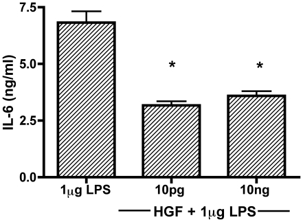
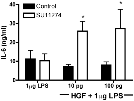
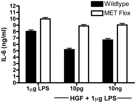
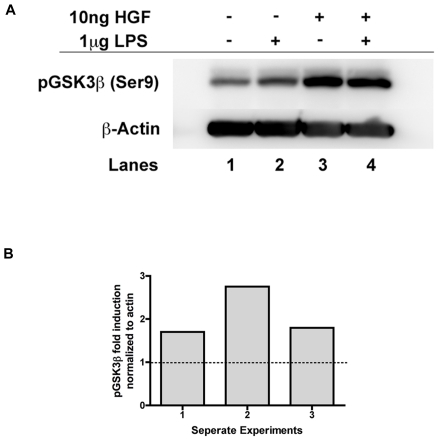
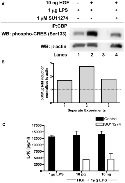
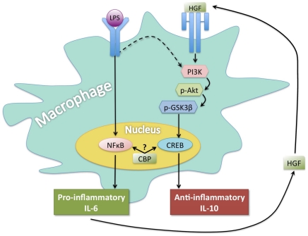
The generation of the pro-inflammatory cytokines IL-6, TNF-α, and IL-1β fuel the acute phase response (APR). To maintain body homeostasis, the increase of inflammatory proteins is resolved by acute phase proteins via presently unknown mechanisms. Hepatocyte growth factor (HGF) is transcribed in response to IL-6. Since IL-6 production promotes the generation of HGF and induces the APR, we posited that accumulating HGF might be a likely candidate for quelling excess inflammation under non-pathological conditions. We sought to assess the role of HGF and how it influences the regulation of inflammation utilizing a well-defined model of inflammatory activation, lipopolysaccharide (LPS)-stimulation of bone marrow derived macrophages (BMM). BMM were isolated from C57BL6 mice and were stimulated with LPS in the presence or absence of HGF. When HGF was present, there was a decrease in production of the pro-inflammatory cytokine IL-6, along with an increase in the anti-inflammatory cytokine IL-10. Altered cytokine production correlated with an increase in phosphorylated GSK3β, increased retention of the phosphorylated NFκB p65 subunit in the cytoplasm, and an enhanced interaction between CBP and phospho-CREB. These changes were a direct result of signaling through the HGF receptor, MET, as effects were reversed in the presence of a selective inhibitor of MET (SU11274) or when using BMM from macrophage-specific conditional MET knockout mice. Combined, these data provide compelling evidence that under normal circumstances, HGF acts to suppress the inflammatory response.
Overview
The claims and statistics of this paper haven’t been checked yet.
Run the check — free, no account needed. It reads the open-access text and checks the sources it cites.
Integrity signals
Registry
Cites
Statistics
Figures

Verification
Claims the paper makes
The claims and statistics of this paper haven’t been checked yet.
Run the check — free, no account needed. It reads the open-access text and checks the sources it cites.
Claims other papers make about this one
Each is checked against this paper’s own text: does it say what the citing paper says it does?
The source backs part of the claim, not all of it — or backs it for a different population, condition or comparison.
In the citing paper
“in cultured macrophages HGF suppresses production of IL-6”
From Synthesis of IL-6 by Hepatocytes Is a Normal Response to Common Hepatic Stimuli (2014) · cites this paper as [10]
Experimental — accuracy not yet measuredThe cited source says what the claim attributes to it.
In the citing paper
“As we have previously shown HGF suppresses LPS-induced production of IL-6 in cultured macrophages via the MET receptor [10]”
From Synthesis of IL-6 by Hepatocytes Is a Normal Response to Common Hepatic Stimuli (2014) · cites this paper as [10]
The cited source says what the claim attributes to it.
In the citing paper
“hepatocellular IL-6 induction in response to HGF is exactly the opposite of what we recently observed with regard to HGF and IL-6 in cultured macrophages [10]”
From Synthesis of IL-6 by Hepatocytes Is a Normal Response to Common Hepatic Stimuli (2014) · cites this paper as [10]
The cited source says what the claim attributes to it.
In the citing paper
“As we have previously shown that HGF can suppress IL-6 production in LPS-stimulated macrophages [10]”
From Synthesis of IL-6 by Hepatocytes Is a Normal Response to Common Hepatic Stimuli (2014) · cites this paper as [10]
The cited source says what the claim attributes to it.
In the citing paper
“In a recent publication we demonstrated that unlike what we now see with hepatocytes, HGF instead suppresses IL-6 production in macrophages via signaling through the MET receptor [10].”
From Synthesis of IL-6 by Hepatocytes Is a Normal Response to Common Hepatic Stimuli (2014) · cites this paper as [10]
Self-check
Percentages, sums and reported differences re-computed from the paper’s own numbers.
The self-check needs the paper’s full text.
Citations
Cites
Janeway, C. A., & Medzhitov, R. (2002) · Annual Review of Immunology · doi:10.1146/annurev.immunol.20.083001.084359 · Cited by 8,394
Acute-Phase Proteins and Other Systemic Responses to Inflammation
Gabay, C., & Kushner, I. (1999) · New England Journal of Medicine · doi:10.1056/nejm199902113400607 · Cited by 6,734
Nuclear Factor-κB — A Pivotal Transcription Factor in Chronic Inflammatory Diseases
Barnes, P. J., & Karin, M. (1997) · New England Journal of Medicine · doi:10.1056/nejm199704103361506 · Cited by 4,622
The regulation of IL-10 production by immune cells
Saraiva, M., & O’Garra, A. (2010) · Nature reviews. Immunology · doi:10.1038/nri2711 · Cited by 3,293
Medzhitov, R. M., & Janeway, C. A. (2000) · New England Journal of Medicine · doi:10.1056/nejm200008033430506 · Cited by 1,827
Xing, Z., Gauldie, J., Cox, G., Baumann, H., Jordana, M., Lei, X.-F., & Achong, M. K. (1998) · Journal of Clinical Investigation · doi:10.1172/jci1368 · Cited by 1,578
The glamour and gloom of glycogen synthase kinase-3
Jope, R. S., & Johnson, G. V. W. (2004) · Trends in Biochemical Sciences · doi:10.1016/j.tibs.2003.12.004 · Cited by 1,535
Interleukin-6 and chronic inflammation
Gabay, C. (2006) · Arthritis Research & Therapy · doi:10.1186/ar1917 · Cited by 1,468
Zhong, H., Voll, R. E., & Ghosh, S. (1998) · Molecular Cell · doi:10.1016/s1097-2765(00)80066-0 · Cited by 1,210
Martin, M., Rehani, K., Jope, R. S., & Michalek, S. M. (2005) · Nature Immunology · doi:10.1038/ni1221 · Cited by 1,099
Cited by
Sorted by citations
Platelet-rich plasma for managing pain and inflammation in osteoarthritis
Andı́a, I., & Maffulli, N. (2013) · Nature Reviews Rheumatology · doi:10.1038/nrrheum.2013.141 · Cited by 476
Known and novel roles of the MET oncogene in cancer: a coherent approach to targeted therapy
Comoglio, P. M., Trusolino, L., & Boccaccio, C. (2018) · Nature reviews. Cancer · doi:10.1038/s41568-018-0002-y · Cited by 385
Dupuis-Maguiraga, L., Noret, M., Brun, S., Grand, R. L., Gras, G. S., & Roques, P. (2012) · PLoS neglected tropical diseases · doi:10.1371/journal.pntd.0001446 · Cited by 317
Good, M. L., Sodhi, C. P., Egan, C. E., Afrazi, A., Jia, H., Yamaguchi, Y., Lü, P., Branca, M. F., Ma, C., Prindle, T., et al. (2015) · Mucosal Immunology · doi:10.1038/mi.2015.30 · Cited by 231
What turns CREB on? And off? And why does it matter?
Steven, A., Friedrich, M., Jank, P., Heimer, N., Budczies, J., Denkert, C., & Seliger, B. (2020) · Cellular and Molecular Life Sciences · doi:10.1007/s00018-020-03525-8 · Cited by 220
Kwilas, A. R., Ardiani, A., Donahue, R. N., Aftab, D. T., & Hodge, J. W. (2014) · Journal of Translational Medicine · doi:10.1186/s12967-014-0294-y · Cited by 193
Cellular and molecular mechanisms of HGF/Met in the cardiovascular system
Gallo, S., Sala, V., Gatti, S., & Crepaldi, T. (2015) · Clinical Science · doi:10.1042/cs20150502 · Cited by 168
Hepatocyte growth factor: A regulator of inflammation and autoimmunity
Molnarfi, N., Benkhoucha, M., Funakoshi, H., Nakamura, T., & Lalive, P. H. (2014) · Autoimmunity Reviews · doi:10.1016/j.autrev.2014.11.013 · Cited by 159
PRP Treatment Efficacy for Tendinopathy: A Review of Basic Science Studies
Zhou, Y., & Wang, J. H. (2016) · BioMed Research International · doi:10.1155/2016/9103792 · Cited by 142
Signals and Cells Involved in Regulating Liver Regeneration
Kang, L., Mars, W. M., & Michalopoulos, G. K. (2012) · Cells · doi:10.3390/cells1041261 · Cited by 137
Self-citationSynthesis of IL-6 by Hepatocytes Is a Normal Response to Common Hepatic Stimuli
Norris, C. A., He, M., Kang, L.-I., Ding, M. Q., Radder, J. E., Haynes, M. M., Yang, Y., Paranjpe, S., Bowen, W. C., Orr, A., et al. (2014) · PLoS ONE · doi:10.1371/journal.pone.0096053 · Cited by 132
“As we have previously shown that HGF can suppress IL-6 production in LPS-stimulated macrophages [10], the combined data suggests that context (i.e. induction of endotoxin signaling in the presence or absence of HGF) may regulate which cells produce the IL-6, as well as the quantity being made.”
BackgroundInfluentialSelf-citationZhou, D. F., Tan, R. J., Lin, L., Zhou, L., & Liu, Y. (2013) · Kidney International · doi:10.1038/ki.2013.102 · Cited by 127
HGF and MET: From Brain Development to Neurological Disorders
Desole, C., Gallo, S., Vitacolonna, A., Montarolo, F., Bertolotto, A., Vivien, D., Comoglio, P. M., & Crepaldi, T. (2021) · Frontiers in Cell and Developmental Biology · doi:10.3389/fcell.2021.683609 · Cited by 129
The cytokines HGF and CXCL13 predict the severity and the mortality in COVID-19 patients
Perreau, M., Suffiotti, M., Marques-Vidal, P. M., Wiedemann, A., Lévy, Y., Laouenan, C., Ghosn, J., Fenwick, C. W., Comte, D., Roger, T., et al. (2021) · Nature Communications · doi:10.1038/s41467-021-25191-5 · Cited by 123
Sánchez, M., Anitua, E., Delgado, D., Sanchez, P., Prado, R., Orive, G., & Padilla, S. (2016) · Expert Opinion on Biological Therapy · doi:10.1080/14712598.2017.1259409 · Cited by 118
Joint pathology and platelet-rich plasma therapies
Andı́a, I., Sánchez, M., & Maffulli, N. (2011) · Expert Opinion on Biological Therapy · doi:10.1517/14712598.2012.632765 · Cited by 119
Autologous fibrin scaffolds: When platelet- and plasma-derived biomolecules meet fibrin
Anitua, E., Nurden, P., do Prado, R. C. P., Nurden, A. T., & Padilla, S. (2018) · Biomaterials · doi:10.1016/j.biomaterials.2018.11.029 · Cited by 124
Li, H., Hicks, J. J., Wang, L., Oyster, N., Philippon, M. J., Hurwitz, S. R., Hogan, M. V., & Huard, J. (2016) · Biomaterials · doi:10.1016/j.biomaterials.2016.02.017 · Cited by 111
Emerging factors affecting peri‐implant bone metabolism
Ínsua, Á., Galindo‐Moreno, P., Miron, R. J., Wang, H., & Monje, A. (2023) · Periodontology 2000 · doi:10.1111/prd.12532 · Cited by 119
Criscimanna, A., Coudriet, G. M., Gittes, G. K., Piganelli, J. D., & Esni, F. (2014) · Gastroenterology · doi:10.1053/j.gastro.2014.08.008 · Cited by 107
Self-citationThe Role of Hepatocyte Growth Factor (HGF) in Insulin Resistance and Diabetes
Oliveira, A. G., Araújo, T. G., Carvalho, B. D. M., Rocha, G. Z., Santos, A., & Saad, M. J. A. (2018) · Frontiers in Endocrinology · doi:10.3389/fendo.2018.00503 · Cited by 106
MET: roles in epithelial-mesenchymal transition and cancer stemness
Jeon, H.-M., & Lee, J. (2017) · Annals of Translational Medicine · doi:10.21037/atm.2016.12.67 · Cited by 101
Ilangumaran, S., Villalobos-Hernandez, A., Bobbala, D., & Ramanathan, S. (2016) · Cytokine · doi:10.1016/j.cyto.2015.12.013 · Cited by 82
Sánchez, M., Anitua, E., Delgado, D., Sanchez, P., do Prado, R. C. P., Goiriena, J. J., Prósper, F., Orive, G., & Padilla, S. (2016) · Expert Opinion on Biological Therapy · doi:10.1517/14712598.2016.1157162 · Cited by 89
Park, G. W., Heo, J., Kang, J. Y., Yang, J. W., Kim, J. S., Kwon, K.-D., Yu, B., & Lee, S. J. (2020) · Scientific Reports · doi:10.1038/s41598-020-69020-z · Cited by 86
Kennelly, H., Mahon, B. P., & English, K. (2016) · Scientific Reports · doi:10.1038/srep38207 · Cited by 86
An, J., Li, Q., Bhang, D., Song, W., & Youn, H. (2020) · Scientific Reports · doi:10.1038/s41598-020-58909-4 · Cited by 82
Sánchez, M., Delgado, D., Sánchez, P., Muiños‐López, E., Paiva, B., Granero‐Moltó, F., Prósper, F., Pompei, O., Pérez, J. C., Azofra, J., et al. (2016) · BioMed Research International · doi:10.1155/2016/4868613 · Cited by 82
Wall, A. A., Luo, L., Hung, Y., Tong, S. J. M., Condon, N. D., Blumenthal, A., Sweet, M. J., & Stow, J. L. (2017) · Journal of Biological Chemistry · doi:10.1074/jbc.m116.766337 · Cited by 78
Hepatocyte Growth Factor Suppresses Inflammation and Promotes Epithelium Repair in Corneal Injury
Omoto, M., Suri, K., Amouzegar, A., Li, M., Katikireddy, K. R., Mittal, S. K., & Chauhan, S. K. (2017) · Molecular Therapy · doi:10.1016/j.ymthe.2017.04.020 · Cited by 78
Fotouhi, A., Maleki, A., Dolati, S., Aghebati‐Maleki, A., & Maleki, L. A. (2018) · Biomedicine & Pharmacotherapy · doi:10.1016/j.biopha.2018.05.019 · Cited by 74
Takafuji, Y., Hori, M., Mizuno, T., & Harada‐Shiba, M. (2018) · Cardiovascular Research · doi:10.1093/cvr/cvy271 · Cited by 73
Skov, V., Larsen, T. S., Thomassen, M., Riley, C. H., Jensen, M. K., Bjerrum, O. W., Kruse, T. A., & Hasselbalch, H. C. (2012) · Leukemia Research · doi:10.1016/j.leukres.2012.07.009 · Cited by 68
Bui, K. H. T., Duong, T. D., Nguyen, N. T., Nguyen, T., Le, V. T., Mai, V. T., Phan, N. L. C., Le, D. M., Phan, N. K., & Pham, P. V. (2014) · Biomedical Research and Therapy · doi:10.7603/s40730-014-0002-9 · Cited by 90
Heuer, L., Braunschweig, D., Ashwood, P., Van de Water, J., & Campbell, D. B. (2011) · Translational Psychiatry · doi:10.1038/tp.2011.48 · Cited by 62
Anitua, E., & Kirchner, F. (2016) · Journal of Craniovertebral Junction and Spine · doi:10.4103/0974-8237.193260 · Cited by 59
Human-Based Biological and Biomimetic Autologous Therapies for Musculoskeletal Tissue Regeneration.
Padilla, S., Sanchez, M., Orive, G., & Anitua, E. (2017) · Trends in Biotechnology · doi:10.1016/j.tibtech.2016.09.008 · Cited by 55
Kusunoki, H., Taniyama, Y., Otsu, R., Rakugi, H., & Morishita, R. (2014) · Hypertension Research · doi:10.1038/hr.2014.41 · Cited by 55
Platelet-rich plasma: combinational treatment modalities for musculoskeletal conditions
Andı́a, I., & Abate, M. (2017) · Frontiers of Medicine · doi:10.1007/s11684-017-0551-6 · Cited by 53
Adaptive homeostasis and the p53 isoform network
Mehta, S. Y., Campbell, H. G., Drummond, C. J., Li, K., Murray, K., Slatter, T. L., Bourdon, J., & Braithwaite, A. W. (2021) · EMBO Reports · doi:10.15252/embr.202153085 · Cited by 50
Jeske, R., Yuan, X., Fu, Q., Bunnell, B. A., Logan, T. M., & Li, Y. (2021) · Frontiers in Immunology · doi:10.3389/fimmu.2021.621744 · Cited by 49
An Eye on Kupffer Cells: Development, Phenotype and the Macrophage Niche
Elchaninov, A. V., Vishnyakova, P., Menyailo, E., Sukhikh, G. T., & Fatkhudinov, T. (2022) · International Journal of Molecular Sciences · doi:10.3390/ijms23179868 · Cited by 48
Raeissadat, S. A., Ahangar, A. G., Rayegani, S. M., Sajjadi, M. M., Ebrahimpour, A., & Yavari, P. (2020) · Journal of Pain Research · doi:10.2147/jpr.s210715 · Cited by 55
NF-κB inhibition reveals a novel role for HGF during skeletal muscle repair
Proto, J. D., Tang, Y. M., Lu, A., Chen, W. C. W., Stahl, E. C., Poddar, M. S., Beckman, S., Robbins, P. D., Niedernhofer, L. J., Imbrogno, K., et al. (2015) · Cell Death and Disease · doi:10.1038/cddis.2015.66 · Cited by 41
Self-citationModulation of Redox Balance Leaves Murine Diabetogenic TH1 T Cells “LAG-3-ing” Behind
Delmastro-Greenwood, M. M., Styche, A. J., Trucco, M., Workman, C. J., Vignali, D. A. A., & Piganelli, J. D. (2012) · Diabetes · doi:10.2337/db11-1591 · Cited by 41
Self-citationGomart, A., Vallée, A., & Lecarpentier, Y. (2021) · Frontiers in Pediatrics · doi:10.3389/fped.2021.713344 · Cited by 41
Delmastro-Greenwood, M. M., Votyakova, T. V., Goetzman, E. S., Marré, M. L., Previte, D. M., Tovmasyan, A. G., Batinic̈‐Haberle, I., Trucco, M., & Piganelli, J. D. (2013) · Antioxidants and Redox Signaling · doi:10.1089/ars.2012.5167 · Cited by 37
Self-citationSaunders, D. C., Aamodt, K. I., Richardson, T. M., Hopkirk, A. L., Aramandla, R., Poffenberger, G., Jenkins, R., Flaherty, D. K., Prasad, N., Levy, S., et al. (2021) · npj Regenerative Medicine · doi:10.1038/s41536-021-00129-z · Cited by 36
Wang, F., He, W., Fanghui, P., Wang, L., & Fan, Q. (2013) · Cell Biology International · doi:10.1002/cbin.10089 · Cited by 33
Basic Science: Molecular and Biological Aspects of Platelet-Rich Plasma Therapies
Andı́a, I., Sánchez, M., & Maffulli, N. (2012) · Operative Techniques in Orthopaedics · doi:10.1053/j.oto.2011.09.005 · Cited by 33
Boissinot, M., Vilaine, M., & Hermouet, S. (2014) · Cancers · doi:10.3390/cancers6031631 · Cited by 34
Anti‐inflammatory effects of platelet biomaterials in a macrophage cellular model
Renn, T.-Y., Kao, Y. ‐., Wang, C., & Burnouf, T. P. R. (2015) · Vox Sanguinis · doi:10.1111/vox.12264 · Cited by 32
Platelet-Rich Plasma and its Use for Cicatricial and Non-Cicatricial Alopecias: A Narrative Review
Alves, R., & Grimalt, R. (2020) · Dermatology and Therapy · doi:10.1007/s13555-020-00408-5 · Cited by 32
Muiños‐López, E., Delgado, D., Sánchez, P., Paiva, B., Anitua, E., Fiz, N., Aizpurua, B., Guadilla, J., Padilla, S., Granero‐Moltó, F., et al. (2016) · Stem Cells International · doi:10.1155/2016/1247950 · Cited by 31
Preclinical and clinical evaluation of MET functions in cancer cells and in the tumor stroma
Finisguerra, V., Prenen, H., & Mazzone, M. (2016) · Oncogene · doi:10.1038/onc.2016.36 · Cited by 30
In vitro evaluation of anti‐fibrotic effects of select cytokines for vocal fold scar treatment
Chen, H., Erndt‐Marino, J. D., Díaz‐Rodríguez, P., Kulwatno, J., Jimenez‐Vergara, A. C., Thibeault, S. L., & Hahn, M. S. (2018) · Journal of Biomedical Materials Research Part B Applied Biomaterials · doi:10.1002/jbm.b.34198 · Cited by 31
Laranjeira, P., Gomes, J., Pedreiro, S., Pedrosa, M. F., Martinho, A., Antunes, B., Ribeiro, T., dos Santos, F., Domingues, R., Abecasis, M. M., et al. (2015) · Stem Cells International · doi:10.1155/2015/819084 · Cited by 30
Karataylı, E., Hall, R. A., Weber, S. N., Dooley, S., & Lammert, F. (2018) · Biochimica et Biophysica Acta (BBA) - Molecular Basis of Disease · doi:10.1016/j.bbadis.2018.11.008 · Cited by 29
Sanghani‐Kerai, A., Black, C. R. M., Cheng, S. O., Collins, L. C., Schneider, N., Blunn, G. W., Watson, F. C. E., & Fitzpatrick, N. (2021) · Bone and Joint Research · doi:10.1302/2046-3758.1010.bjr-2020-0540.r1 · Cited by 29
Shintani, Y., Aoki, H., Nishihara, M., Ohno‐Urabe, S., Furusho, A., Hiromatsu, S., Akashi, H., Imaizumi, T., & Aoyagi, S. (2011) · Atherosclerosis · doi:10.1016/j.atherosclerosis.2011.02.025 · Cited by 28
Mendoza‐Marí, Y., Ojalvo, A. G., Fernández‐Mayola, M., Rodríguez-Rodríguez, N., Martínez-Jiménez, I., & Berlanga‐Acosta, J. (2022) · Scars Burns & Healing · doi:10.1177/20595131211067380 · Cited by 28
Wang, J., Liu, B., Sun, F., Xu, Y. X., Luan, H., Yang, M., Wang, C., Zhang, T., Zhou, Z., & Yan, H. (2022) · International Immunopharmacology · doi:10.1016/j.intimp.2022.109045 · Cited by 29
Rossi, N., Lee, K. A., Bermudez, M. V., Visconti, A., Thomas, A. M., Bolte, L. A., Björk, J. R., de Ruijter, L. K., Newton‐Bishop, J., Harland, M., et al. (2022) · EBioMedicine · doi:10.1016/j.ebiom.2022.104235 · Cited by 29
Serum biomarkers for Modic changes in patients with chronic low back pain
Karppinen, J. I., Koivisto, K., Ketola, J., Haapea, M., Paananen, M., Herzig, K., Alini, M., Lotz, J. C., Dudli, S., Samartzis, D., et al. (2021) · European Spine Journal · doi:10.1007/s00586-020-06713-z · Cited by 28
Novel Role for Endogenous Hepatocyte Growth Factor in the Pathogenesis of Intracranial Aneurysms
Silva, R. A. P., Chalouhi, N., Wegman‐Points, L., Ali, M. S., Mitchell, I., Pierce, G. L., Chu, Y., Ballas, Z. K., Heistad, D. D., & Hasan, D. M. (2014) · Hypertension · doi:10.1161/hypertensionaha.114.04681 · Cited by 27
Elucidating the mechanism of corneal epithelial cell repair: unraveling the impact of growth factors
Gong, J., Ding, G., Hao, Z., Li, Y., Deng, A., & Zhang, C. (2024) · Frontiers in Medicine · doi:10.3389/fmed.2024.1384500 · Cited by 26
Innate immune responses of equine monocytes cultured in equine platelet lysate
Naskou, M. C., Norton, N. A., Copland, I. B., Galipeau, J., & Peroni, J. F. (2017) · Veterinary Immunology and Immunopathology · doi:10.1016/j.vetimm.2017.11.005 · Cited by 25
Cross-talk between lung cancer and bones results in neutrophils that promote tumor progression
de Azevedo, P. O., Paiva, A. E., Santos, G. S. P., Lousado, L., Andreotti, J. P., Sena, I. F. G., Tagliati, C. A., Mintz, A., & Birbrair, A. (2018) · Cancer and Metastasis Reviews · doi:10.1007/s10555-018-9759-4 · Cited by 26
Abd-Elgawad, H., Abu‐Elsaad, N., El-Karef, A., & Ibrahim, T. M. (2016) · Canadian Journal of Physiology and Pharmacology · doi:10.1139/cjpp-2016-0001 · Cited by 24
Vaquerizo, V., Padilla, S., Aguirre, J. J., Begoña, L., Orive, G., & Anitua, E. (2017) · Knee Surgery Sports Traumatology Arthroscopy · doi:10.1007/s00167-017-4565-z · Cited by 23
Zhen, R., Yang, J., Wang, Y., Li, Y., Chen, B., Song, Y., Ma, G., & Yang, B. (2018) · Molecular Medicine Reports · doi:10.3892/mmr.2018.8559 · Cited by 23
Lamblin, N., Bauters, A., Fertin, M. J., de Groote, P., Pinet, F., & Bauters, C. (2011) · European Journal of Heart Failure · doi:10.1093/eurjhf/hfr137 · Cited by 23
Hu, Z.-P., Fang, X., Sheng, B., Guo, Y., & Yu, Y. (2020) · Vascular Pharmacology · doi:10.1016/j.vph.2020.106659 · Cited by 22
Met/HGFR triggers detrimental reactive microglia in TBI
Rehman, R., Miller, M., Krishnamurthy, S. S., Kjell, J., Elsayed, L., Hauck, S. M., Heuvel, F. O., Conquest, A., Chandrasekar, A., Ludolph, A. C., et al. (2022) · Cell Reports · doi:10.1016/j.celrep.2022.111867 · Cited by 23
Tivantinib: critical review with a focus on hepatocellular carcinoma
Caremoli, E. R., & Labianca, R. (2014) · Expert Opinion on Investigational Drugs · doi:10.1517/13543784.2014.949339 · Cited by 20
Multipotent Neurotrophic Effects of Hepatocyte Growth Factor in Spinal Cord Injury
Yamane, K., Misawa, H., Takigawa, T., Ito, Y., Ozaki, T., & Matsukawa, A. (2019) · International Journal of Molecular Sciences · doi:10.3390/ijms20236078 · Cited by 20
Interleukin-6 Receptor (IL-6R) Expression in Human Gastric Carcinoma and its Clinical Significance
Simondurairaj, C., Krishnakumar, R., Sundaram, S., & Venkatraman, G. (2019) · Cancer Investigation · doi:10.1080/07357907.2019.1638395 · Cited by 20
Molecular Survey of Cell Source Usage during Subtotal Hepatectomy-Induced Liver Regeneration in Rats
Elchaninov, A. V., Fatkhudinov, T., Usman, N. Y., Kananykhina, E. Y., Arutyunyan, I. V., Макаров, А. В., Bolshakova, G. B., Goldshtein, D. V., & Sukhikh, G. T. (2016) · PLoS ONE · doi:10.1371/journal.pone.0162613 · Cited by 19
A Noncanonical Role for Plasminogen Activator Inhibitor Type 1 in Obesity-Induced Diabetes
Coudriet, G. M., Stoops, J. W., Orr, A. V., Bhushan, B., Koral, K., Lee, S., Previte, D. M., Dong, H. H., Michalopoulos, G. K., Mars, W. M., et al. (2019) · American Journal Of Pathology · doi:10.1016/j.ajpath.2019.04.004 · Cited by 19
Self-citationSánchez, M., Beitia, M., Pompei, O., Jorquera, C., Sánchez, P., Knörr, J., Soldado, F., López, L., Oraa, J., Bilbao, A. M., et al. (2020) · IntechOpen eBooks · doi:10.5772/intechopen.90543 · Cited by 19
Esteban, E., Expósito, F., Crespo, G., Lambea, J., Pinto, Á., Puente, J., Arranz, J. Á., Redrado, M., Rodríguez‐Antona, C., de Andrea, C., et al. (2021) · Cancers · doi:10.3390/cancers13112849 · Cited by 18
Mizuno, S., & Nakamura, T. (2012) · Gastroenterology Research and Practice · doi:10.1155/2012/909350 · Cited by 18
Platelet-rich plasma injections for tendinopathy and osteoarthritis
Andı́a, I., & Abate, M. (2012) · International Journal of Clinical Rheumatology · doi:10.2217/ijr.12.36 · Cited by 16
IL10 deficiency promotes alveolar enlargement and lymphoid dysmorphogenesis in the aged murine lung
Malinina, A., Dikeman, D. A., Westbrook, R., Moats, M., Gidner, S., Poonyagariyagorn, H. K., Walston, J. D., & Neptune, E. (2020) · Aging Cell · doi:10.1111/acel.13130 · Cited by 16
The Role of Growth Factors in the Pathogenesis of Dengue: A Scoping Review
Solórzano, V. F., de Lima, R. C., & de Azeredo, E. L. (2022) · Pathogens · doi:10.3390/pathogens11101179 · Cited by 14
Suzuka, T., Kotani, T., Saito, T., Matsuda, S., Sato, T., & Takeuchi, T. (2022) · Arthritis Research & Therapy · doi:10.1186/s13075-022-02915-6 · Cited by 15
Yoon, Y. J., Shin, H.-S., & Lim, J. (2019) · Radiotherapy and Oncology · doi:10.1016/j.radonc.2019.05.012 · Cited by 15
Khuu, S., Fernandez, J., & Handsfield, G. G. (2023) · PLoS Computational Biology · doi:10.1371/journal.pcbi.1011042 · Cited by 16
Causal role of circulating inflammatory cytokines in cardiac diseases, structure and function
Ruan, W., Zhou, X., Liu, H., Wang, T., Zhang, G., & Lin, K. (2024) · Heart & Lung · doi:10.1016/j.hrtlng.2024.04.018 · Cited by 13
Bang, Y., Hwang, S., Kim, Y., Sung, D. K., Yang, M., Ahn, S. Y., Sung, S. I., Joo, K. M., & Chang, Y. S. (2024) · Respiratory Research · doi:10.1186/s12931-024-02908-w · Cited by 13
Protective effect of PRP against testicular oxidative stress on D-galactose induced male rats
Rizal, D. M., Puspitasari, I., & Yuliandari, A. (2020) · AIP conference proceedings · doi:10.1063/5.0015830 · Cited by 13
Liu, D., Zhong, M., Zhan, D., Zhang, Y., & Liu, S. (2020) · Oncology Reports · doi:10.3892/or.2020.7799 · Cited by 13
Escherichia coli flagellin stimulates pro-inflammatory immune response
Zgair, A. K. (2012) · World Journal of Microbiology and Biotechnology · doi:10.1007/s11274-012-1019-0 · Cited by 12
Nimsanor, N., Phetfong, J., Kitiyanant, N., Kamprom, W., & Supokawej, A. (2019) · BioScience Trends · doi:10.5582/bst.2019.01191 · Cited by 11
Chen, C.-L., Kao, C., Yang, M.-H., Fan, G.-Y., Cherng, J., Tsao, C., Wu, S., Cha, T., & Meng, E. (2021) · Frontiers in Pharmacology · doi:10.3389/fphar.2021.755615 · Cited by 12
Freestanding Magnetic Microtissues for Tissue Engineering Applications
Santos, L. F., Patrício, S. G., Silva, A. S., & Mano, J. F. (2021) · Advanced Healthcare Materials · doi:10.1002/adhm.202101532 · Cited by 11
Ziegler, M. E., Claytor, B., Bell, M., Casas, L. A., & Widgerow, A. D. (2020) · Aesthetic Surgery Journal Open Forum · doi:10.1093/asjof/ojaa033 · Cited by 11
Understanding the Renal Fibrotic Process in Leptospirosis
Prado, L. G., & Barbosa, A. S. (2021) · International Journal of Molecular Sciences · doi:10.3390/ijms221910779 · Cited by 10
Gantier, M., Rispal, R., Fourrier, A., Ménoret, S., Delbos, F., Anegón, I., & Nguyen, T. H. (2024) · Stem Cell Research & Therapy · doi:10.1186/s13287-024-03673-9 · Cited by 9
Ceruso, A., Gonzalez‐Pujana, A., Igartua, M., Santos‐Vizcaíno, E., & Hernández, R. M. (2021) · Drug Delivery and Translational Research · doi:10.1007/s13346-021-00934-5 · Cited by 9
Metadata
- Authors
- Coudriet, G. M., He, J., Trucco, M., Mars, W. M., & Piganelli, J. D.
- Affiliations
- University of Pittsburgh (US)
- Corresponding author
- Piganelli, Jon D.
- Venue
- PLoS ONE
- Publisher
- Public Library of Science (PLoS)
- Published
- 2010-11-02
- Volume
- 5
- Issue
- 11
- Pages
- e15384
- ISSN
- 1932-6203 (ISSN-L)
- PMC
- PMC2970559
- OpenAlex
- W2164624420
- Cited by
- 155
- References
- 39
- Citations by year
- 2026: 2 · 2025: 6 · 2024: 17 · 2023: 6 · 2022: 13 · 2021: 14 · 2020: 12 · 2019: 9 · 2018: 10 · 2017: 8 · 2016: 17 · 2015: 6 · 2014: 11 · 2013: 9 · 2012: 10
- FWCI
- 5.04
- Type
- journal article
- Language
- en
- Open access
- gold
- doi.org
- Locations
- PLoS ONE (publishedVersion, cc-by)
- PubMed Central (publishedVersion, cc-by)
- PubMed (publishedVersion)
- D-Scholarship@Pitt (University of Pittsburgh) (acceptedVersion)
- DOAJ (DOAJ: Directory of Open Access Journals) (submittedVersion)
- Funders
- American Diabetes Association (CDA 7.07)
- American Heart Association
- University of Pittsburgh
- Medical Center, University of Pittsburgh
- Children's Hospital of Pittsburgh
- Topics
- Health Sciences › Medicine › Hepatology › Liver physiology and pathology
- Life Sciences › Immunology and Microbiology › Immunology › Cytokine Signaling Pathways and Interactions
- Life Sciences › Biochemistry, Genetics and Molecular Biology › Cancer Research › NF-κB Signaling Pathways
- Keywords
- hepatocyte growth factor, interleukin-6, bone marrow-derived macrophages, lipopolysaccharide stimulation, acute phase response, inflammation resolution, GSK3β, NF-κB p65, CREB, CBP, IL-10, anti-inflammatory cytokines
- MeSH terms
- Glycogen Synthase Kinase 3 beta, Animals, Bone Marrow Cells, Cells, Cultured, Dose-Response Relationship, Drug, Female, Indoles, Inflammation, Lipopolysaccharides, Macrophages, Male, Membrane Proteins, Mice, Inbred C57BL, Phosphoproteins, Phosphorylation, Piperazines, Protein Binding, Sulfonamides, Blotting, Western, Interleukin-6, Hepatocyte Growth Factor, Cyclic AMP Response Element-Binding Protein, Mice, Knockout, Proto-Oncogene Proteins c-met, Glycogen Synthase Kinase 3, Immunoprecipitation, Mice, Transcription Factor RelA, Mice, 129 Strain
About these checks
Registry, retraction, Cites and statistics results are computed, not judged, with one exception: a printed reference that no index lists is read once by a model before it counts as a citation error. Claims are extracted by a model and kept only when their quote appears verbatim in the text; each cited source is then checked, with a model’s help, for whether it says what the claim relies on. Those verdicts can be wrong — read the claim’s quote and the cited source before relying on one.
Statistics and arithmetic are recomputed, not judged.
Confidence bands are shown only for fields calibrated on our annotated test set. None is calibrated yet, so no band is shown.
What counts as a claim ›How we measure confidence ›How we check this ›
Are you an author, or do you see a problem with this page? Request a review
