

The DOI matches its Crossref record; 57 of the 57 references it cites were found in a registry or an index.
Microglial Morphology and Dynamic Behavior Is Regulated by Ionotropic Glutamatergic and GABAergic Neurotransmission
Open accessAbstract
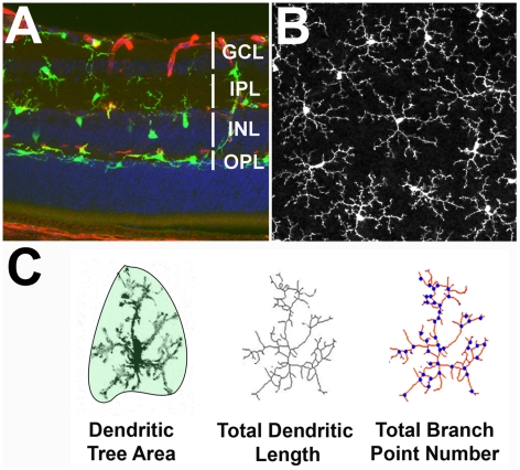
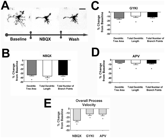
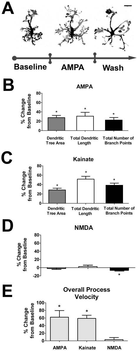
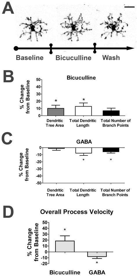
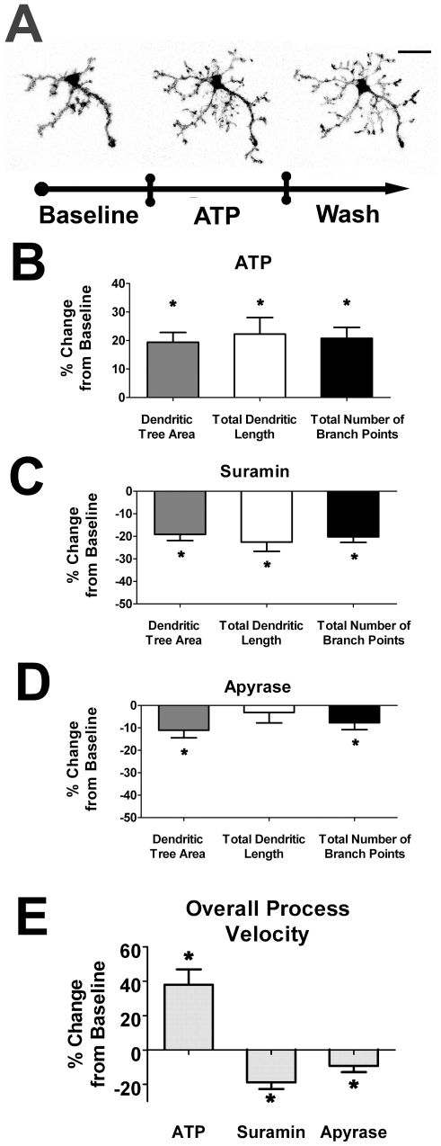
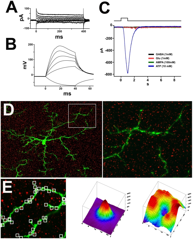
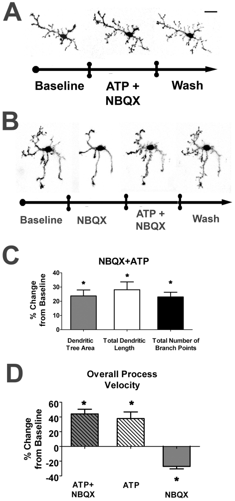
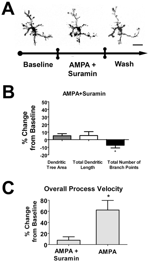
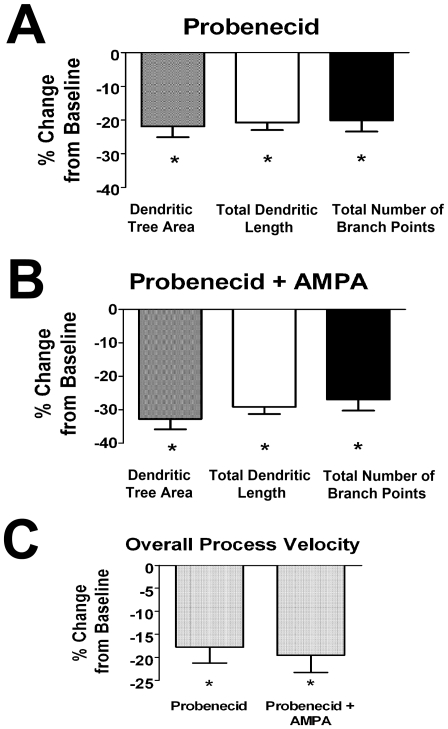
PURPOSE: Microglia represent the primary resident immune cells in the CNS, and have been implicated in the pathology of neurodegenerative diseases. Under basal or "resting" conditions, microglia possess ramified morphologies and exhibit dynamic surveying movements in their processes. Despite the prominence of this phenomenon, the function and regulation of microglial morphology and dynamic behavior are incompletely understood. We investigate here whether and how neurotransmission regulates "resting" microglial morphology and behavior. METHODS: We employed an ex vivo mouse retinal explant system in which endogenous neurotransmission and dynamic microglial behavior are present. We utilized live-cell time-lapse confocal imaging to study the morphology and behavior of GFP-labeled retinal microglia in response to neurotransmitter agonists and antagonists. Patch clamp electrophysiology and immunohistochemical localization of glutamate receptors were also used to investigate direct-versus-indirect effects of neurotransmission by microglia. RESULTS: Retinal microglial morphology and dynamic behavior were not cell-autonomously regulated but are instead modulated by endogenous neurotransmission. Morphological parameters and process motility were differentially regulated by different modes of neurotransmission and were increased by ionotropic glutamatergic neurotransmission and decreased by ionotropic GABAergic neurotransmission. These neurotransmitter influences on retinal microglia were however unlikely to be directly mediated; local applications of neurotransmitters were unable to elicit electrical responses on microglia patch-clamp recordings and ionotropic glutamatergic receptors were not located on microglial cell bodies or processes by immunofluorescent labeling. Instead, these influences were mediated indirectly via extracellular ATP, released in response to glutamatergic neurotransmission through probenecid-sensitive pannexin hemichannels. CONCLUSIONS: Our results demonstrate that neurotransmission plays an endogenous role in regulating the morphology and behavior of "resting" microglia in the retina. These findings illustrate a mode of constitutive signaling between the neural and immune compartments of the CNS through which immune cells may be regulated in concert with levels of neural activity.
Overview
The claims and statistics of this paper haven’t been checked yet.
Run the check — free, no account needed. It reads the open-access text and checks the sources it cites.
Integrity signals
Registry
Cites
Statistics
Figures
Verification
Claims the paper makes
The claims and statistics of this paper haven’t been checked yet.
Run the check — free, no account needed. It reads the open-access text and checks the sources it cites.
Claims other papers make about this one
Each is checked against this paper’s own text: does it say what the citing paper says it does?
The source backs part of the claim, not all of it — or backs it for a different population, condition or comparison.
In the citing paper
“Previous work has demonstrated that microglia have the capacity to interact with synaptic elements in response to neurotransmitter release and/or sensory experience (Biber et al., 2007; Fontainhas et al., 2011; Nimmerjahn et al., 2005; Ransohoff and Perry, 2009; Tremblay et al., 2010a; Wake et al., 2009).”
From Microglia Sculpt Postnatal Neural Circuits in an Activity and Complement-Dependent Manner (2012) · cites this paper as Fontainhas et al., 2011
Experimental — accuracy not yet measuredIn the citing paper
“and that microglia respond to changes in ionotropic neurotransmission via the intermediary release of ATP by astrocytes20.”
From Microglial P2Y12 is necessary for synaptic plasticity in mouse visual cortex (2016) · cites this paper as [20]
Self-check
Percentages, sums and reported differences re-computed from the paper’s own numbers.
The self-check needs the paper’s full text.
Citations
Cites
Resting microglial cells are highly dynamic surveillants of brain parenchyma in vivo
Nimmerjahn, A., Kirchhoff, F., & Helmchen, F. (2005) · Science · doi:10.1126/science.1110647 · Cited by 5,827
ATP mediates rapid microglial response to local brain injury in vivo
Davalos, D., Grutzendler, J., Yang, G., Kim, J. V., Zuo, Y., Jung, S., Littman, D. R., Dustin, M. L., & Gan, W.-B. (2005) · Nature Neuroscience · doi:10.1038/nn1472 · Cited by 3,913
Microglia: Active sensor and versatile effector cells in the normal and pathologic brain
Hanisch, U.-K., & Kettenmann, H. (2007) · Nature Neuroscience · doi:10.1038/nn1997 · Cited by 3,676
Jung, S., Aliberti, J., Graemmel, P., Sunshine, M. J., Kreutzberg, G. W., Sher, A., & Littman, D. R. (2000) · Molecular and Cellular Biology · doi:10.1128/mcb.20.11.4106-4114.2000 · Cited by 2,632
Heterogeneity in the distribution and morphology of microglia in the normal adult mouse brain
Lawson, L. J., Perry, V. H., Dri, P., & Gordon, S. (1990) · Neuroscience · doi:10.1016/0306-4522(90)90229-w · Cited by 2,045
Microglial physiology: unique stimuli, specialized responses
Ransohoff, R. M., & Perry, V. H. (2009) · Annual Review of Immunology · doi:10.1146/annurev.immunol.021908.132528 · Cited by 1,858
Wake, H., Moorhouse, A. J., Jinno, S., Kohsaka, S., & Nabekura, J. (2009) · The Journal of Neuroscience · doi:10.1523/jneurosci.4363-08.2009 · Cited by 1,619
The P2Y12 receptor regulates microglial activation by extracellular nucleotides
Haynes, S. E., Hollopeter, G., Yang, G., Kurpius, D., Dailey, M. E., Gan, W.-B., & Julius, D. (2006) · Nature Neuroscience · doi:10.1038/nn1805 · Cited by 1,542
Neuronal ‘On’ and ‘Off’ signals control microglia
Biber, K., Neumann, H., Inoue, K., & Boddeke, H. W. G. M. (2007) · Trends in Neurosciences · doi:10.1016/j.tins.2007.08.007 · Cited by 787
Neurotransmitter receptors on microglia
Pocock, J. M., & Kettenmann, H. (2007) · Trends in Neurosciences · doi:10.1016/j.tins.2007.07.007 · Cited by 650
Cited by
Sorted by citations
Microglia Sculpt Postnatal Neural Circuits in an Activity and Complement-Dependent Manner
Schafer, D. P., Lehrman, E. K., Kautzman, A. G., Koyama, R., Mardinly, A. R., Yamasaki, R., Ransohoff, R. M., Greenberg, M. E., Barres, B. A., & Stevens, B. (2012) · Neuron · doi:10.1016/j.neuron.2012.03.026 · Cited by 4,089
“…work has demonstrated that microglia have the capacity to interact with synaptic elements in response to neurotransmitter release and/or sensory experience (Biber et al., 2007; Fontainhas et al., 2011; Nimmerjahn et al., 2005; Ransohoff and Perry, 2009; Tremblay et al., 2010a; Wake et al., 2009).”
Microglia emerge as central players in brain disease
Salter, M. W., & Stevens, B. (2017) · Nature Medicine · doi:10.1038/nm.4397 · Cited by 1,767
Microglia: new roles for the synaptic stripper
Kettenmann, H., Kirchhoff, F., & Verkhratsky, A. (2013) · Neuron · doi:10.1016/j.neuron.2012.12.023 · Cited by 1,173
The Role of Microglia in the Healthy Brain: Figure 1.
Tremblay, M.-È., Stevens, B., Sierra, A., Wake, H., Bessis, A., & Nimmerjahn, A. (2011) · The Journal of Neuroscience · doi:10.1523/jneurosci.4158-11.2011 · Cited by 992
Microglia Heterogeneity in the Single-Cell Era
Masuda, T., Sankowski, R., Staszewski, O., & Prinz, M. (2020) · Cell Reports · doi:10.1016/j.celrep.2020.01.010 · Cited by 784
Metadata
- Authors
- Fontainhas, A. M., Wang, M., Liang, K. J., Chen, S., Mettu, P., Damani, M., Fariss, R. N., Li, W., & Wong, W. T.
- Affiliations
- National Institutes of Health (US)
- Corresponding author
- Wong, Wai T.
- Venue
- PLoS ONE
- Publisher
- Public Library of Science (PLoS)
- Published
- 2011-01-25
- Volume
- 6
- Issue
- 1
- Pages
- e15973
- ISSN
- 1932-6203 (ISSN-L)
- PMC
- PMC3026789
- OpenAlex
- W1997602206
- Cited by
- 334
- References
- 57
- Citations by year
- 2026: 8 · 2025: 12 · 2024: 15 · 2023: 18 · 2022: 20 · 2021: 44 · 2020: 30 · 2019: 28 · 2018: 19 · 2017: 27 · 2016: 28 · 2015: 21 · 2014: 20 · 2013: 24 · 2012: 12
- FWCI
- 11.32
- Type
- journal article
- Language
- en
- Open access
- gold
- doi.org
- Licence
- Locations
- PLoS ONE (publishedVersion, public-domain)
- PubMed Central (publishedVersion, public-domain)
- PubMed (publishedVersion)
- DOAJ (DOAJ: Directory of Open Access Journals) (submittedVersion, cc-by-sa)
- Figshare (submittedVersion, cc-by)
- Funders
- National Eye Institute
- Topics
- Life Sciences › Neuroscience › Neurology › Neuroinflammation and Neurodegeneration Mechanisms
- Life Sciences › Neuroscience › Cellular and Molecular Neuroscience › Neuroscience and Neuropharmacology Research
- Health Sciences › Medicine › Radiology, Nuclear Medicine and Imaging › Retinal Imaging and Analysis
- Keywords
- microglia, microglial morphology, microglial motility, resting microglia, retinal microglia, extracellular ATP, glutamate receptors, neuroimmune signaling, neurodegenerative diseases
- MeSH terms
- Animals, Immune System, Synaptic Transmission, Retina, Microglia, Receptors, GABA, Microscopy, Video, Cell Shape, Green Fluorescent Proteins, Mice, Receptors, Ionotropic Glutamate, In Vitro Techniques
About these checks
Registry, retraction, Cites and statistics results are computed, not judged, with one exception: a printed reference that no index lists is read once by a model before it counts as a citation error. Claims are extracted by a model and kept only when their quote appears verbatim in the text; each cited source is then checked, with a model’s help, for whether it says what the claim relies on. Those verdicts can be wrong — read the claim’s quote and the cited source before relying on one.
Statistics and arithmetic are recomputed, not judged.
Confidence bands are shown only for fields calibrated on our annotated test set. None is calibrated yet, so no band is shown.
What counts as a claim ›How we measure confidence ›How we check this ›
Are you an author, or do you see a problem with this page? Request a review
