

The DOI matches its Crossref record; 47 of the 47 references it cites were found in a registry or an index.
The Origin of GPCRs: Identification of Mammalian like Rhodopsin, Adhesion, Glutamate and Frizzled GPCRs in Fungi
Open accessAbstract
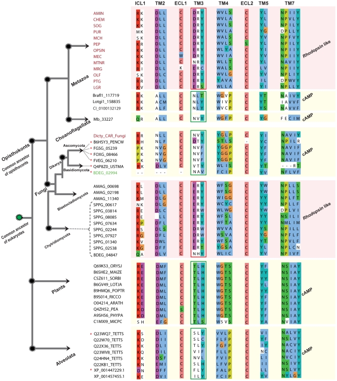
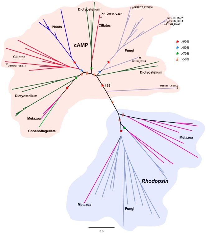
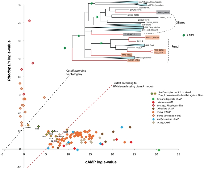
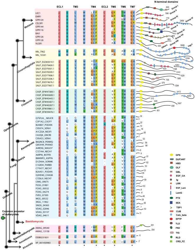
G protein-coupled receptors (GPCRs) in humans are classified into the five main families named Glutamate, Rhodopsin, Adhesion, Frizzled and Secretin according to the GRAFS classification. Previous results show that these mammalian GRAFS families are well represented in the Metazoan lineages, but they have not been shown to be present in Fungi. Here, we systematically mined 79 fungal genomes and provide the first evidence that four of the five main mammalian families of GPCRs, namely Rhodopsin, Adhesion, Glutamate and Frizzled, are present in Fungi and found 142 novel sequences between them. Significantly, we provide strong evidence that the Rhodopsin family emerged from the cAMP receptor family in an event close to the split of Opisthokonts and not in Placozoa, as earlier assumed. The Rhodopsin family then expanded greatly in Metazoans while the cAMP receptor family is found in 3 invertebrate species and lost in the vertebrates. We estimate that the Adhesion and Frizzled families evolved before the split of Unikonts from a common ancestor of all major eukaryotic lineages. Also, the study highlights that the fungal Adhesion receptors do not have N-terminal domains whereas the fungal Glutamate receptors have a broad repertoire of mammalian-like N-terminal domains. Further, mining of the close unicellular relatives of the Metazoan lineage, Salpingoeca rosetta and Capsaspora owczarzaki, obtained a rich group of both the Adhesion and Glutamate families, which in particular provided insight to the early emergence of the N-terminal domains of the Adhesion family. We identified 619 Fungi specific GPCRs across 79 genomes and revealed that Blastocladiomycota and Chytridiomycota phylum have Metazoan-like GPCRs rather than the GPCRs specific for Fungi. Overall, this study provides the first evidence of the presence of four of the five main GRAFS families in Fungi and clarifies the early evolutionary history of the GPCR superfamily.
Overview
The claims and statistics of this paper haven’t been checked yet.
Run the check — free, no account needed. It reads the open-access text and checks the sources it cites.
Integrity signals
Registry
Cites
Statistics
Figures
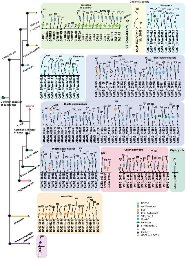
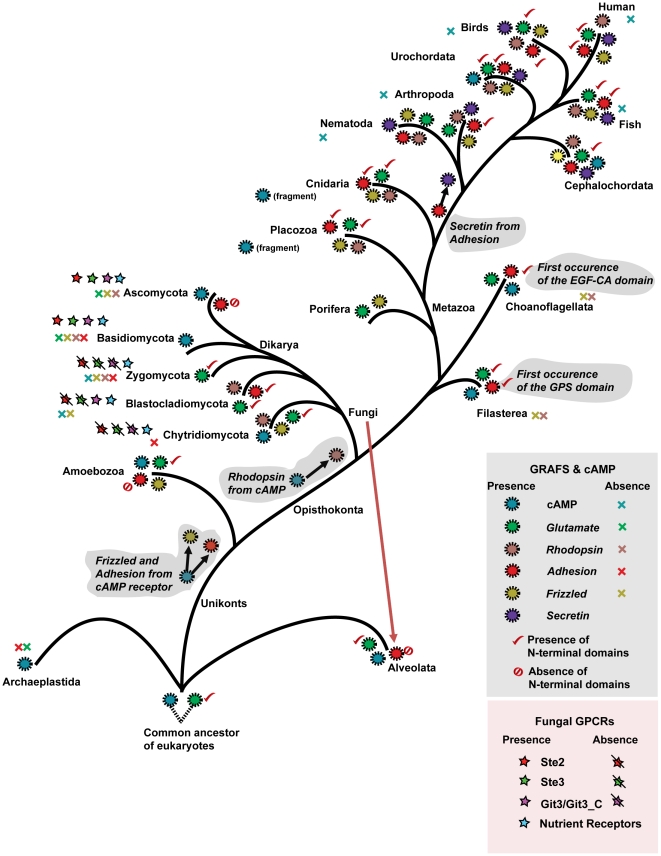
Verification
Claims the paper makes
The claims and statistics of this paper haven’t been checked yet.
Run the check — free, no account needed. It reads the open-access text and checks the sources it cites.
Claims other papers make about this one
Each is checked against this paper’s own text: does it say what the citing paper says it does?
In the citing paper
“Following from this Sun et al. present experimental evidence that nerve cells can also conduct biophotons, and that this effect can be inhibited by the application of a neural conduction block anaesthetic 124,133 .”
From Quantum effects in the brain: A review (2020) · cites this paper as [133]
Self-check
Percentages, sums and reported differences re-computed from the paper’s own numbers.
The self-check needs the paper’s full text.
Citations
Cites
MrBayes 3: Bayesian phylogenetic inference under mixed models
Ronquist, F., & Huelsenbeck, J. P. (2003) · Bioinformatics · doi:10.1093/bioinformatics/btg180 · Cited by 29,194
Guindon, S., Dufayard, J.-F., Lefort, V., Anisimova, M., Hordijk, W., & Gascuel, O. (2010) · Systematic Biology · doi:10.1093/sysbio/syq010 · Cited by 18,376
The Pfam Protein Families Database
Bateman, A. G. (2002) · Nucleic Acids Research · doi:10.1093/nar/30.1.276 · Cited by 14,075
Cd-hit: a fast program for clustering and comparing large sets of protein or nucleotide sequences
Li, W.-Z., & Godzik, A. (2006) · Bioinform. · doi:10.1093/bioinformatics/btl158 · Cited by 10,996
Eddy, S. R. (1998) · Bioinformatics · doi:10.1093/bioinformatics/14.9.755 · Cited by 5,515
MAFFT version 5: improvement in accuracy of multiple sequence alignment
Katoh, K., Kuma, K., Toh, H., & Miyata, T. (2005) · Nucleic Acids Research · doi:10.1093/nar/gki198 · Cited by 5,063
Genome Sequence of the Nematode C. elegans : A Platform for Investigating Biology
Berg, J. (1998) · Science · doi:10.1126/science.282.5396.2012 · Cited by 4,458
Fredriksson, R., Lagerström, M. C., Lundin, L.-G., & Schiöth, H. B. (2003) · Molecular Pharmacology · doi:10.1124/mol.63.6.1256 · Cited by 2,941
Self-citationThe Pfam protein families database
Finn, R. D., Mistry, J., Tate, J., Coggill, P., Heger, A., Pollington, J. E., Gavin, O. L., Gunasekaran, P., Ceric, G., Forslund, K., et al. (2010) · Nucleic Acids Research · doi:10.1093/nar/gkp985 · Cited by 2,694
The structure and function of G-protein-coupled receptors
Rosenbaum, D. M., Rasmussen, S. G. F., & Kobilka, B. K. (2009) · Nature · doi:10.1038/nature08144 · Cited by 2,534
Cited by
Sorted by citations
Allosteric sodium in class A GPCR signaling
Katritch, V., Fenalti, G., Abola, E. E., Roth, B. L., Cherezov, V., & Stevens, R. C. (2014) · Trends in Biochemical Sciences · doi:10.1016/j.tibs.2014.03.002 · Cited by 525
International Union of Basic and Clinical Pharmacology. XCIV. Adhesion G Protein–Coupled Receptors
Hamann, J., Aust, G., Araç, D., Engel, F. B., Formstone, C. J., Fredriksson, R., Hall, R. A., Harty, B. L., Kirchhoff, C., Knapp, B., et al. (2015) · Pharmacological Reviews · doi:10.1124/pr.114.009647 · Cited by 489
Self-citationThe Wnt Pathway: From Signaling Mechanisms to Synthetic Modulators
Rim, E. Y., Clevers, H. C., & Nusse, R. (2022) · Annual Review of Biochemistry · doi:10.1146/annurev-biochem-040320-103615 · Cited by 507
Structure of the human smoothened receptor bound to an antitumour agent
Wang, C., Wu, H., Katritch, V., Han, G. W., Huang, X., Liu, W., Siu, F., Roth, B. L., Cherezov, V., & Stevens, R. C. (2013) · Nature · doi:10.1038/nature12167 · Cited by 479
Molecular Biology of Atherosclerosis
Hopkins, P. N. (2013) · Physiological Reviews · doi:10.1152/physrev.00004.2012 · Cited by 469
Molecular evolution of peptidergic signaling systems in bilaterians
Mirabeau, O., & Joly, J. (2013) · Proceedings of the National Academy of Sciences · doi:10.1073/pnas.1219956110 · Cited by 429
Evolution of phototransduction, vertebrate photoreceptors and retina
Lamb, T. D. (2013) · Progress in Retinal and Eye Research · doi:10.1016/j.preteyeres.2013.06.001 · Cited by 400
Selectivity determinants of GPCR–G-protein binding
Flock, T., Hauser, A. S., Lund, N., Gloriam, D. E., Balaji, S., & Babu, M. M. (2017) · Nature · doi:10.1038/nature22070 · Cited by 384
Loh, K. M., van Amerongen, R., & Nusse, R. (2016) · Developmental Cell · doi:10.1016/j.devcel.2016.08.011 · Cited by 335
Heterotrimeric G protein signalling in the plant kingdom
Urano, D., Chen, J., Botella, J. R., & Jones, A. M. (2013) · Open Biology · doi:10.1098/rsob.120186 · Cited by 281
Sticky Signaling—Adhesion Class G Protein–Coupled Receptors Take the Stage
Langenhan, T., Aust, G., & Hamann, J. (2013) · Science Signaling · doi:10.1126/scisignal.2003825 · Cited by 263
de Mendoza, A., Sebé-Pedrós, A., & Ruiz‐Trillo, I. (2014) · Genome Biology and Evolution · doi:10.1093/gbe/evu038 · Cited by 202
Mechanisms of adhesion G protein–coupled receptor activation
Vizurraga, A., Adhikari, R., Yeung, J., Yu, M., & Tall, G. G. (2020) · Journal of Biological Chemistry · doi:10.1074/jbc.rev120.007423 · Cited by 179
Membrane composition and dynamics: A target of bioactive virgin olive oil constituents
López, S., Bermúdez, B., la Paz, S. M., Jaramillo, S., Varela, L. M., Ortega‐Gómez, A., Abia, R., & Muriana, F. J. G. (2014) · Biochimica et Biophysica Acta (BBA) - Biomembranes · doi:10.1016/j.bbamem.2014.01.007 · Cited by 175
Neuropeptide GPCRs in C. elegans
Frooninckx, L., Rompay, L. V., Temmerman, L., Sinay, E. V., Beets, I., Janssen, T., Husson, S. J., & Schoofs, L. (2012) · Frontiers in Endocrinology · doi:10.3389/fendo.2012.00167 · Cited by 166
Fluorescent proteins and genetically encoded biosensors
Wang, M., Da, Y., & Tian, Y. (2023) · Chemical Society Reviews · doi:10.1039/d2cs00419d · Cited by 156
The origin and evolution of Wnt signalling
Holzem, M., Boutros, M., & Holstein, T. W. (2024) · Nature Reviews Genetics · doi:10.1038/s41576-024-00699-w · Cited by 110
GPCRs: Lipid-Dependent Membrane Receptors That Act as Drug Targets
Chattopadhyay, A. (2014) · Advances in Biology · doi:10.1155/2014/143023 · Cited by 101
Matching structure with function: the GAIN domain of Adhesion-GPCR and PKD1-like proteins
Prömel, S., Langenhan, T., & Araç, D. (2013) · Trends in Pharmacological Sciences · doi:10.1016/j.tips.2013.06.002 · Cited by 94
What We Do Not Know about Fungal Cell Adhesion Molecules
Lipke, P. N. (2018) · Journal of Fungi · doi:10.3390/jof4020059 · Cited by 99
Cvicek, V., Goddard, W. A., & Abrol, R. (2016) · PLoS Computational Biology · doi:10.1371/journal.pcbi.1004805 · Cited by 99
Krishnan, A., & Schiöth, H. B. (2015) · Journal of Experimental Biology · doi:10.1242/jeb.110312 · Cited by 81
Self-citationJacobson, K. A., & Costanzi, S. (2012) · Molecular Pharmacology · doi:10.1124/mol.112.079335 · Cited by 85
Quantum effects in the brain: A review
Adams, B., & Petruccione, F. (2020) · AVS Quantum Science · doi:10.1116/1.5135170 · Cited by 80
Eukaryotic G Protein Signaling Evolved to Require G Protein–Coupled Receptors for Activation
Bradford, W. S., Buckholz, A. P., Morton, J., Price, C. M., Jones, A. M., & Urano, D. (2013) · Science Signaling · doi:10.1126/scisignal.2003768 · Cited by 76
The evolutionary history and spectral tuning of vertebrate visual opsins
Hagen, J. F. D., Roberts, N. S., & Johnston, R. J. (2022) · Developmental Biology · doi:10.1016/j.ydbio.2022.10.014 · Cited by 76
Evidence for a Transient Additional Ligand Binding Site in the TAS2R46 Bitter Taste Receptor
Sandal, M., Behrens, M., Brockhoff, A., Musiani, F., Giorgetti, A., Carloni, P., & Meyerhof, W. (2015) · Journal of Chemical Theory and Computation · doi:10.1021/acs.jctc.5b00472 · Cited by 73
The genetic factors of bilaterian evolution
Heger, P., Zheng, W. L., Rottmann, A., Panfilio, K. A., & Wiehe, T. (2020) · eLife · doi:10.7554/elife.45530 · Cited by 76
The Origin and Evolution of G Protein-Coupled Receptor Kinases
Mushegian, A. R., Gurevich, V. V., & Gurevich, E. V. (2012) · PLoS ONE · doi:10.1371/journal.pone.0033806 · Cited by 68
Glycoprotein Hormones and Their Receptors Emerged at the Origin of Metazoans
Roch, G. J., & Sherwood, N. M. (2014) · Genome Biology and Evolution · doi:10.1093/gbe/evu118 · Cited by 69
Adhesion GPCRs in Regulating Immune Responses and Inflammation
Lin, H., Hsiao, C., Pabst, C., Hébert, J., Schöneberg, T., & Hamann, J. (2017) · Advances in immunology · doi:10.1016/bs.ai.2017.05.005 · Cited by 70
Peeters, M. C., Fokkelman, M., Boogaard, B., Egerod, K. L., van de Water, B., IJzerman, A. P., & Schwartz, T. W. (2015) · Cellular Signalling · doi:10.1016/j.cellsig.2015.08.015 · Cited by 67
Towards the Idea of Molecular Brains
Timsit, Y., & Sergeant-Perthuis, G. (2021) · International Journal of Molecular Sciences · doi:10.3390/ijms222111868 · Cited by 77
Do Plants Contain G Protein-Coupled Receptors?
Taddese, B., UPTON, G. J. G., Bailey, G. R., Jordan, S. R. D., Abdulla, N. Y., Reeves, P. J., & Reynolds, C. A. (2013) · PLANT PHYSIOLOGY · doi:10.1104/pp.113.228874 · Cited by 68
Hwang, J., Moon, M. J., Park, S., Kim, D., Cho, E. B., Ha, N., Son, G. H., Kim, K., Vaudry, H., & Seong, J. Y. (2013) · Molecular Biology and Evolution · doi:10.1093/molbev/mst031 · Cited by 65
An Empirical Test of Convergent Evolution in Rhodopsins
Mackin, K. A., Roy, R. A., & Theobald, D. L. (2013) · Molecular Biology and Evolution · doi:10.1093/molbev/mst171 · Cited by 61
The role of GPR56/ADGRG1 in health and disease
Singh, A. K., & Lin, H. (2021) · Biomedical Journal · doi:10.1016/j.bj.2021.04.012 · Cited by 60
A Comprehensive Mutagenesis Screen of the Adhesion GPCR Latrophilin-1/ADGRL1
Nazarko, O. V., Kibrom, A., Winkler, J., Leon, K., Stoveken, H. M., Salzman, G., Merdas, K., Lu, Y., Narkhede, P., Tall, G. G., et al. (2018) · iScience · doi:10.1016/j.isci.2018.04.019 · Cited by 57
Shpakov, A. O. (2023) · International Journal of Molecular Sciences · doi:10.3390/ijms24076187 · Cited by 57
Structural Architecture and Functional Evolution of Wnts
Bazán, J. F., Janda, C. Y., & García, K. C. (2012) · Developmental Cell · doi:10.1016/j.devcel.2012.07.011 · Cited by 56
The G protein-coupled receptor GPR34 – The past 20 years of a grownup
Schöneberg, T., Meister, J., Knierim, A. B., & Schulz, A. (2018) · Pharmacology & Therapeutics · doi:10.1016/j.pharmthera.2018.04.008 · Cited by 54
Krishnan, A., Dnyansagar, R., Almén, M. S., Williams, M. J., Fredriksson, R., Manoj, N., & Schiöth, H. B. (2014) · BMC Evolutionary Biology · doi:10.1186/s12862-014-0270-4 · Cited by 57
Self-citationNagy, L. G., Varga, T., Csernetics, Á., & Virágh, M. (2020) · Fungal Biology Reviews · doi:10.1016/j.fbr.2020.07.002 · Cited by 59
“Round Up the Usual Suspects”: A Comment on Nonexistent Plant G Protein-Coupled Receptors
Urano, D., & Jones, A. M. (2013) · PLANT PHYSIOLOGY · doi:10.1104/pp.112.212324 · Cited by 53
Prediction of G Protein-Coupled Receptors with SVM-Prot Features and Random Forest
Liao, Z., Ju, Y., & Zou, Q. (2016) · Scientifica · doi:10.1155/2016/8309253 · Cited by 53
Receptors Mediating Host-Microbiota Communication in the Metaorganism: The Invertebrate Perspective
Dierking, K., & Pita, L. (2020) · Frontiers in Immunology · doi:10.3389/fimmu.2020.01251 · Cited by 57
The transporter–opsin– G protein‐coupled receptor ( TOG ) superfamily
Yee, D. C., Shlykov, M. A., Västermark, Å., Reddy, V. S., Arora, S., Sun, E. I., & Saier, M. H. (2013) · FEBS Journal · doi:10.1111/febs.12499 · Cited by 51
Wolf, S., & Grünewald, S. (2015) · PLoS ONE · doi:10.1371/journal.pone.0123533 · Cited by 52
Reconciling the deep homology of neuromodulation with the evolution of behavior
Katz, P. S., & Lillvis, J. L. (2014) · Current Opinion in Neurobiology · doi:10.1016/j.conb.2014.05.002 · Cited by 54
Eukaryotic G protein-coupled receptors as descendants of prokaryotic sodium-translocating rhodopsins
Shalaeva, D. N., Galperin, M. Y., & Mulkidjanian, A. Y. (2015) · Biology Direct · doi:10.1186/s13062-015-0091-4 · Cited by 49
P2Y Receptors in Immune Response and Inflammation
Duc, D. L., Schulz, A., Lede, V., Schulze, A., Thor, D., Brüser, A., & Schöneberg, T. (2017) · Advances in immunology · doi:10.1016/bs.ai.2017.05.006 · Cited by 51
Martı́n, J. F., van den Berg, M. A., van Themaat, E. V. L., & Liras, P. (2019) · Biotechnology Advances · doi:10.1016/j.biotechadv.2019.04.014 · Cited by 52
Cabrera, I. E., Pacentine, I. V., Lim, A., Guerrero, N., Kryštofová, S., Li, L., Michkov, A. V., Servin, J. A., Ahrendt, S. R., Carrillo, A. J., et al. (2015) · G3 Genes Genomes Genetics · doi:10.1534/g3.115.020974 · Cited by 47
Evolutionary hierarchy of vertebrate-like heterotrimeric G protein families
Krishnan, A., Mustafa, A., Almén, M. S., Fredriksson, R., Williams, M. J., & Schiöth, H. B. (2015) · Molecular Phylogenetics and Evolution · doi:10.1016/j.ympev.2015.05.009 · Cited by 46
Self-citationDissecting signaling and functions of adhesion G protein–coupled receptors
Araç, D., Aust, G., Calebiro, D., Engel, F. B., Formstone, C. J., Goffinet, A. M., Hamann, J., Kittel, R. J., Liebscher, I., Lin, H., et al. (2012) · Annals of the New York Academy of Sciences · doi:10.1111/j.1749-6632.2012.06820.x · Cited by 45
Self-citationFeeding and the Rhodopsin Family G-Protein Coupled Receptors in Nematodes and Arthropods
Cardoso, J. C. R., Félix, R. C., Fonseca, V. G., & Power, D. M. (2012) · Frontiers in Endocrinology · doi:10.3389/fendo.2012.00157 · Cited by 42
Retracing the path of planar cell polarity
Schenkelaars, Q., Fierro‐Constaín, L., Renard, E., & Borchiellini, C. (2016) · BMC Evolutionary Biology · doi:10.1186/s12862-016-0641-0 · Cited by 40
Prevertebrate Local Gene Duplication Facilitated Expansion of the Neuropeptide GPCR Superfamily
Yun, S., Furlong, M., Sim, M., Cho, M., Park, S., Cho, E. B., Reyes‐Alcaraz, A., Hwang, J., Kim, J., & Seong, J. Y. (2015) · Molecular Biology and Evolution · doi:10.1093/molbev/msv179 · Cited by 41
Bondarev, A. D., Attwood, M. M., Jonsson, J., Chubarev, V. N., Tarasov, V. V., & Schiöth, H. B. (2020) · Expert Opinion on Drug Discovery · doi:10.1080/17460441.2020.1791075 · Cited by 40
Self-citationConvergent evolution of animal and microbial rhodopsins
Kojima, K., & Sudo, Y. (2023) · RSC Advances · doi:10.1039/d2ra07073a · Cited by 41
The nervous system of Xenacoelomorpha: a genomic perspective
Perea-Atienza, E., Gavilán, B., Chiodin, M., Abril, J. F., Hoff, K. J., Poustka, A. J., & Martı́nez, P. (2015) · Journal of Experimental Biology · doi:10.1242/jeb.110379 · Cited by 39
Chapter One - Ubiquitination and Deubiquitination of G Protein-Coupled Receptors
Jean-Charles, P.-Y., Snyder, J. C., & Shenoy, S. K. (2016) · Progress in molecular biology and translational science · doi:10.1016/bs.pmbts.2016.05.001 · Cited by 39
Hahnel, S. R., Wheeler, N. J., Lu, Z., Wangwiwatsin, A., McVeigh, P., Maule, A. G., Berriman, M., Day, T. A., Ribeiro, P., & Grevelding, C. G. (2018) · PLoS Pathogens · doi:10.1371/journal.ppat.1006718 · Cited by 38
Whole Genome Wide Expression Profiles on Germination of Verticillium dahliae Microsclerotia
Hu, D., Wang, C., Tao, F., Cui, Q., Xu, X., Shang, W., & Hu, X. (2014) · PLoS ONE · doi:10.1371/journal.pone.0100046 · Cited by 38
Krishnan, A., Almén, M. S., Fredriksson, R., & Schiöth, H. B. (2013) · Gene · doi:10.1016/j.gene.2013.05.005 · Cited by 36
Self-citationViriyakosol, S., Singhania, A., Fierer, J., Goldberg, J. M., Kirkland, T. N., & Woelk, C. H. (2013) · BMC Microbiology · doi:10.1186/1471-2180-13-121 · Cited by 38
Vibrational resonance, allostery, and activation in rhodopsin-like G protein-coupled receptors
Woods, K. N., Pfeffer, J., Dutta, A., & Klein‐Seetharaman, J. (2016) · Scientific Reports · doi:10.1038/srep37290 · Cited by 32
Computational studies to predict or explain G protein coupled receptor polypharmacology
Jacobson, K. A., Costanzi, S., & Paoletta, S. (2014) · Trends in Pharmacological Sciences · doi:10.1016/j.tips.2014.10.009 · Cited by 31
Daddam, J. R., Sreenivasulu, B., Peddanna, K., & Katike, U. (2020) · RSC Advances · doi:10.1039/c9ra09245e · Cited by 30
New functions and signaling mechanisms for the class of adhesion G protein–coupled receptors
Liebscher, I., Ackley, B. D., Araç, D., Ariestanti, D. M., Aust, G., Bae, B.-I., Bista, B. R., Bridges, J. P., Duman, J. G., Engel, F. B., et al. (2014) · Annals of the New York Academy of Sciences · doi:10.1111/nyas.12580 · Cited by 30
Self-citationHow to bake a brain: yeast as a model neuron
Sarto‐Jackson, I., & Tomáška, Ľ. (2016) · Current Genetics · doi:10.1007/s00294-015-0554-2 · Cited by 29
The evolutionary analysis reveals domain fusion of proteins with Frizzled-like CRD domain
Yan, J., Jia, H., Ma, Z., Ye, H., Zhou, M., Su, L., Liu, J., & Guo, A. (2013) · Gene · doi:10.1016/j.gene.2013.09.083 · Cited by 27
16 Sexual Development in Fungi
Peraza‐Reyes, L., Brun, S., Grognet, P., & Malagnac, F. (2016) · The mycota · doi:10.1007/978-3-032-15548-1_13 · Cited by 27
Plattner, H. (2015) · Biological reviews/Biological reviews of the Cambridge Philosophical Society · doi:10.1111/brv.12218 · Cited by 27
Seppälä, S., Solomon, K., Gilmore, S. P., Henske, J. K., & O’Malley, M. (2016) · Microbial Cell Factories · doi:10.1186/s12934-016-0611-7 · Cited by 29
Effect of nitroprusside on arteriolar constriction after retinal branch vein occlusion.
Donati, G., Pournaras, C. J., & Tsacopoulos, M. (1998) · PubMed · Cited by 26
Alzugaray, M. E., Bruno, M. C., Sambucaro, M. J. V., & Ronderos, J. R. (2019) · Scientific Reports · doi:10.1038/s41598-019-46712-9 · Cited by 25
Gao, J. Q., Xu, X., Huang, K., & Liang, Z. (2021) · Frontiers in Microbiology · doi:10.3389/fmicb.2021.631392 · Cited by 27
Mickael, M., Rajput, A. B., Steyn, J. S., Wiemerslage, L., & Bürglin, T. R. (2016) · Comparative Biochemistry and Physiology Part D Genomics and Proteomics · doi:10.1016/j.cbd.2016.08.005 · Cited by 24
Comprehensive, structurally-informed alignment and phylogeny of vertebrate biogenic amine receptors
Spielman, S. J., Kumar, K., & Wilke, C. O. (2015) · PeerJ · doi:10.7717/peerj.773 · Cited by 24
Dynamics and mechanistic underpinnings to pharmacology of class A GPCRs: an NMR perspective
Huang, S. K., & Prosser, R. S. (2022) · American Journal of Physiology-Cell Physiology · doi:10.1152/ajpcell.00044.2022 · Cited by 22
A brief review on the evolution of GPCR: conservation and diversification
Zhang, Z., Wu, J., Yu, J. M., & Xiao, J. (2012) · Open Journal of Genetics · doi:10.4236/ojgen.2012.24b003 · Cited by 22
Powell, M. J. (2017) · Springer eBooks · doi:10.1007/978-3-319-28149-0_17 · Cited by 22
Ren, J., Chung‐Davidson, Y., Jia, L., & Li, W. (2019) · BMC Evolutionary Biology · doi:10.1186/s12862-019-1463-7 · Cited by 22
Mojib, N., & Kubanek, J. (2019) · Limnology and Oceanography · doi:10.1002/lno.11345 · Cited by 19
HTR6 and SSTR3 targeting to primary cilia
Barbeito, P., & Garcia-Gonzalo, F. R. (2021) · Biochemical Society Transactions · doi:10.1042/bst20191005 · Cited by 20
An Ancient Autoproteolytic Domain Found in GAIN, ZU5 and Nucleoporin98
Liao, Y., Pei, J., Cheng, H., & Grishin, N. V. (2014) · Journal of Molecular Biology · doi:10.1016/j.jmb.2014.10.011 · Cited by 20
Kerekes, K., Bányai, L., Trexler, M., & Patthy, L. (2019) · Growth Factors · doi:10.1080/08977194.2019.1626380 · Cited by 20
Rump, M. T., Kozma, M. T., Pawar, S., & Derby, C. D. (2021) · PLoS ONE · doi:10.1371/journal.pone.0252066 · Cited by 20
Bendena, W. G., Campbell, J. C., Zara, L. C., Tobe, S. S., & Chin-Sang, I. D. (2012) · Frontiers in Endocrinology · doi:10.3389/fendo.2012.00093 · Cited by 19
The Role of Neuropeptide-Stimulated cAMP-EPACs Signalling in Cancer Cells
Gao, Z., Lei, W. I., & Lee, L. T. O. (2022) · Molecules · doi:10.3390/molecules27010311 · Cited by 18
Peeters, M. C., Mos, I., Lenselink, E. B., Lucchesi, M., IJzerman, A. P., & Schwartz, T. W. (2016) · The FASEB Journal · doi:10.1096/fj.201500110 · Cited by 18
Cartography of rhodopsin-like G protein-coupled receptors across vertebrate genomes
Rinne, M. K., Tanoli, Z.-U.-R., Khan, A., & Xhaard, H. (2019) · Scientific Reports · doi:10.1038/s41598-018-33120-8 · Cited by 15
Modulating vertebrate physiology by genomic fine-tuning of GPCR functions
Schöneberg, T. (2024) · Physiological Reviews · doi:10.1152/physrev.00017.2024 · Cited by 18
The Relevance of Genomic Signatures at Adhesion GPCR Loci in Humans
Kovacs, P. L., & Schöneberg, T. (2016) · Handbook of experimental pharmacology · doi:10.1007/978-3-319-41523-9_9 · Cited by 18
G-protein-coupled Receptors in Fungi
El-Defrawy, M. M. H., & Hesham, A. E. (2020) · Fungal biology · doi:10.1007/978-3-030-41870-0_3 · Cited by 18
Photoreceptors mapping from past history till date
Parihar, P., Singh, R., Singh, S., Tripathi, D. K., Chauhan, D. K., Singh, V. P., & Prasad, S. M. (2016) · Journal of Photochemistry and Photobiology B Biology · doi:10.1016/j.jphotobiol.2016.06.020 · Cited by 19
Highly diversified expansions shaped the evolution of membrane bound proteins in metazoans
Attwood, M. M., Krishnan, A., Almén, M. S., & Schiöth, H. B. (2017) · Scientific Reports · doi:10.1038/s41598-017-11543-z · Cited by 14
Self-citationGu, X., Chen, Z., & Wang, D. (2020) · Frontiers in Bioengineering and Biotechnology · doi:10.3389/fbioe.2020.00635 · Cited by 15
Kim, D., Park, J. C., & Lee, J. (2022) · Comparative Biochemistry and Physiology Part C Toxicology & Pharmacology · doi:10.1016/j.cbpc.2022.109297 · Cited by 15
Metadata
- Authors
- Krishnan, A., Almén, M. S., Fredriksson, R., & Schiöth, H. B.
- Affiliations
- Uppsala University (SE)
- Corresponding author
- Schiöth, Helgi B.
- Venue
- PLoS ONE
- Publisher
- Public Library of Science (PLoS)
- Published
- 2012-01-04
- Volume
- 7
- Issue
- 1
- Pages
- e29817
- ISSN
- 1932-6203 (ISSN-L)
- PMC
- PMC3251606
- OpenAlex
- W1983820999
- Cited by
- 204
- References
- 47
- Citations by year
- 2026: 6 · 2025: 11 · 2024: 16 · 2023: 11 · 2022: 11 · 2021: 7 · 2020: 16 · 2019: 14 · 2018: 8 · 2017: 14 · 2016: 23 · 2015: 20 · 2014: 18 · 2013: 18 · 2012: 10
- FWCI
- 8.48
- Type
- journal article
- Language
- en
- Open access
- gold
- doi.org
- Locations
- PLoS ONE (publishedVersion, cc-by)
- Europe PMC (PubMed Central) (publishedVersion, cc-by)
- PubMed Central (publishedVersion, cc-by)
- PubMed (publishedVersion)
- Uppsala University Publications (Uppsala University) (submittedVersion, other-oa)
- RePEc: Research Papers in Economics (submittedVersion)
- DOAJ (DOAJ: Directory of Open Access Journals) (submittedVersion, cc-by-sa)
- Figshare (submittedVersion, cc-by)
- Funders
- Novo Nordisk
- Vetenskapsrådet
- Magnus Bergvalls Stiftelse
- Uppsala Universitet
- Novo Nordisk Fonden
- Topics
- Life Sciences › Biochemistry, Genetics and Molecular Biology › Molecular Biology › Receptor Mechanisms and Signaling
- Life Sciences › Biochemistry, Genetics and Molecular Biology › Molecular Biology › Fungal and yeast genetics research
- Life Sciences › Biochemistry, Genetics and Molecular Biology › Molecular Biology › Genomics and Phylogenetic Studies
- Keywords
- G protein-coupled receptors, GPCR evolution, rhodopsin family, cell adhesion receptors, glutamate receptors, Frizzled receptors, N-terminal domain, Salpingoeca rosetta, Capsaspora owczarzaki, Blastocladiomycota, Chytridiomycota, fungal genome
- MeSH terms
- Amino Acid Sequence, Animals, Cloning, Molecular, Fungi, Genes, Fungal, Humans, Mammals, Molecular Sequence Data, Phylogeny, Rhodopsin, Cell Adhesion Molecules, Genome, Fungal, Receptors, Glutamate, Evolution, Molecular, Receptors, G-Protein-Coupled, Frizzled Receptors
About these checks
Registry, retraction, Cites and statistics results are computed, not judged, with one exception: a printed reference that no index lists is read once by a model before it counts as a citation error. Claims are extracted by a model and kept only when their quote appears verbatim in the text; each cited source is then checked, with a model’s help, for whether it says what the claim relies on. Those verdicts can be wrong — read the claim’s quote and the cited source before relying on one.
Statistics and arithmetic are recomputed, not judged.
Confidence bands are shown only for fields calibrated on our annotated test set. None is calibrated yet, so no band is shown.
What counts as a claim ›How we measure confidence ›How we check this ›
Are you an author, or do you see a problem with this page? Request a review
