

The DOI matches its Crossref record; 35 of the 35 references it cites were found in a registry or an index.
Correlation Functions Quantify Super-Resolution Images and Estimate Apparent Clustering Due to Over-Counting
Open accessAbstract
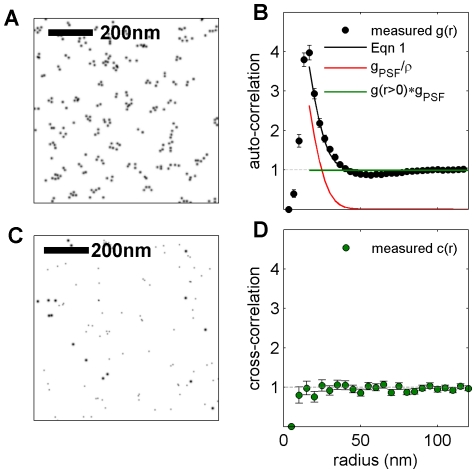
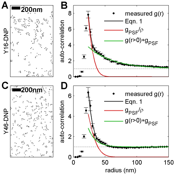
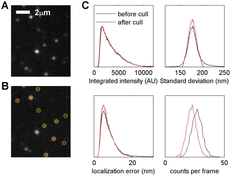
We present an analytical method using correlation functions to quantify clustering in super-resolution fluorescence localization images and electron microscopy images of static surfaces in two dimensions. We use this method to quantify how over-counting of labeled molecules contributes to apparent self-clustering and to calculate the effective lateral resolution of an image. This treatment applies to distributions of proteins and lipids in cell membranes, where there is significant interest in using electron microscopy and super-resolution fluorescence localization techniques to probe membrane heterogeneity. When images are quantified using pair auto-correlation functions, the magnitude of apparent clustering arising from over-counting varies inversely with the surface density of labeled molecules and does not depend on the number of times an average molecule is counted. In contrast, we demonstrate that over-counting does not give rise to apparent co-clustering in double label experiments when pair cross-correlation functions are measured. We apply our analytical method to quantify the distribution of the IgE receptor (FcεRI) on the plasma membranes of chemically fixed RBL-2H3 mast cells from images acquired using stochastic optical reconstruction microscopy (STORM/dSTORM) and scanning electron microscopy (SEM). We find that apparent clustering of FcεRI-bound IgE is dominated by over-counting labels on individual complexes when IgE is directly conjugated to organic fluorophores. We verify this observation by measuring pair cross-correlation functions between two distinguishably labeled pools of IgE-FcεRI on the cell surface using both imaging methods. After correcting for over-counting, we observe weak but significant self-clustering of IgE-FcεRI in fluorescence localization measurements, and no residual self-clustering as detected with SEM. We also apply this method to quantify IgE-FcεRI redistribution after deliberate clustering by crosslinking with two distinct trivalent ligands of defined architectures, and we evaluate contributions from both over-counting of labels and redistribution of proteins.
Overview
The claims and statistics of this paper haven’t been checked yet.
Run the check — free, no account needed. It reads the open-access text and checks the sources it cites.
Integrity signals
Registry
Cites
Statistics
Figures
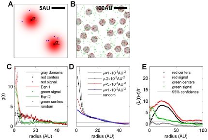
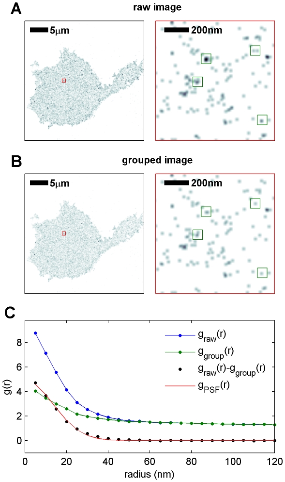
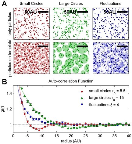
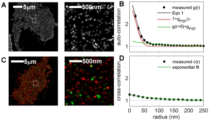
Verification
Claims the paper makes
The claims and statistics of this paper haven’t been checked yet.
Run the check — free, no account needed. It reads the open-access text and checks the sources it cites.
Claims other papers make about this one
Each is checked against this paper’s own text: does it say what the citing paper says it does?
The source backs part of the claim, not all of it — or backs it for a different population, condition or comparison.
In the citing paper
“Different from the ACF, overcounting has no effect on the PCF39.”
From A trans-synaptic nanocolumn aligns neurotransmitter release to receptors (2016) · cites this paper as [39]
Experimental — accuracy not yet measuredThe cited source says what the claim attributes to it.
In the citing paper
“The extent of ga(r) over 1, i.e. r0 for ga(r0) =1, is related to the pattern size of the internal heterogeneity (Extended Data Figure 2b–c)39.”
From A trans-synaptic nanocolumn aligns neurotransmitter release to receptors (2016) · cites this paper as [39]
Self-check
Percentages, sums and reported differences re-computed from the paper’s own numbers.
The self-check needs the paper’s full text.
Citations
Cites
The Fluid Mosaic Model of the Structure of Cell Membranes
Singer, S. J., & Nicolson, G. L. (1972) · Science · doi:10.1126/science.175.4023.720 · Cited by 9,158
Imaging Intracellular Fluorescent Proteins at Nanometer Resolution
Betzig, E., Patterson, G. H., Sougrat, R., Lindwasser, O. W., Olenych, S. G., Bonifacino, J. S., Davidson, M. W., Lippincott‐Schwartz, J., & Hess, H. F. (2006) · Science · doi:10.1126/science.1127344 · Cited by 8,941
Sub-diffraction-limit imaging by stochastic optical reconstruction microscopy (STORM)
Rust, M., Bates, M., & Zhuang, X. (2006) · Nature Methods · doi:10.1038/nmeth929 · Cited by 7,823
Crystal Statistics. I. A Two-Dimensional Model with an Order-Disorder Transition
Onsager, L. (1944) · Physical Review · doi:10.1103/physrev.65.117 · Cited by 6,634
Ultra-High Resolution Imaging by Fluorescence Photoactivation Localization Microscopy
Hess, S. T., Girirajan, T. P. K., & Mason, M. D. (2006) · Biophysical Journal · doi:10.1529/biophysj.106.091116 · Cited by 3,684
Subdiffraction‐Resolution Fluorescence Imaging with Conventional Fluorescent Probes
Heilemann, M., van de Linde, S., Schüttpelz, M., Kasper, R. T., Seefeldt, B., Mukherjee, A., Tinnefeld, P., & Sauer, M. (2008) · Angewandte Chemie International Edition · doi:10.1002/anie.200802376 · Cited by 1,870
Fluorescence microscopy with diffraction resolution barrier broken by stimulated emission
Klar, T. A., Jakobs, S., Dyba, M., Egner, A., & Hell, S. W. (2000) · Proceedings of the National Academy of Sciences · doi:10.1073/pnas.97.15.8206 · Cited by 1,869
Fast, three-dimensional super-resolution imaging of live cells
Jones, S. A., Shim, S., He, J., & Zhuang, X. (2011) · Nature Methods · doi:10.1038/nmeth.1605 · Cited by 757
Probing protein heterogeneity in the plasma membrane using PALM and pair correlation analysis
Sengupta, P., Jovanović‐Talisman, T., Skoko, D., Renz, M., Veatch, S. L., & Lippincott‐Schwartz, J. (2011) · Nature Methods · doi:10.1038/nmeth.1704 · Cited by 588
Self-citationStatistical Mechanics: Entropy, Order Parameters, and Complexity
Sethna, J. P. (2021) · doi:10.1093/oso/9780198865247.001.0001 · Cited by 495
Cited by
Sorted by citations
Single-molecule localization microscopy
Lelek, M., Gyparaki, M. T., Beliu, G., Schueder, F., Griffié, J., Manley, S., Jungmann, R., Sauer, M., Lakadamyali, M., & Zimmer, C. (2021) · Nature Reviews Methods Primers · doi:10.1038/s43586-021-00038-x · Cited by 1,017
Measuring image resolution in optical nanoscopy
Nieuwenhuizen, R. P. J., Lidke, K. A., Bates, M., Leyton-Puig, D., Grünwald, D., Stallinga, S., & Rieger, B. (2013) · Nature Methods · doi:10.1038/nmeth.2448 · Cited by 804
A trans-synaptic nanocolumn aligns neurotransmitter release to receptors
Tang, A.-H., Chen, H., Li, T. P., Metzbower, S. R., MacGillavry, H. D., & Blanpied, T. A. (2016) · Nature · doi:10.1038/nature19058 · Cited by 677
Phosphatidylserine Vesicles Enable Efficient En Bloc Transmission of Enteroviruses
Chen, Y.-H., Du, W., Hagemeijer, M. C., Takvorian, P. M., Pau, C., Cali, A., Brantner, C. A., Stempinski, E., Connelly, P. S., Ma, H.-C., et al. (2015) · Cell · doi:10.1016/j.cell.2015.01.032 · Cited by 470
ecDNA hubs drive cooperative intermolecular oncogene expression
Hung, K. L., Yost, K. E., Xie, L., Shi, Q., Helmsauer, K., Luebeck, J., Schöpflin, R., Lange, J. T., González, R. C., Weiser, N. E., et al. (2021) · Nature · doi:10.1038/s41586-021-04116-8 · Cited by 364
Pessina, F., Giavazzi, F., Yin, Y., Gioia, U., Vitelli, V., Galbiati, A., Barozzi, S., Garre, M., Oldani, A., Flaus, A., et al. (2019) · Nature Cell Biology · doi:10.1038/s41556-019-0392-4 · Cited by 360
Tracking single molecules at work in living cells
Kusumi, A., Tsunoyama, T. A., Hirosawa, K. M., Kasai, R. S., & Fujiwara, T. K. (2014) · Nature Chemical Biology · doi:10.1038/nchembio.1558 · Cited by 339
Force loading explains spatial sensing of ligands by cells
Oria, R., Wiegand, T., Escribano, J. M. G., Elósegui-Artola, A., Uriarte, J. J., Moreno-Pulido, C., Platzman, I., Delcanale, P., Albertazzi, L., Navajas, D., et al. (2017) · Nature · doi:10.1038/nature24662 · Cited by 330
Khater, I. M., Nabi, I. R., & Hamarneh, G. (2020) · Patterns · doi:10.1016/j.patter.2020.100038 · Cited by 288
Probing Exchange Pathways in One-Dimensional Aggregates with Super-Resolution Microscopy
Albertazzi, L., van der Zwaag, D., Leenders, C. M. A., Fitzner, R., van der Hofstad, R., & Meijer, E. W. (2014) · Science · doi:10.1126/science.1250945 · Cited by 277
The Continuing Mystery of Lipid Rafts
Levental, I., & Veatch, S. L. (2016) · Journal of Molecular Biology · doi:10.1016/j.jmb.2016.08.022 · Cited by 273
Self-citationSuper-Resolution Microscopy: From Single Molecules to Supramolecular Assemblies
Sydor, A. M., Czymmek, K. J., Puchner, E. M., & Mennella, V. (2015) · Trends in Cell Biology · doi:10.1016/j.tcb.2015.10.004 · Cited by 262
3D imaging of Sox2 enhancer clusters in embryonic stem cells
Liu, Z. J., Legant, W. R., Chen, B., Li, L., Grimm, J. B., Lavis, L. D., Betzig, E., & Tjian, R. (2014) · eLife · doi:10.7554/elife.04236 · Cited by 252
Synaptic weight set by Munc13-1 supramolecular assemblies
Sakamoto, H., Ariyoshi, T., Kimpara, N., Sugao, K., Taiko, I., Takikawa, K., Asanuma, D., Namiki, S., & Hirose, K. (2018) · Nature Neuroscience · doi:10.1038/s41593-017-0041-9 · Cited by 235
Cell-specific STORM super-resolution imaging reveals nanoscale organization of cannabinoid signaling
Dudok, B., Barna, L., Ledri, M., Szabó, S. I., Szabadits, E., Pintér, B., Woodhams, S. G., Henstridge, C. M., Balla, G. Y., Nyilas, R., et al. (2015) · Nature Neuroscience · doi:10.1038/nn.3892 · Cited by 227
Zhang, H., Christensen, C. L., Dries, R., Oser, M. G., Deng, J., Diskin, B., Li, F., Pan, Y., Zhang, X., Yin, Y., et al. (2019) · Cancer Cell · doi:10.1016/j.ccell.2019.11.003 · Cited by 222
Stone, M. B., Shelby, S. A., Núñez, M. F., Wisser, K. C., & Veatch, S. L. (2017) · eLife · doi:10.7554/elife.19891 · Cited by 205
Self-citationAndronov, L. P., Orlov, I. M., Lutz, Y., Vonesch, J., & Klaholz, B. P. (2016) · Scientific Reports · doi:10.1038/srep24084 · Cited by 194
How to switch a fluorophore: from undesired blinking to controlled photoswitching
van de Linde, S., & Sauer, M. (2013) · Chemical Society Reviews · doi:10.1039/c3cs60195a · Cited by 191
Baddeley, D., & Bewersdorf, J. (2017) · Annual Review of Biochemistry · doi:10.1146/annurev-biochem-060815-014801 · Cited by 190
Puchner, E. M., Walter, J. M., Kasper, R. T., Huang, B., & Lim, W. A. (2013) · Proceedings of the National Academy of Sciences · doi:10.1073/pnas.1309676110 · Cited by 182
Turning single-molecule localization microscopy into a quantitative bioanalytical tool
Nicovich, P. R., Owen, D. M., & Gaus, K. (2017) · Nature Protocols · doi:10.1038/nprot.2016.166 · Cited by 164
Super-Resolution Microscopy: Shedding Light on the Cellular Plasma Membrane
Stone, M. B., Shelby, S. A., & Veatch, S. L. (2017) · Chemical Reviews · doi:10.1021/acs.chemrev.6b00716 · Cited by 168
Self-citationOwen, D. M., Magenau, A., Williamson, D. J., & Gaus, K. (2012) · BioEssays · doi:10.1002/bies.201200044 · Cited by 162
Fricke, F., Beaudouin, J., Eils, R., & Heilemann, M. (2015) · Scientific Reports · doi:10.1038/srep14072 · Cited by 158
Defining the rate-limiting processes of bacterial cytokinesis
Coltharp, C., Buss, J. A., Plumer, T., & Xiao, J. (2016) · Proceedings of the National Academy of Sciences · doi:10.1073/pnas.1514296113 · Cited by 157
da Silva, R. M. P., van der Zwaag, D., Albertazzi, L., Lee, S. S., Meijer, E. W., & Stupp, S. I. (2016) · Nature Communications · doi:10.1038/ncomms11561 · Cited by 158
Pathogenic Tau Impairs Axon Initial Segment Plasticity and Excitability Homeostasis
Sohn, P. D., Huang, C. T.-L., Yan, R., Fan, L., Tracy, T. E., Camargo, C. M., Montgomery, K. M., Arhar, T., Mok, S.-A., Freilich, R., et al. (2019) · Neuron · doi:10.1016/j.neuron.2019.08.008 · Cited by 148
“Two-dimensional cross-correlation analysis (Figures 4G–H) between two color channels (Stone and Veatch, 2015; Veatch et al., 2012) was done with a custom MATLAB program.”
MethodSengupta, P., Jovanović‐Talisman, T., & Lippincott‐Schwartz, J. (2013) · Nature Protocols · doi:10.1038/nprot.2013.005 · Cited by 148
Superresolution Imaging of Biological Systems Using Photoactivated Localization Microscopy
Sengupta, P., van Engelenburg, S. B., & Lippincott‐Schwartz, J. (2014) · Chemical Reviews · doi:10.1021/cr400614m · Cited by 146
Membrane phase separation drives responsive assembly of receptor signaling domains
Shelby, S., Castello-Serrano, I., Wisser, K. C., Levental, I., & Veatch, S. (2023) · Nature Chemical Biology · doi:10.1038/s41589-023-01268-8 · Cited by 123
Self-citationZhao, Z., Roy, R., Gebhardt, J. C. M., Suter, D. M., Chapman, A. R., & Xie, X. S. (2013) · Proceedings of the National Academy of Sciences · doi:10.1073/pnas.1318496111 · Cited by 143
Pageon, S. V., Nicovich, P. R., Mollazade, M., Tabarin, T., & Gaus, K. (2016) · Molecular Biology of the Cell · doi:10.1091/mbc.e16-07-0478 · Cited by 133
Kinetic disruption of lipid rafts is a mechanosensor for phospholipase D
Petersen, E. N., Chung, H.-W., Nayebosadri, A., & Hansen, S. B. (2016) · Nature Communications · doi:10.1038/ncomms13873 · Cited by 129
Maity, P. C., Blount, A. L., Jumaa, H., Ronneberger, O., Lillemeier, B. F., & Reth, M. G. (2015) · Science Signaling · doi:10.1126/scisignal.2005887 · Cited by 124
Superresolution microscopy for microbiology
Coltharp, C., & Xiao, J. (2012) · Cellular Microbiology · doi:10.1111/cmi.12024 · Cited by 132
Bayesian cluster identification in single-molecule localization microscopy data
Rubin‐Delanchy, P., Burn, G. L., Griffié, J., Williamson, D. J., Heard, N. A., Cope, A. P., & Owen, D. M. (2015) · Nature Methods · doi:10.1038/nmeth.3612 · Cited by 122
Buss, J. A., Coltharp, C., Shtengel, G. E., Yang, X., Hess, H. F., & Xiao, J. (2015) · PLoS Genetics · doi:10.1371/journal.pgen.1005128 · Cited by 112
XLF and APLF bind Ku80 at two remote sites to ensure DNA repair by non-homologous end joining
Nemoz, C., Ropars, V., Frit, P., Gontier, A., Drevet, P., Yu, J., Guérois, R., Pitois, A., Comte, A., Delteil, C., et al. (2018) · Nature Structural & Molecular Biology · doi:10.1038/s41594-018-0133-6 · Cited by 121
Lee, W. T. C., Yin, Y., Morten, M. J., Tonzi, P., Gwo, P. P., Odermatt, D. C., Modesti, M., Cantor, S. B., Gari, K., Huang, T. T., et al. (2021) · Nature Communications · doi:10.1038/s41467-021-22830-9 · Cited by 107
Gudheti, M. V., Curthoys, N. M., Gould, T. J., Kim, D., Gunewardene, M. S., Gabor, K. A., Gosse, J. A., Kim, C. H., Zimmerberg, J., & Hess, S. T. (2013) · Biophysical Journal · doi:10.1016/j.bpj.2013.03.054 · Cited by 106
Multi-Color Quantum Dot Tracking Using a High-Speed Hyperspectral Line-Scanning Microscope
Cutler, P. J., Malik, M. D., Liu, S., Byars, J. M., Lidke, D. S., & Lidke, K. A. (2013) · PLoS ONE · doi:10.1371/journal.pone.0064320 · Cited by 105
Martínez‐Muñoz, L., Rodrı́guez-Frade, J. M., Barroso, R., Sorzano, C. Ó. S., Torreno-Pina, J. A., Santiago, C. A., Manzo, C., Lucas, P., García‐Cuesta, E. M., Gutierrez, E., et al. (2018) · Molecular Cell · doi:10.1016/j.molcel.2018.02.034 · Cited by 104
Gradients Of Rac1 Nanoclusters Support Spatial Patterns of Rac1 Signaling
Remorino, A., de Beco, S., Cayrac, F., Federico, F. D., Cornilleau, G., Gautreau, A. M., Parrini, M. C., Masson, J., Dahan, M., & Coppey, M. (2017) · Cell Reports · doi:10.1016/j.celrep.2017.10.069 · Cited by 98
Saliba, D. G., Céspedes, P. F., Bálint, Š., Compeer, E. B., Korobchevskaya, K., Valvo, S., Mayya, V., Kvalvaag, A. S., Peng, Y., Dong, T., et al. (2019) · eLife · doi:10.7554/elife.47528 · Cited by 98
Localization microscopy coming of age: from concepts to biological impact
Sauer, M. (2013) · Journal of Cell Science · doi:10.1242/jcs.123612 · Cited by 99
Visualizing Cell Structure and Function with Point-Localization Superresolution Imaging
Sengupta, P., van Engelenburg, S. B., & Lippincott‐Schwartz, J. (2012) · Developmental Cell · doi:10.1016/j.devcel.2012.09.022 · Cited by 96
Valley, C. C., Arndt‐Jovin, D. J., Karedla, N., Steinkamp, M. P., Chizhik, A. I., Hlavacek, W. S., Wilson, B. S., Lidke, K. A., & Lidke, D. S. (2015) · Molecular Biology of the Cell · doi:10.1091/mbc.e15-05-0269 · Cited by 93
Roy, N. H., Chan, J., Lambelé, M., & Thali, M. (2013) · Journal of Virology · doi:10.1128/jvi.00790-13 · Cited by 89
3D ATAC-PALM: super-resolution imaging of the accessible genome
Xie, L., Dong, P., Chen, X., Hsieh, T.-H. S., Banala, S., Marzio, M. D., English, B. P., Qi, Y., Jung, S. K., Kieffer-Kwon, K.-R., et al. (2020) · Nature Methods · doi:10.1038/s41592-020-0775-2 · Cited by 86
Sequence-Independent Self-Assembly of Germ Granule mRNAs into Homotypic Clusters
Trcek, T., Douglas, T. E., Grosch, M., Yin, Y., Eagle, W. V. I., Gavis, E. R., Shroff, H., Rothenberg, E., & Lehmann, R. (2020) · Molecular Cell · doi:10.1016/j.molcel.2020.05.008 · Cited by 86
Direct Stochastic Optical Reconstruction Microscopy (dSTORM)
Endesfelder, U., & Heilemann, M. (2014) · Methods in molecular biology · doi:10.1007/978-1-4939-2080-8_14 · Cited by 85
Adhikari, S., Moscatelli, J., Smith, E. M., Banerjee, C., & Puchner, E. M. (2019) · Nature Communications · doi:10.1038/s41467-019-11384-6 · Cited by 85
Bademosi, A. T., Lauwers, E., Padmanabhan, P., Odierna, G. L., Chai, Y. J., Papadopulos, A., Goodhill, G. J., Verstreken, P., van Swinderen, B., & Meunier, F. A. (2016) · Nature Communications · doi:10.1038/ncomms13660 · Cited by 87
Progress in quantitative single-molecule localization microscopy
Deschout, H. G., Shivanandan, A., Annibale, P., Scarselli, M., & Rađenović, A. (2014) · Histochemistry and Cell Biology · doi:10.1007/s00418-014-1217-y · Cited by 83
Direct imaging of liquid domains in membranes by cryo-electron tomography
Cornell, C. E., Mileant, A., Thakkar, N., Lee, K. K., & Keller, S. L. (2020) · Proceedings of the National Academy of Sciences · doi:10.1073/pnas.2002245117 · Cited by 82
Quantitative super-resolution microscopy: pitfalls and strategies for image analysis
Durisic, N., Laparra-Cuervo, L., & Lakadamyali, M. (2014) · Current Opinion in Chemical Biology · doi:10.1016/j.cbpa.2014.04.005 · Cited by 82
Deschamps, J. R., Rowald, A., & Ries, J. (2016) · Optics Express · doi:10.1364/oe.24.028080 · Cited by 82
Steady-state cross-correlations for live two-color super-resolution localization data-sets
Stone, M., & Veatch, S. (2015) · Nature Communications · doi:10.1038/ncomms8347 · Cited by 68
Self-citationCrystal Structure and Molecular Imaging of the Nav Channel β3 Subunit Indicates a Trimeric Assembly
Namadurai, S., Balasuriya, D., Rajappa, R., Wiemhöfer, M., Stott, K. M., Klingauf, J., Edwardson, J. M., Chirgadze, D. Y., & Jackson, A. P. (2014) · Journal of Biological Chemistry · doi:10.1074/jbc.m113.527994 · Cited by 78
Kabbani, A. M., Raghunathan, K., Lencer, W. I., Kenworthy, A. K., & Kelly, C. V. (2020) · Proceedings of the National Academy of Sciences · doi:10.1073/pnas.2001119117 · Cited by 73
Method for co-cluster analysis in multichannel single-molecule localisation data
Rossy, J., Cohen, E. A. K., Gaus, K., & Owen, D. M. (2014) · Histochemistry and Cell Biology · doi:10.1007/s00418-014-1208-z · Cited by 75
Resolving mixed mechanisms of protein subdiffusion at the T cell plasma membrane
Golan, Y., & Sherman, E. (2017) · Nature Communications · doi:10.1038/ncomms15851 · Cited by 75
Mondal, S., Narayan, K. B., Botterbusch, S., Powers, I., Zheng, J., James, H. P., Jin, R., & Baumgart, T. (2022) · Nature Communications · doi:10.1038/s41467-022-32529-0 · Cited by 69
Quantitative analysis of single-molecule superresolution images
Coltharp, C., Yang, X., & Xiao, J. (2014) · Current Opinion in Structural Biology · doi:10.1016/j.sbi.2014.08.008 · Cited by 71
Sengupta, P., & Lippincott‐Schwartz, J. (2012) · BioEssays · doi:10.1002/bies.201200022 · Cited by 70
Termini, C. M., Cotter, M. L., Marjon, K. D., Buranda, T., Lidke, K. A., & Gillette, J. M. (2014) · Molecular Biology of the Cell · doi:10.1091/mbc.e13-11-0660 · Cited by 69
Translesion polymerase kappa-dependent DNA synthesis underlies replication fork recovery
Tonzi, P., Yin, Y., Lee, C. W. T., Rothenberg, E., & Huang, T. T. (2018) · eLife · doi:10.7554/elife.41426 · Cited by 69
Delcanale, P., Miret-Ontiveros, B., Arista-Romero, M., Pujals, S., & Albertazzi, L. (2018) · ACS Nano · doi:10.1021/acsnano.7b09063 · Cited by 68
Shelby, S. A., Holowka, D., Baird, B. A., & Veatch, S. L. (2013) · Biophysical Journal · doi:10.1016/j.bpj.2013.09.049 · Cited by 67
Self-citationSequential Superresolution Imaging of Multiple Targets Using a Single Fluorophore
Valley, C. C., Liu, S., Lidke, D. S., & Lidke, K. A. (2015) · PLoS ONE · doi:10.1371/journal.pone.0123941 · Cited by 64
Unveiling the Inner Workings of Live Bacteria Using Super-Resolution Microscopy
Tuson, H. H., & Biteen, J. S. (2014) · Analytical Chemistry · doi:10.1021/ac5041346 · Cited by 63
Extracting microtubule networks from superresolution single-molecule localization microscopy data
Zhang, Z., Nishimura, Y., & Kanchanawong, P. (2016) · Molecular Biology of the Cell · doi:10.1091/mbc.e16-06-0421 · Cited by 59
Purshouse, K. R., Friman, E. T., Boyle, S., Dewari, P. S., Grant, V., Hamdan, A., Morrison, G. M., Brennan, P. M., Beentjes, S. V., Pollard, S. M., et al. (2022) · eLife · doi:10.7554/elife.80207 · Cited by 62
Investigating Cellular Structures at the Nanoscale with Organic Fluorophores
van de Linde, S., Aufmkolk, S., Franke, C., Holm, T., Klein, T., Löschberger, A., Proppert, S., Wolter, S., & Sauer, M. (2013) · Chemistry & Biology · doi:10.1016/j.chembiol.2012.11.004 · Cited by 61
Pike, J. A., Khan, A. O., Pallini, C., Thomas, S. G., Mund, M., Ries, J., Poulter, N. S., & Styles, I. B. (2019) · Bioinformatics · doi:10.1093/bioinformatics/btz788 · Cited by 61
A Bayesian cluster analysis method for single-molecule localization microscopy data
Griffié, J., Shannon, M. J., Bromley, C. L., Boelen, L., Burn, G. L., Williamson, D. J., Heard, N. A., Cope, A. P., Owen, D. M., & Rubin‐Delanchy, P. (2016) · Nature Protocols · doi:10.1038/nprot.2016.149 · Cited by 59
Coltharp, C., Kessler, R. P., & Xiao, J. (2012) · PLoS ONE · doi:10.1371/journal.pone.0051725 · Cited by 56
Trapping of Syntaxin1a in Presynaptic Nanoclusters by a Clinically Relevant General Anesthetic
Bademosi, A. T., Steeves, J. B., Karunanithi, S., Zalucki, O. H., Gormal, R. S., Liu, S., Lauwers, E., Verstreken, P., Anggono, V., Meunier, F. A., et al. (2018) · Cell Reports · doi:10.1016/j.celrep.2017.12.054 · Cited by 57
Contributed Review: Review of integrated correlative light and electron microscopy
Timmermans, F. J., & Otto, C. (2015) · Review of Scientific Instruments · doi:10.1063/1.4905434 · Cited by 56
Éltes, T., Kirizs, T., Nusser, Z., & Holderith, N. B. (2017) · Journal of Neuroscience · doi:10.1523/jneurosci.2024-16.2017 · Cited by 53
Róna, G., Roberti, D., Yin, Y., Pagan, J. K., Homer, H., Sassani, E., Zeke, A., Busino, L., Rothenberg, E., & Pagano, M. (2018) · eLife · doi:10.7554/elife.38771 · Cited by 61
Superresolution Imaging of Aquaporin-4 Cluster Size in Antibody-Stained Paraffin Brain Sections
Smith, A. J., & Verkman, A. S. (2015) · Biophysical Journal · doi:10.1016/j.bpj.2015.10.047 · Cited by 54
Quantitative Localization Microscopy: Effects of Photophysics and Labeling Stoichiometry
Nieuwenhuizen, R. P. J., Bates, M., Szymborska, A., Lidke, K. A., Rieger, B., & Stallinga, S. (2015) · PLoS ONE · doi:10.1371/journal.pone.0127989 · Cited by 53
Single-Molecule Insights into Retention at a Reversed-Phase Chromatographic Interface
Mabry, J. N., Skaug, M. J., & Schwartz, D. K. (2014) · Analytical Chemistry · doi:10.1021/ac5026418 · Cited by 53
Tracking Low-Copy Transcription Factors in Living Bacteria: The Case of the lac Repressor
de León, F. G., Sellars, L. E., Stracy, M., Busby, S. J. W., & Kapanidis, A. N. (2017) · Biophysical Journal · doi:10.1016/j.bpj.2017.02.028 · Cited by 52
Wang, F., Liu, Y., Zhang, T., Gao, J., Xu, Y., Xie, G., Zhao, W., Wang, H., & Yang, Y. (2020) · Aging Cell · doi:10.1111/acel.13224 · Cited by 51
Quantitative Single-Molecule Localization Microscopy
Hugelier, S., Colosi, P., & Lakadamyali, M. (2023) · Annual Review of Biophysics · doi:10.1146/annurev-biophys-111622-091212 · Cited by 51
Superresolution Imaging Captures Carbohydrate Utilization Dynamics in Human Gut Symbionts
Karunatilaka, K. S., Cameron, E. A., Martens, E. C., Koropatkin, N. M., & Biteen, J. S. (2014) · mBio · doi:10.1128/mbio.02172-14 · Cited by 53
Kasula, R., Chai, Y. J., Bademosi, A. T., Harper, C. B., Gormal, R. S., Morrow, I. C., Hosy, E., Collins, B. M., Choquet, D., Papadopulos, A., et al. (2016) · The Journal of Cell Biology · doi:10.1083/jcb.201508118 · Cited by 51
Dynamic pattern generation in cell membranes: Current insights into membrane organization
Raghunathan, K., & Kenworthy, A. K. (2018) · Biochimica et Biophysica Acta (BBA) - Biomembranes · doi:10.1016/j.bbamem.2018.05.002 · Cited by 51
Modified Pearson correlation coefficient for two-color imaging in spherocylindrical cells
Mohapatra, S., & Weisshaar, J. C. (2018) · BMC Bioinformatics · doi:10.1186/s12859-018-2444-3 · Cited by 51
USP1-Trapping Lesions as a Source of DNA Replication Stress and Genomic Instability
Coleman, K. E., Yin, Y., Lui, S. K. L., Keegan, S., Fenyo, D., Smith, D. J., Rothenberg, E., & Huang, T. T. (2022) · Nature Communications · doi:10.1038/s41467-022-29369-3 · Cited by 50
Loss of PTEN promotes formation of signaling-capable clathrin-coated pits
Rosselli‐Murai, L. K., Yates, J. A., Yoshida, S., Bourg, J. T., Ho, K. K. Y., White, M., Prisby, J., Tan, X., Altemus, M., Bao, L., et al. (2018) · Journal of Cell Science · doi:10.1242/jcs.208926 · Cited by 48
Self-citationDiffraction-unlimited imaging: from pretty pictures to hard numbers
Vandenberg, W., Leutenegger, M., Lasser, T., Hofkens, J., & Dedecker, P. (2015) · Cell and Tissue Research · doi:10.1007/s00441-014-2109-0 · Cited by 49
Caetano, F. A., Dirk, B. S., Tam, J. H. K., Cavanagh, P. C., Goiko, M., Ferguson, S. S. G., Pasternak, S., Dikeakos, J. D., de Bruyn, J. R., & Heit, B. (2015) · PLoS Computational Biology · doi:10.1371/journal.pcbi.1004634 · Cited by 50
Lee, J., Sengupta, P., Brzostowski, J. A., Lippincott‐Schwartz, J., & Pierce, S. K. (2016) · Molecular Biology of the Cell · doi:10.1091/mbc.e16-06-0452 · Cited by 49
Dietz, M. S., & Heilemann, M. (2019) · Nanoscale · doi:10.1039/c9nr06364a · Cited by 47
Bohrer, C. H., Yang, X., Thakur, S., Weng, X., Tenner, B., McQuillen, R. J., Ross, B. L., Wooten, M., Chen, X., Zhang, J., et al. (2021) · Nature Methods · doi:10.1038/s41592-021-01154-y · Cited by 47
Test Samples for Optimizing STORM Super-Resolution Microscopy
Metcalf, D. J., Edwards, R., Kumarswami, N., & Knight, A. E. (2013) · Journal of Visualized Experiments · doi:10.3791/50579 · Cited by 46
Metadata
- Authors
- Veatch, S. L., Machta, B. B., Shelby, S. A., Chiang, E. N., Holowka, D. A., & Baird, B. A.
- Affiliations
- Cornell University (US)
- Corresponding author
- Veatch, Sarah L.
- Venue
- PLoS ONE
- Publisher
- Public Library of Science (PLoS)
- Published
- 2012-02-27
- Volume
- 7
- Issue
- 2
- Pages
- e31457
- ISSN
- 1932-6203 (ISSN-L)
- PMC
- PMC3288038
- OpenAlex
- W2073576660
- Cited by
- 290
- References
- 35
- Citations by year
- 2026: 6 · 2025: 11 · 2024: 12 · 2023: 13 · 2022: 19 · 2021: 22 · 2020: 16 · 2019: 24 · 2018: 24 · 2017: 23 · 2016: 33 · 2015: 21 · 2014: 36 · 2013: 23 · 2012: 7
- FWCI
- 12.73
- Type
- journal article
- Language
- en
- Open access
- gold
- doi.org
- Locations
- PLoS ONE (publishedVersion, cc-by)
- PubMed Central (publishedVersion, cc-by)
- PubMed (publishedVersion)
- arXiv (Cornell University) (submittedVersion)
- RePEc: Research Papers in Economics (submittedVersion)
- DOAJ (DOAJ: Directory of Open Access Journals) (submittedVersion, cc-by-sa)
- arXiv (Cornell University)
- Funders
- National Institutes of Health
- National Institute of General Medical Sciences (R00GM087810)
- National Institute of Allergy and Infectious Diseases (R01-AI18306, R01AI018306)
- Topics
- Life Sciences › Biochemistry, Genetics and Molecular Biology › Biophysics › Advanced Fluorescence Microscopy Techniques
- Life Sciences › Biochemistry, Genetics and Molecular Biology › Biophysics › Cell Image Analysis Techniques
- Life Sciences › Biochemistry, Genetics and Molecular Biology › Molecular Biology › Lipid Membrane Structure and Behavior
- Keywords
- super-resolution imaging, storms, scanning electron microscopy, FcεRI, IgE receptor, mast cells, membrane protein clustering, protein redistribution, crosslinking
- MeSH terms
- Animals, Cell Line, Cell Membrane, Fluorescent Dyes, Fourier Analysis, Immunoglobulin E, Image Processing, Computer-Assisted, Ligands, Microscopy, Electron, Microscopy, Electron, Scanning, Stochastic Processes, Models, Statistical, Cluster Analysis, Normal Distribution, Poisson Distribution, Receptors, IgE, Rats
About these checks
Registry, retraction, Cites and statistics results are computed, not judged, with one exception: a printed reference that no index lists is read once by a model before it counts as a citation error. Claims are extracted by a model and kept only when their quote appears verbatim in the text; each cited source is then checked, with a model’s help, for whether it says what the claim relies on. Those verdicts can be wrong — read the claim’s quote and the cited source before relying on one.
Statistics and arithmetic are recomputed, not judged.
Confidence bands are shown only for fields calibrated on our annotated test set. None is calibrated yet, so no band is shown.
What counts as a claim ›How we measure confidence ›How we check this ›
Are you an author, or do you see a problem with this page? Request a review
