

The DOI matches its Crossref record; 44 of the 45 references it cites were found in a registry or an index.
Candida albicans-Epithelial Interactions: Dissecting the Roles of Active Penetration, Induced Endocytosis and Host Factors on the Infection Process
Open accessAbstract
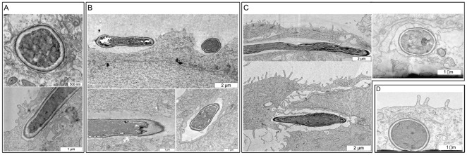
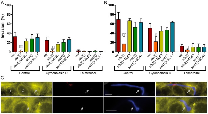
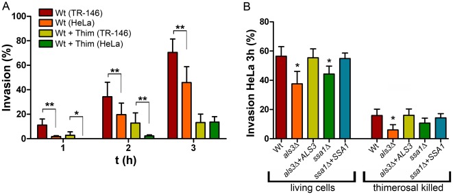
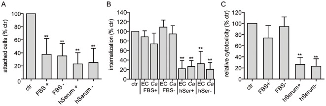
Candida albicans frequently causes superficial infections by invading and damaging epithelial cells, but may also cause systemic infections by penetrating through epithelial barriers. C. albicans is a remarkable pathogen because it can invade epithelial cells via two distinct mechanisms: induced endocytosis, analogous to facultative intracellular enteropathogenic bacteria, and active penetration, similar to plant pathogenic fungi. Here we investigated the contributions of the two invasion routes of C. albicans to epithelial invasion. Using selective cellular inhibition approaches and differential fluorescence microscopy, we demonstrate that induced endocytosis contributes considerably to the early time points of invasion, while active penetration represents the dominant epithelial invasion route. Although induced endocytosis depends mainly on Als3-E-cadherin interactions, we observed E-cadherin independent induced endocytosis. Finally, we provide evidence of a protective role for serum factors in oral infection: human serum strongly inhibited C. albicans adhesion to, invasion and damage of oral epithelial cells.
Overview
The claims and statistics of this paper haven’t been checked yet.
Run the check — free, no account needed. It reads the open-access text and checks the sources it cites.
Integrity signals
Registry
Cites
Statistics
Figures
Verification
Claims the paper makes
The claims and statistics of this paper haven’t been checked yet.
Run the check — free, no account needed. It reads the open-access text and checks the sources it cites.
Claims other papers make about this one
Each is checked against this paper’s own text: does it say what the citing paper says it does?
The source backs part of the claim, not all of it — or backs it for a different population, condition or comparison.
In the citing paper
“This result is in contrast to what has been found for Als3, which is required for both induced endocytosis and active penetration [29], and provides a likely explanation for why Hyr1 is dispensable for induction of epithelial cell damage.”
From Candida albicans stimulates formation of a multi-receptor complex that mediates epithelial cell invasion during oropharyngeal infection (2023) · cites this paper as [29]
Experimental — accuracy not yet measured
Self-check
Percentages, sums and reported differences re-computed from the paper’s own numbers.
The self-check needs the paper’s full text.
Citations
Cites
Isogenic strain construction and gene mapping in Candida albicans.
Fonzi, W. A., & Irwin, M. Y. (1993) · Genetics · doi:10.1093/genetics/134.3.717 · Cited by 1,695
Gillum, A. M., Tsay, E. Y. H., & Kirsch, D. R. (1984) · Molecular and General Genetics MGG · doi:10.1007/bf00328721 · Cited by 1,253
Bacterial Adhesion and Entry into Host Cells
Pizarro-Cerdá, J., & Cossart, P. (2006) · Cell · doi:10.1016/j.cell.2006.02.012 · Cited by 977 · cited 1×
“Bacteria, on the other hand, cannot actively penetrate host cells, but have developed several strategies to cause uptake by induced endocytosis [25,26].”
BackgroundOn the Trail of a Cereal Killer: Exploring the Biology of Magnaporthe grisea
Talbot, N. J. (2003) · Annual Review of Microbiology · doi:10.1146/annurev.micro.57.030502.090957 · Cited by 956
STUDIES ON THE PROPAGATION IN VITRO OF POLIOMYELITIS VIRUSES
Scherer, W. F., Syverton, J. T., & Gey, G. O. (1953) · Journal of Experimental Medicine · doi:10.1084/jem.97.5.695 · Cited by 856
Als3 Is a Candida albicans Invasin That Binds to Cadherins and Induces Endocytosis by Host Cells
Phan, Q. T., Myers, C. L., Fu, Y., Sheppard, D. C., Yeaman, M. R., Welch, W. H., Ibrahim, A. S., Edwards, J. E., & Filler, S. G. (2007) · PLoS Biology · doi:10.1371/journal.pbio.0050064 · Cited by 641
Candida albicans Cell Wall Proteins
Chaffin, W. L. (2008) · Microbiology and Molecular Biology Reviews · doi:10.1128/mmbr.00032-07 · Cited by 515
Cellular interactions of Candida albicans with human oral epithelial cells and enterocytes
Dalle, F., Wächtler, B., L’Ollivier, C., Holland, G., Bannert, N., Wilson, D., Labruère, C., Bonnin, A., & Hube, B. (2009) · Cellular Microbiology · doi:10.1111/j.1462-5822.2009.01394.x · Cited by 371
Self-citationSundstrom, P. (2002) · Cellular Microbiology · doi:10.1046/j.1462-5822.2002.00206.x · Cited by 336
Malaria Parasite Development in the Mosquito and Infection of the Mammalian Host
Aly, A. S. I., Vaughan, A. M., & Kappe, S. H. I. (2009) · Annual Review of Microbiology · doi:10.1146/annurev.micro.091208.073403 · Cited by 317
CIp10, an efficient and convenient integrating vector for Candida albicans
Murad, A. M. A., Lee, P. R., Broadbent, I. D., Barelle, C. J., & Brown, A. J. P. (2000) · Yeast · doi:10.1002/1097-0061(20000315)16:4 · Cited by 305
Zakikhany, K., Naglik, J. R., Schmidt-Westhausen, A., Holland, G., Schaller, M., & Hube, B. (2007) · Cellular Microbiology · doi:10.1111/j.1462-5822.2007.01009.x · Cited by 288
Self-citationWächtler, B., Wilson, D., Haedicke, K., Dalle, F., & Hube, B. (2011) · PLoS ONE · doi:10.1371/journal.pone.0017046 · Cited by 282
Self-citationFungal Invasion of Normally Non-Phagocytic Host Cells
Filler, S. G., & Sheppard, D. C. (2006) · PLoS Pathogens · doi:10.1371/journal.ppat.0020129 · Cited by 275
Zhao, X., Oh, S.-H., Cheng, G., Green, C. B., Nuessen, J. A., Yeater, K., Leng, R. P., Brown, A. J. P., & Hoyer, L. L. (2004) · Microbiology · doi:10.1099/mic.0.26943-0 · Cited by 254
Mavor, A., Thewes, S., & Hube, B. (2005) · Current Drug Targets · doi:10.2174/138945005774912735 · Cited by 252 · cited 1×
“albicans is a normal member of the human microbiota and colonizes the oral cavity, gastrointestinal tract and urinogenitary tract of up to 70% or more of the population [1,2,3].”
BackgroundSelf-citationStevenson, B., Choy, H. A., Pinne, M., Rotondi, M. L., Miller, M. C., DeMoll, E., Kraiczy, P., Cooley, A. E., Creamer, T. P., Suchard, M. A., et al. (2007) · PLoS ONE · doi:10.1371/journal.pone.0001188 · Cited by 250
Gozalbo, D., Gil-Navarro, I., Azorín, I., Renau-Piqueras, J., Martínez, J. P., & Gil, M. (1998) · Infection and Immunity · doi:10.1128/iai.66.5.2052-2059.1998 · Cited by 231
Schaller, M., Korting, H. C., Schäfer, W., Bastert, J., Chen, W., & Hube, B. (1999) · Molecular Microbiology · doi:10.1046/j.1365-2958.1999.01590.x · Cited by 230
Self-citationHost Cell Invasion and Virulence Mediated by Candida albicans Ssa1
Sun, J. N., Solis, N. V., Phan, Q. T., Bajwa, J. S., Kashleva, H., Thompson, A., Liu, Y., Dongari-Bagtzoglou, A., Edgerton, M., & Filler, S. G. (2010) · PLoS Pathogens · doi:10.1371/journal.ppat.1001181 · Cited by 220
Pathogenesis of Candida infections
Odds, F. C. (1994) · Journal of the American Academy of Dermatology · doi:10.1016/s0190-9622(08)81257-1 · Cited by 220
Park, H., Myers, C. L., Sheppard, D. C., Phan, Q. T., Sanchez, A. A., E. Edwards, J., & Filler, S. G. (2004) · Cellular Microbiology · doi:10.1111/j.1462-5822.2004.00476.x · Cited by 207
Rupniak, H. T., Rowlatt, C., Lane, E. B., Steele, J. G., Trejdosiewicz, L. K., Laskiewicz, B., Povey, S., & Hill, B. T. (1985) · JNCI Journal of the National Cancer Institute · doi:10.1093/jnci/75.4.621 · Cited by 192
STUDIES ON THE PROPAGATION IN VITRO OF POLIOMYELITIS VIRUSES
Syverton, J. T., & SCHERER, W. F. (1952) · The Journal of Experimental Medicine · doi:10.1084/jem.96.4.355 · Cited by 170
Molecular mechanisms of mechanosensing and their roles in fungal contact sensing
Kumamoto, C. A. (2008) · Nature Reviews Microbiology · doi:10.1038/nrmicro1960 · Cited by 153
Eitel, J., & Dersch, P. (2002) · Infection and Immunity · doi:10.1128/iai.70.9.4880-4891.2002 · Cited by 145
Adherence mechanisms in human pathogenic fungi
Tronchin, G., Pihet, M., Lopes-Bezerra, L. M., & Bouchara, J. (2008) · Medical Mycology · doi:10.1080/13693780802206435 · Cited by 139
Candida albicans internalization by host cells is mediated by a clathrin-dependent mechanism
Moreno-Ruiz, E., Galán-Díez, M., Zhu, W., Fernández-Ruiz, E., d'Enfert, C., Filler, S. G., Cossart, P., & Veiga, E. (2009) · Cellular Microbiology · doi:10.1111/j.1462-5822.2009.01319.x · Cited by 133
Bergmann, S., Lang, A., Rohde, M., Agarwal, V., Rennemeier, C., Grashoff, C., Preissner, K. T., & Hammerschmidt, S. (2008) · Journal of Cell Science · doi:10.1242/jcs.035600 · Cited by 120
N-cadherin Mediates Endocytosis of Candida albicans by Endothelial Cells
Phan, Q. T., Fratti, R. A., Prasadarao, N. V., Edwards, J. E., & Filler, S. G. (2005) · Journal of Biological Chemistry · doi:10.1074/jbc.m412592200 · Cited by 113
Apoptosis of Airway Epithelial Cells
Lau, Y. E., Bowdish, D. M. E., Cosseau, C., Hancock, R. E. W., & Davidson, D. J. (2005) · American Journal of Respiratory Cell and Molecular Biology · doi:10.1165/rcmb.2005-0170oc · Cited by 99
Naidu, A. S., Paulsson, M., & Wadström, T. (1988) · Journal of Clinical Microbiology · doi:10.1128/jcm.26.8.1549-1554.1988 · Cited by 76
Zipfel, P. F., Skerka, C., Kupka, D., & Luo, S. (2011) · International Journal of Medical Microbiology · doi:10.1016/j.ijmm.2011.04.010 · Cited by 67
Bacterial entry into cells: A role for the endocytic machinery
Bonazzi, M., & Cossart, P. (2006) · FEBS Letters · doi:10.1016/j.febslet.2006.04.010 · Cited by 65
Hgc1 Mediates Dynamic Candida albicans-Endothelium Adhesion Events during Circulation
Wilson, D., & Hube, B. (2009) · Eukaryotic Cell · doi:10.1128/ec.00307-09 · Cited by 42
Self-citationSpreghini, E., Gismondi, A., Piccoli, M., & Santoni, G. (1999) · The Journal of Infectious Diseases · doi:10.1086/314822 · Cited by 40
Reduced gene expression of E-cadherin and associated catenins in human cervical carcinoma cell lines
Denk, C., Hülsken, J., & Schwarz, E. (1997) · Cancer Letters · doi:10.1016/s0304-3835(97)00308-x · Cited by 40
Candida albicans cellular internalization: a new pathogenic factor?
Drago, L., Mombelli, B., Vecchi, E. D., Bonaccorso, C., Fassina, M. C., & Gismondo, M. R. (2000) · International Journal of Antimicrobial Agents · doi:10.1016/s0924-8579(00)00296-x · Cited by 37
Santoni, G., Spreghini, E., Lucciarini, R., Amantini, C., & Piccoli, M. (2001) · Microbial Pathogenesis · doi:10.1006/mpat.2001.0459 · Cited by 36
Characterization of the invasion of porcine endothelial cells by Streptococcus suis serotype 2.
Vanier, G., Segura, M., & Gottschalk, M. G. (2007) · PubMed Central · Cited by 29
N-Terminal E-Cadherin Peptides Act as Decoy Receptors for Listeria monocytogenes
Tatley, F. D. S., Aldwell, F. E., Dunbier, A. K., & Guilford, P. (2003) · Infection and Immunity · doi:10.1128/iai.71.3.1580-1583.2003 · Cited by 23
Fibronectin inhibits endocytosis of calcium oxalate crystals by renal tubular cells
Tsujihata, M., Yoshimura, K., TSUJIKAWA, K., Tei, N., & Okuyama, Ä. (2006) · International Journal of Urology · doi:10.1111/j.1442-2042.2006.01396.x · Cited by 15
Molecular and Cellular Mechanisms of Bacterial Entry into Host Cells
Dersch, P. (2002) · KARGER eBooks · doi:10.1159/000068128 · Cited by 14
Homologous serum‐stimulated fibronectin synthesis in human retinal pigmented epithelial cells
Stocks, S. Z., Shiels, I. A., & Taylor, S. M. (1999) · Australian and New Zealand Journal of Ophthalmology · doi:10.1046/j.1440-1606.1999.00199.x · Cited by 5
Cited by
Sorted by citations
Candida albicans pathogenicity mechanisms
Mayer, F. L., Wilson, D., & Hube, B. (2013) · Virulence · doi:10.4161/viru.22913 · Cited by 1,848
Self-citationCandidalysin is a fungal peptide toxin critical for mucosal infection
Moyes, D. L., Wilson, D., Richardson, J. P., Mogavero, S., Tang, S. X., Wernecke, J., Hoefs, S., Gratacap, R. L., Robbins, J., Runglall, M., et al. (2016) · Nature · doi:10.1038/nature17625 · Cited by 928
Self-citationCandida albicans cell-type switching and functional plasticity in the mammalian host
Noble, S. M., Gianetti, B. A., & Witchley, J. N. (2016) · Nature Reviews Microbiology · doi:10.1038/nrmicro.2016.157 · Cited by 548
Santos, G. C. D. O., Vasconcelos, C. C., Lopes, A. J. O., Cartágenes, M. D. S. D. S., Filho, A. K. D. B., do Nascimento, F. R. F., Ramos, R. M., Pires, E. R. R. B., de Andrade, M. S., Rocha, F. M. G., et al. (2018) · Frontiers in Microbiology · doi:10.3389/fmicb.2018.01351 · Cited by 381
d’Enfert, C., Kaune, A.-K., Alaban, L. R., Chakraborty, S., Cole, N. L., Delavy, M., Kosmala, D., Marsaux, B., Fróis-Martins, R., Morelli, M., et al. (2020) · FEMS Microbiology Reviews · doi:10.1093/femsre/fuaa060 · Cited by 377
Self-citation
Metadata
- Authors
- Wächtler, B., Citiulo, F., Jablonowski, N., Förster, S., Dalle, F., Schaller, M., Wilson, D., & Hube, B.
- Affiliations
- Leibniz-Institut für Naturstoff-Forschung und Infektionsbiologie e. V. - Hans-Knöll-Institut (HKI) (DE)
- University of Tübingen (DE)
- Corresponding author
- Hube, Bernhard
- Venue
- PLoS ONE
- Publisher
- Public Library of Science (PLoS)
- Published
- 2012-05-14
- Volume
- 7
- Issue
- 5
- Pages
- e36952
- ISSN
- 1932-6203 (ISSN-L)
- PMC
- PMC3351431
- OpenAlex
- W1995203663
- Cited by
- 250
- References
- 45
- Citations by year
- 2026: 11 · 2025: 19 · 2024: 19 · 2023: 29 · 2022: 23 · 2021: 25 · 2020: 21 · 2019: 15 · 2018: 19 · 2017: 17 · 2016: 12 · 2015: 11 · 2014: 15 · 2013: 10 · 2012: 4
- FWCI
- 8.87
- Type
- journal article
- Language
- en
- Open access
- gold
- doi.org
- Locations
- PLoS ONE (publishedVersion, cc-by)
- Europe PMC (PubMed Central) (publishedVersion, cc-by)
- PubMed Central (publishedVersion, cc-by)
- PubMed (publishedVersion)
- Aberdeen University Research Archive (Aberdeen University) (submittedVersion)
- HAL (submittedVersion, other-oa)
- DOAJ (DOAJ: Directory of Open Access Journals) (submittedVersion)
- Funders
- Deutsche Forschungsgemeinschaft (Hu528/10, SPP1160)
- Bundesministerium für Bildung und Forschung (0315901B)
- Topics
- Health Sciences › Medicine › Infectious Diseases › Antifungal resistance and susceptibility
- Life Sciences › Biochemistry, Genetics and Molecular Biology › Molecular Biology › Fungal and yeast genetics research
- Health Sciences › Medicine › Infectious Diseases › Fungal Infections and Studies
- Keywords
- Candida albicans, epithelial invasion, ALS3, E-cadherin, oral epithelial cells, serum factors, fungal adhesion, host-pathogen interaction, oral candidiasis
- MeSH terms
- Blood Physiological Phenomena, Candida albicans, Candidiasis, Oral, Cell Adhesion, Cell Line, Endocytosis, Epithelial Cells, Fungal Proteins, Humans, Mouth Mucosa, Cadherins, Microscopy, Electron, Transmission, Host-Pathogen Interactions
About these checks
Registry, retraction, Cites and statistics results are computed, not judged, with one exception: a printed reference that no index lists is read once by a model before it counts as a citation error. Claims are extracted by a model and kept only when their quote appears verbatim in the text; each cited source is then checked, with a model’s help, for whether it says what the claim relies on. Those verdicts can be wrong — read the claim’s quote and the cited source before relying on one.
Statistics and arithmetic are recomputed, not judged.
Confidence bands are shown only for fields calibrated on our annotated test set. None is calibrated yet, so no band is shown.
What counts as a claim ›How we measure confidence ›How we check this ›
Are you an author, or do you see a problem with this page? Request a review
