

The DOI matches its Crossref record; 60 of the 61 references it cites were found in a registry or an index.
LGR6 Is a High Affinity Receptor of R-Spondins and Potentially Functions as a Tumor Suppressor
Open accessAbstract
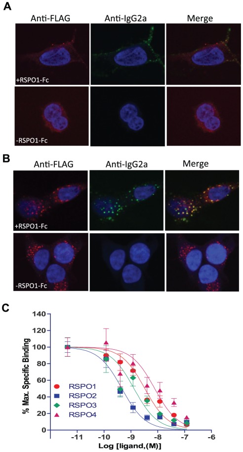
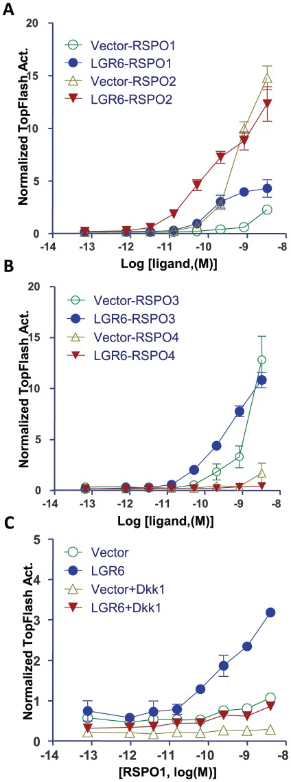
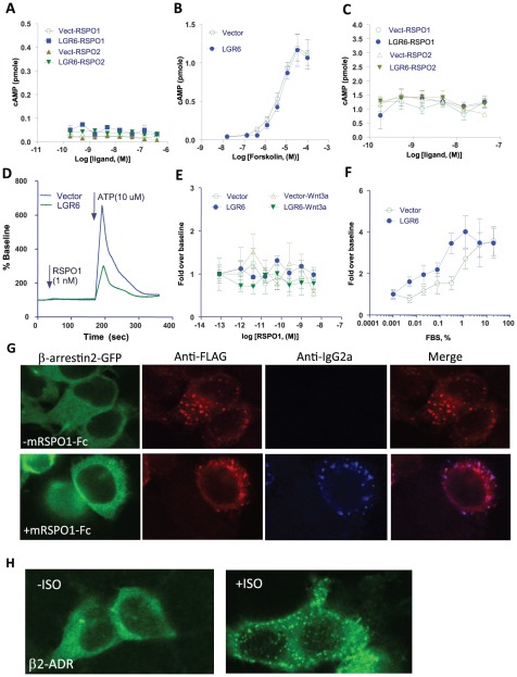
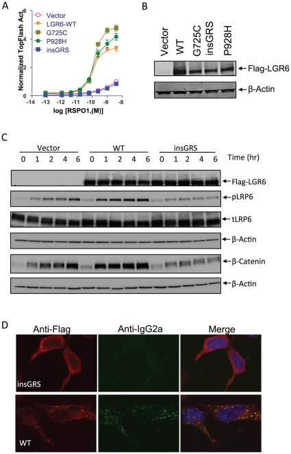
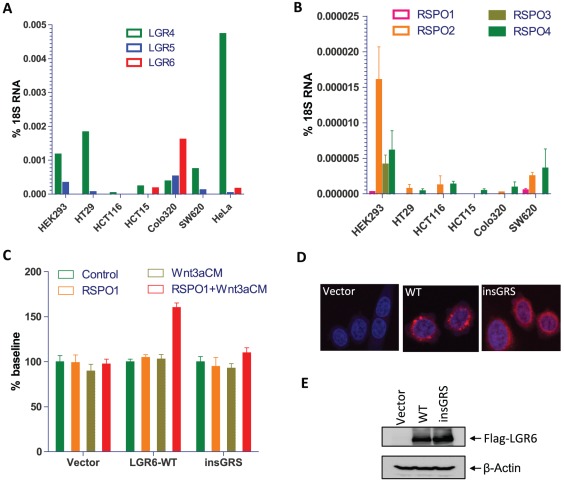
BACKGROUND: LGR6 (leucine-rich repeat containing, G protein-coupled receptor 6) is a member of the rhodopsin-like seven transmembrane domain receptor superfamily with the highest homology to LGR4 and LGR5. LGR6 was found as one of the novel genes mutated in colon cancer through total exon sequencing and its promoter region is hypermethylated in 20-50% of colon cancer cases. In the skin, LGR6 marks a population of stem cells that can give rise to all cell lineages. Recently, we and others demonstrated that LGR4 and LGR5 function as receptors of R-spondins to potentiate Wnt/β-catenin signaling. However, the binding affinity and functional response of LGR6 to R-spondins, and the activity of colon cancer mutants of LGR6 have not been determined. PRINCIPAL FINDINGS: We found that LGR6 also binds and responds to R-spondins 1-3 with high affinity to enhance Wnt/β-catenin signaling through increased LRP6 phosphorylation. Similar to LGR4 and LGR5, LGR6 is not coupled to heterotrimeric G proteins or to β-arrestin following R-spondin stimulation. Functional and expression analysis of three somatic mutations identified in colon cancer samples indicates that one mutant fails to bind and respond to R-spondin (loss-of-function), but the other two have no significant effect on receptor function. Overexpression of wild-type LGR6 in HeLa cells leads to increased cell migration following co-treatment with R-spondin1 and Wnt3a when compared to vector control cells or cells overexpressing the loss-of-function mutant. CONCLUSIONS: LGR6 is a high affinity receptor for R-spondins 1-3 and potentially functions as a tumor suppressor despite its positive effect on Wnt/β-catenin signaling.
Overview
The claims and statistics of this paper haven’t been checked yet.
Run the check — free, no account needed. It reads the open-access text and checks the sources it cites.
Integrity signals
Registry
Cites
Statistics
Figures
Verification
Claims the paper makes
The claims and statistics of this paper haven’t been checked yet.
Run the check — free, no account needed. It reads the open-access text and checks the sources it cites.
Claims other papers make about this one
Each is checked against this paper’s own text: does it say what the citing paper says it does?
The cited source says what the claim attributes to it.
In the citing paper
“Stimulation of Lgr6 by R-spondins potentiates Wnt/-catenin signaling (Gong et al., 2012).”
From An RNA-Sequencing Transcriptome and Splicing Database of Glia, Neurons, and Vascular Cells of the Cerebral Cortex (2014) · cites this paper as Gong et al., 2012
Self-check
Percentages, sums and reported differences re-computed from the paper’s own numbers.
The self-check needs the paper’s full text.
Citations
Cites
Identification of stem cells in small intestine and colon by marker gene Lgr5
Barker, N., van Es, J. H., Kuipers, J., Kujala, P., van den Born, M., Cozijnsen, M., Haegebarth, A., Korving, J., Begthel, H., Peters, P. J., et al. (2007) · Nature · doi:10.1038/nature06196 · Cited by 5,687
Wnt/β-Catenin Signaling: Components, Mechanisms, and Diseases
MacDonald, B. T., Tamai, K., & He, X. (2009) · Developmental Cell · doi:10.1016/j.devcel.2009.06.016 · Cited by 5,583
Cancer genes and the pathways they control
Vogelstein, B., & Kinzler, K. W. (2004) · Nature Medicine · doi:10.1038/nm1087 · Cited by 4,346
The Consensus Coding Sequences of Human Breast and Colorectal Cancers
Sjöblom, T., Jones, S., Wood, L. D., Parsons, D. W., Lin, J., Barber, T. D., Mandelker, D. L., Leary, R. J., Ptak, J., Silliman, N., et al. (2006) · Science · doi:10.1126/science.1133427 · Cited by 3,161
Crystal structure of the β2 adrenergic receptor–Gs protein complex
Rasmussen, S. G. F., DeVree, B. T., Zou, Y., Kruse, A. C., Chung, K. Y., Kobilka, T. S., Thian, F. S., Chae, P. S., Pardon, E., Calinski, D., et al. (2011) · Nature · doi:10.1038/nature10361 · Cited by 3,009
Constitutive Transcriptional Activation by a β-Catenin-Tcf Complex in APC −/− Colon Carcinoma
Korinek, V., Barker, N., Morin, P. J., van Wichen, D., de Weger, R., Kinzler, K. W., Vogelstein, B., & Clevers, H. (1997) · Science · doi:10.1126/science.275.5307.1784 · Cited by 2,905
Distant metastasis occurs late during the genetic evolution of pancreatic cancer
Yachida, S., Jones, S., Bozic, I., Antal, T., Leary, R., Fu, B., Kamiyama, M., Hruban, R. H., Eshleman, J. R., Nowak, M. A., et al. (2010) · Nature · doi:10.1038/nature09515 · Cited by 2,502
COSMIC: mining complete cancer genomes in the Catalogue of Somatic Mutations in Cancer
Forbes, S. A., Bindal, N., Bamford, S., Cole, C., Kok, C. Y., Beare, D., Jia, M., Shepherd, R., Leung, K., Menzies, A., et al. (2011) · Nucleic Acids Research · doi:10.1093/nar/gkq929 · Cited by 2,278
Dickkopf-1 is a member of a new family of secreted proteins and functions in head induction
Glinka, A., Wu, W., Delius, H., Monaghan, A. P., Blumenstock, C., & Niehrs, C. (1998) · Nature · doi:10.1038/34848 · Cited by 1,608
Lgr5 homologues associate with Wnt receptors and mediate R-spondin signalling
de Lau, W., Barker, N., Low, T. Y., Koo, B., Li, V. S. W., Teunissen, H., Kujala, P., Haegebarth, A., Peters, P. J., van de Wetering, M., et al. (2011) · Nature · doi:10.1038/nature10337 · Cited by 1,302
Cited by
Sorted by citations
Zhang, Y., Chen, K., Sloan, S. A., Bennett, M. L., Scholze, A. R., O'Keeffe, S., Phatnani, H. P., Guarnieri, P., Caneda, C., Ruderisch, N., et al. (2014) · The Journal of Neuroscience · doi:10.1523/jneurosci.1860-14.2014 · Cited by 4,875
“Stimulation of Lgr6 by R-spondins potentiates Wnt/ -catenin signaling (Gong et al., 2012).”
BackgroundDavenport, A. P., Alexander, S. P. H., Sharman, J. L., Pawson, A. J., Benson, H. E., Monaghan, A. E., Liew, W. C., Mpamhanga, C. P., Bonner, T. I., Neubig, R. R., et al. (2013) · Pharmacological Reviews · doi:10.1124/pr.112.007179 · Cited by 329
Greicius, G., Kabiri, Z., Sigmundsson, K., Liang, C., Bunte, R., Singh, M. K., & Virshup, D. M. (2018) · Proceedings of the National Academy of Sciences · doi:10.1073/pnas.1713510115 · Cited by 286
Maresin 1 activates LGR6 receptor promoting phagocyte immunoresolvent functions
Chiang, N., Libreros, S., Norris, P. C., de la Rosa, X., & Serhan, C. N. (2019) · Journal of Clinical Investigation · doi:10.1172/jci129448 · Cited by 259
Organoids for Drug Discovery and Personalized Medicine
Takahashi, T. (2018) · The Annual Review of Pharmacology and Toxicology · doi:10.1146/annurev-pharmtox-010818-021108 · Cited by 210
Lgr5-Expressing Cells Are Sufficient and Necessary for Postnatal Mammary Gland Organogenesis
Plaks, V., Brenot, A., Lawson, D. A., Linnemann, J. R., van Kappel, E. C., Wong, K., de Sauvage, F. J., Klein, O. D., & Werb, Z. (2013) · Cell Reports · doi:10.1016/j.celrep.2012.12.017 · Cited by 206
Homeostasis of the sebaceous gland and mechanisms of acne pathogenesis
Clayton, R. W., Göbel, K., Niessen, C. M., Paus, R., Steensel, M. A. M. V., & Lim, X. (2019) · British Journal of Dermatology · doi:10.1111/bjd.17981 · Cited by 187
The structural basis of R-spondin recognition by LGR5 and RNF43Preprint
Chen, P., Chen, X., Lin, Z., Fang, D., & He, X. L. (2013) · Genes & Development · doi:10.1101/gad.219915.113 · Cited by 178
Wnt Signaling in Vertebrate Neural Development and Function
Mulligan, K. A., & Cheyette, B. N. R. (2012) · Journal of Neuroimmune Pharmacology · doi:10.1007/s11481-012-9404-x · Cited by 177
Wnt/β-Catenin Signaling and Obesity
Chen, N., & Wang, J. (2018) · Frontiers in Physiology · doi:10.3389/fphys.2018.00792 · Cited by 173
Targeting LGR5 in Colorectal Cancer: therapeutic gold or too plastic?
Morgan, R., Mortensson, E., & Williams, A. C. (2018) · British Journal of Cancer · doi:10.1038/s41416-018-0118-6 · Cited by 154
The R-spondin family of proteins: Emerging regulators of WNT signaling
Jin, Y.-R., & Yoon, J. K. (2012) · The International Journal of Biochemistry & Cell Biology · doi:10.1016/j.biocel.2012.09.006 · Cited by 138
Structural basis for R-spondin recognition by LGR4/5/6 receptorsPreprint
Wang, D., Huang, B., Zhang, S., Yu, X., Wu, W., & Wang, X. (2013) · Genes & Development · doi:10.1101/gad.219360.113 · Cited by 135
Neuroendocrinology and neurobiology of sebaceous glands
Clayton, R. W., Langan, E. A., Ansell, D. M., de Vos, I. J. H. M., Göbel, K., Schneider, M. R., Picardo, M., Lim, X., Steensel, M. A. M. V., & Paus, R. (2020) · Biological reviews/Biological reviews of the Cambridge Philosophical Society · doi:10.1111/brv.12579 · Cited by 126
Scopelliti, A., Cordero, J. B., Diao, F., Strathdee, K. E., White, B. H., Sansom, O. J., & Vidal, M. (2014) · Current Biology · doi:10.1016/j.cub.2014.04.007 · Cited by 124
Mammary Development and Breast Cancer: A Wnt Perspective
Yu, Q. C., Verheyen, E. M., & Zeng, Y. A. (2016) · Cancers · doi:10.3390/cancers8070065 · Cited by 122
Novel Insights into the Bovine Polled Phenotype and Horn Ontogenesis in Bovidae
Allais‐Bonnet, A., Grohs, C., Međugorac, I., Krebs, S., Djari, A., Graf, A., Fritz, S., Seichter, D., Baur, A., Ruß, I., et al. (2013) · PLoS ONE · doi:10.1371/journal.pone.0063512 · Cited by 116
Lgr6 marks nail stem cells and is required for digit tip regeneration
Lehoczky, J. A., & Tabin, C. J. (2015) · Proceedings of the National Academy of Sciences · doi:10.1073/pnas.1518874112 · Cited by 116
R-spondin1 is a novel hormone mediator for mammary stem cell self-renewalPreprint
Cai, C., Yu, Q. C., Jiang, W., Liu, W., Song, W., Yu, H., Zhang, L., Yang, Y., & Zeng, Y. A. (2014) · Genes & Development · doi:10.1101/gad.245142.114 · Cited by 100
Stem Cell Signaling Pathways in the Small Intestine
Takahashi, T., & Shiraishi, A. (2020) · International Journal of Molecular Sciences · doi:10.3390/ijms21062032 · Cited by 83
Frizzleds as GPCRs – More Conventional Than We Thought!
Schulte, G., & Wright, S. C. (2018) · Trends in Pharmacological Sciences · doi:10.1016/j.tips.2018.07.001 · Cited by 83
Carmon, K. S., Gong, X. W., Yi, J., Wu, L., Thomas, A., Moore, C. M., Masuho, I., Timson, D. J., Martemyanov, K. A., & Liu, Q. J. (2017) · Journal of Biological Chemistry · doi:10.1074/jbc.m117.786798 · Cited by 73
Self-citationHGF/R-spondin1 rescues liver dysfunction through the induction of Lgr5+ liver stem cells
Yuan, L., Fang, Z., Liu, H., Wang, L., Cheng, Z., Tang, N., Li, T., Liu, T., Han, H.-X., Cao, G., et al. (2017) · Nature Communications · doi:10.1038/s41467-017-01341-6 · Cited by 67
Analysis of LGR4 Receptor Distribution in Human and Mouse Tissues
Yi, J., Xiong, W., Gong, X. W., Bellister, S. A., Ellis, L. M., & Liu, Q. (2013) · PLoS ONE · doi:10.1371/journal.pone.0078144 · Cited by 65
Self-citationLi, X., Larsson, P. G., Ljuslinder, I., Öhlund, D., Myte, R., Löfgren‐Burström, A., Zingmark, C., Ling, A., Edin, S., & Palmqvist, R. (2020) · Cancers · doi:10.3390/cancers12040923 · Cited by 64
R-spondins engage heparan sulfate proteoglycans to potentiate WNT signaling
Dubey, R., van Kerkhof, P., Jordens, I., Malinauskas, T., Pusapati, G. V., McKenna, J. K., Li, D., Carette, J. E., Ho, M., Siebold, C., et al. (2020) · eLife · doi:10.7554/elife.54469 · Cited by 64
Snyder, J. C., Rochelle, L. K., Lyerly, H. K., Caron, M. G., & Barak, L. S. (2013) · Journal of Biological Chemistry · doi:10.1074/jbc.m112.447540 · Cited by 62
Lgr6 is a stem cell marker in mouse skin squamous cell carcinoma
Huang, P., Kandyba, E. E., Jabouille, A., Sjölund, J., Kumar, A., Halliwill, K. D., McCreery, M. Q., Delrosario, R., Kang, H. C., Wong, C. E., et al. (2017) · Nature Genetics · doi:10.1038/ng.3957 · Cited by 59
BMP9 Reduces Bone Loss in Ovariectomized Mice by Dual Regulation of Bone Remodeling
Zhou, Y., Yang, Y., Jing, Y., Yuan, T.-J., Sun, L., Tao, B., Liu, J., & Zhao, H. (2020) · Journal of Bone and Mineral Research · doi:10.1002/jbmr.3957 · Cited by 59
Mismatch Repair Proteins Initiate Epigenetic Alterations during Inflammation-Driven Tumorigenesis
Maiuri, A. R., Peng, M., Podicheti, R., Sriramkumar, S., Kamplain, C. M., Rusch, D. B., Shields, C. E. D., Sears, C. L., & O’Hagan, H. M. (2017) · Cancer Research · doi:10.1158/0008-5472.can-17-0056 · Cited by 57
Ruan, X., Liu, A., Zhong, M., Wei, J., Zhang, W., Rong, Y., Liu, W., Li, M., Qing, X., Chen, G., et al. (2019) · Molecular Therapy — Oncolytics · doi:10.1016/j.omto.2019.04.002 · Cited by 56
Self-citationFukuma, M., Tanese, K., Effendi, K., Yamazaki, K., Masugi, Y., Suda, M., & Sakamoto, M. (2012) · Experimental Cell Research · doi:10.1016/j.yexcr.2012.10.011 · Cited by 52
Exploring the Wnt Pathway as a Therapeutic Target for Prostate Cancer
Koushyar, S., Méniel, V. S., Phesse, T. J., & Pearson, H. B. (2022) · Biomolecules · doi:10.3390/biom12020309 · Cited by 48
R-spondin signaling as a pivotal regulator of tissue development and homeostasis
Nagano, K. (2019) · Japanese Dental Science Review · doi:10.1016/j.jdsr.2019.03.001 · Cited by 47
Maresin-1 resolution with RORα and LGR6
Im, D. (2020) · Progress in Lipid Research · doi:10.1016/j.plipres.2020.101034 · Cited by 47
LGR5 is Expressed by Ewing Sarcoma and Potentiates Wnt/β-Catenin Signaling
Scannell, C. A., Pedersen, E. A., Mosher, J. T., Krook, M. A., Nicholls, L. A., Wilky, B. A., Loeb, D. M., & Lawlor, E. R. (2013) · Frontiers in Oncology · doi:10.3389/fonc.2013.00081 · Cited by 46
Hunting for the function of orphan GPCRs – beyond the search for the endogenous ligand
Ahmad, R., Wojciech, S., & Jockers, R. (2014) · British Journal of Pharmacology · doi:10.1111/bph.12942 · Cited by 45
Epidermal expression of L gr6 is dependent on nerve endings and S chwann cells
Liao, X., & Nguyen, H. (2014) · Experimental Dermatology · doi:10.1111/exd.12340 · Cited by 44
LGR4: A New Receptor Member in Endocrine and Metabolic Diseases
Zhang, N., Yuan, M., & Wang, J. (2023) · Endocrine Reviews · doi:10.1210/endrev/bnad003 · Cited by 43
LGR6 promotes osteogenesis by activating the Wnt/β-catenin signaling pathway
Liu, S.-L., Zhou, Y., Tang, D.-B., Zhou, N., Zheng, W., Tang, Z.-H., Duan, C., Zheng, L., & Chen, J. (2019) · Biochemical and Biophysical Research Communications · doi:10.1016/j.bbrc.2019.08.122 · Cited by 42
Etet, P. F. S., Vecchio, L., Kamga, P. B., Nukenine, E. N., Krampera, M., & Kamdje, A. H. N. (2012) · Biochimica et Biophysica Acta (BBA) - Reviews on Cancer · doi:10.1016/j.bbcan.2012.08.002 · Cited by 40
Novel Regulation of Wnt Signaling at the Proximal Membrane Level
Jiang, X., & Cong, F. (2016) · Trends in Biochemical Sciences · doi:10.1016/j.tibs.2016.06.003 · Cited by 36
Wnt and Vitamin D at the Crossroads in Solid Cancer
González‐Sancho, J. M., Larriba, M. J., & Múñoz, A. (2020) · Cancers · doi:10.3390/cancers12113434 · Cited by 35
Kong, Y., Ou, X., Li, X., Zeng, Y., Gao, G., Lyu, N., & Liu, P. (2020) · Molecular Therapy — Oncolytics · doi:10.1016/j.omto.2020.06.020 · Cited by 34
Geng, A., Wu, T. H., Cai, C., Song, W., Wang, J., Yu, Q. C., & Zeng, Y. A. (2020) · eLife · doi:10.7554/elife.56434 · Cited by 34
Identification of the Prognostic Signatures of Glioma With Different PTEN Status
Zhang, P., Meng, X., Liu, L., Li, S., Li, Y., Ali, S., Li, S., Xiong, J., Liu, X., Li, S., et al. (2021) · Frontiers in Oncology · doi:10.3389/fonc.2021.633357 · Cited by 32
Maresin 1 Alleviates Diabetic Kidney Disease via LGR6‐Mediated cAMP‐SOD2‐ROS Pathway
Li, X., Xu, B.-T., Wu, J. T., Pu, Y., Wan, S., Zeng, Y., Wang, M., Luo, L., Zhang, F., Jiang, Z., et al. (2022) · Oxidative Medicine and Cellular Longevity · doi:10.1155/2022/7177889 · Cited by 32
Liu, Q., Zhao, Y., Xing, H., Li, L., Li, R., Dai, J., Li, Q., & Fang, S. (2018) · Gene · doi:10.1016/j.gene.2018.11.098 · Cited by 31
Self-citationDevelopment and Maintenance of Epidermal Stem Cells in Skin Adnexa
Mokrý, J., & Pisal, R. V. (2020) · International Journal of Molecular Sciences · doi:10.3390/ijms21249736 · Cited by 30
Roles of nAChR and Wnt signaling in intestinal stem cell function and inflammation
Takahashi, T. (2020) · International Immunopharmacology · doi:10.1016/j.intimp.2020.106260 · Cited by 28
Mouse LGR6 regulates osteogenesis in vitro and in vivo through differential ligand use
Khedgikar, V., Charles, J. F., & Lehoczky, J. A. (2021) · Bone · doi:10.1016/j.bone.2021.116267 · Cited by 28
Cheverud, J. M., Lawson, H. A., Bouckaert, K., Kossenkov, A. V., Showe, L. C., Cort, L., Blankenhorn, E. P., Bedelbaeva, K., Gourevitch, D., Zhang, Y., et al. (2014) · Heredity · doi:10.1038/hdy.2013.133 · Cited by 27
LGR6 promotes the progression of gastric cancer through PI3K/AKT/mTOR pathway
Ke, J., Ma, P., Chen, J., Qin, J., & Qian, H. (2018) · OncoTargets and Therapy · doi:10.2147/ott.s149303 · Cited by 27
Macyczko, J. R., Wang, N., Zhao, J., Ren, Y., Lü, W., Ikezu, T. C., Zhao, N., Liu, C., Bu, G., & Li, Y. (2022) · Molecular Neurobiology · doi:10.1007/s12035-022-03065-1 · Cited by 25
Xu, J., Sun, T., & Hu, X. (2015) · Molecular Medicine Reports · doi:10.3892/mmr.2015.3913 · Cited by 24
R-Spondin-2 Is Upregulated in Idiopathic Pulmonary Fibrosis and Affects Fibroblast Behavior
Munguía-Reyes, A., Balderas-Martínez, Y. I., Becerril, C., Checa, M., Ramı́rez, R., Ortiz, B., Meléndez-Zajgla, J., Pardo, A., & Selman, M. (2018) · American Journal of Respiratory Cell and Molecular Biology · doi:10.1165/rcmb.2017-0115oc · Cited by 24
Established and In-trial GPCR Families in Clinical Trials: A Review for Target Selection
Saikia, S., Bordoloi, M., & Sarmah, R. (2018) · Current Drug Targets · doi:10.2174/1389450120666181105152439 · Cited by 23
Tu, M., Wei, T., Jia, Y., Wang, Y., & Wu, J. (2022) · Journal of Thoracic Disease · doi:10.21037/jtd-22-886 · Cited by 22
WNT11, a new gene associated with early onset osteoporosis, is required for osteoblastogenesis
da Silva, C. C., Édouard, T., Fradin, M., Aubert‐Mucca, M., Ricquebourg, M., Raman, R., Salles, J., Charon, V., Guggenbuhl, P., Müller, M., et al. (2021) · Human Molecular Genetics · doi:10.1093/hmg/ddab349 · Cited by 21
Wnt-associated adult stem cell marker Lgr6 is required for osteogenesis and fracture healing
Doherty, L., Wan, M., Peterson, A., Youngstrom, D. W., King, J. S., Kalajzić, I., Hankenson, K. D., & Sanjay, A. (2022) · Bone · doi:10.1016/j.bone.2023.116681 · Cited by 21
In a Class of Their Own – RXFP1 and RXFP2 are Unique Members of the LGR Family
Petrie, E. J., Lagaida, S., Sethi, A., Bathgate, R. A. D., & Gooley, P. R. (2015) · Frontiers in Endocrinology · doi:10.3389/fendo.2015.00137 · Cited by 21
DNA methyltransferase inhibition reduces inflammation-induced colon tumorigenesis
Maiuri, A. R., Savant, S. S., Podicheti, R., Rusch, D. B., & O’Hagan, H. M. (2019) · Epigenetics · doi:10.1080/15592294.2019.1634986 · Cited by 21
Amphiregulin mediates the hormonal regulation on Rspondin-1 expression in the mammary gland
Cai, C., Geng, A., Wang, M., Yang, L. C., Yu, Q. C., & Zeng, Y. A. (2019) · Developmental Biology · doi:10.1016/j.ydbio.2019.10.006 · Cited by 20
Evidence for Lgr6 as a Novel Marker of Osteoblastic Progenitors in Mice
Khedgikar, V., & Lehoczky, J. A. (2018) · JBMR Plus · doi:10.1002/jbm4.10075 · Cited by 20
LGRs in Skeletal Tissues: An Emerging Role for Wnt‐Associated Adult Stem Cell Markers in Bone
Doherty, L., & Sanjay, A. (2020) · JBMR Plus · doi:10.1002/jbm4.10380 · Cited by 20
LGR5 constitutively activates NF‐κB signaling to regulate the growth of intestinal crypts
Lai, S., Cheng, R., Gao, D., Chen, Y., & Deng, C. (2020) · The FASEB Journal · doi:10.1096/fj.202001329r · Cited by 20
Nasr, T., Holderbaum, A. M., Chaturvedi, P., Agarwal, K., Kinney, J. L., Daniels, K., Trisno, S. L., Ustiyan, V., Shannon, J. M., Wells, J. M., et al. (2020) · Disease Models & Mechanisms · doi:10.1242/dmm.046573 · Cited by 20
Down-regulation of LGR6 promotes bone fracture recovery using bone marrow stromal cells
Cui, Y., Huang, R.-C., Wang, Y., Zhu, L., & Zhang, X. (2018) · Biomedicine & Pharmacotherapy · doi:10.1016/j.biopha.2017.12.109 · Cited by 20
Multiple Roles for Cholinergic Signaling from the Perspective of Stem Cell Function
Takahashi, T. (2021) · International Journal of Molecular Sciences · doi:10.3390/ijms22020666 · Cited by 19
Rogers, M. A., Kalter, V., Marcias, G., Zapatka, M., Barbus, S., & Lichter, P. (2015) · Journal of Cancer Research and Clinical Oncology · doi:10.1007/s00432-015-2011-5 · Cited by 19
The Lgr5 transgene is expressed specifically in glycinergic amacrine cells in the mouse retina
Sukhdeo, K., Koch, C. E., Miller, T. E., Zhou, H., Rivera, M., Yan, K. K.-L., Cepko, C. L., Lathia, J. D., & Rich, J. N. (2013) · Experimental Eye Research · doi:10.1016/j.exer.2013.11.003 · Cited by 18
Wang, F., Dai, C., Zhang, L., Bing, C., Qin, J., & Liu, Y. (2018) · International Journal of Molecular Medicine · doi:10.3892/ijmm.2018.3633 · Cited by 18
Molecular determinants of WNT9b responsiveness in nephron progenitor cells
Dickinson, K., Hammond, L. C., Karner, C. M., Hastie, N. D., Carroll, T. J., & Goodyer, P. (2019) · PLoS ONE · doi:10.1371/journal.pone.0215139 · Cited by 18
Lehoczky, J. A., & Tabin, C. J. (2018) · Nature · doi:10.1038/d41586-018-04820-y · Cited by 18
Di, D., Li, C., Dai, Y., Wei, M., Wang, S., Song, W., Zhou, H., Cui, Y., Zhang, R., Huang, Q., et al. (2021) · Frontiers in Microbiology · doi:10.3389/fmicb.2021.765008 · Cited by 17
Association of genetic variants in migraineurs with and without restless legs syndrome
Lin, G., Lin, Y., Liang, C., Lee, J., Tsai, C., Hung, K., Luo, W., Tsai, C., Hsu, Y., Ho, T., et al. (2020) · Annals of Clinical and Translational Neurology · doi:10.1002/acn3.51186 · Cited by 16
A novel autoantibody test for the detection of pre-neoplastic lung lesions
Lowe, F., Shen, W., Zu, J., Li, J., Wang, H., Zhang, X., & Zhong, L. (2014) · Molecular Cancer · doi:10.1186/1476-4598-13-78 · Cited by 16
Cortesi, E. E., Meeusen, B., Vanstapel, A., Verleden, S. E., Vanaudenaerde, B. M., Wuyts, W. A., Janssens, W., Janssens, V., Roskams, T., & Ventura, J. (2021) · Cells · doi:10.3390/cells10123437 · Cited by 16
Snyder, J. C., Rochelle, L. K., Barak, L. S., & Caron, M. G. (2013) · PLoS ONE · doi:10.1371/journal.pone.0084476 · Cited by 16
Discovery of polypeptide ligand‐receptor pairs based on their co‐evolution
Hsueh, A. J. W., & Feng, Y. (2020) · The FASEB Journal · doi:10.1096/fj.202000779r · Cited by 15
Digit Tip Injuries: Current Treatment and Future Regenerative Paradigms
Miller, T. J., Deptula, P. L., Buncke, G. M., & Maan, Z. N. (2019) · Stem Cells International · doi:10.1155/2019/9619080 · Cited by 15
Characterizing the cellular attachment receptor for Langat virus
Rodrigues, R., Danskog, K., Överby, A. K., & Arnberg, N. (2019) · PLoS ONE · doi:10.1371/journal.pone.0217359 · Cited by 14
Cui, J., Toh, Y., Park, S.-H., Yu, W., Tu, J., Wu, L., Li, L., Jacob, J., Pan, S., Carmon, K. S., et al. (2021) · Journal of Medicinal Chemistry · doi:10.1021/acs.jmedchem.1c00395 · Cited by 14
Self-citationIntrinsic disorder in spondins and some of their interacting partners
Alowolodu, O., Johnson, G., Alashwal, L., Addou, I., Zhdanova, I. V., & Uversky, V. N. (2016) · Intrinsically Disordered Proteins · doi:10.1080/21690707.2016.1255295 · Cited by 13
Class A Orphans (version 2019.5) in the IUPHAR/BPS Guide to Pharmacology Database
Alexander, S. P. H., Battey, J., Benson, H. E., Benya, R. V., Bonner, T. I., Davenport, A. P., Eguchi, S., Harmar, A. J., Holliday, N. D., Jensen, R. T., et al. (2019) · IUPHAR/BPS Guide to Pharmacology CITE · doi:10.2218/gtopdb/f16/2019.5 · Cited by 12
An "R‐spondin code" for multimodal signaling ON‐OFF states
Niehrs, C., Seidl, C., & Lee, H. (2024) · BioEssays · doi:10.1002/bies.202400144 · Cited by 10
LGR5 enhances the osteoblastic differentiation of MC3T3‑E1 cells through the Wnt/β‑catenin pathway
Yu, W. H., Xie, C., Chen, F., Cheng, P. F., Yang, L., & Pan, X. (2021) · Experimental and Therapeutic Medicine · doi:10.3892/etm.2021.10321 · Cited by 10
Impaired Mitochondria Promote Aging-Associated Sebaceous Gland Dysfunction and Pathology
Ahmed, N. S., Foote, J. B., & Singh, K. K. (2022) · American Journal Of Pathology · doi:10.1016/j.ajpath.2022.07.006 · Cited by 10
Overexpression of NT-3 in the hippocampus suppresses the early phase of the adult neurogenic process
Kasakura, N., Murata, Y., Shindo, A., Kitaoka, S., Furuyashiki, T., Suzuki, K., & Segi‐Nishida, E. (2023) · Frontiers in Neuroscience · doi:10.3389/fnins.2023.1178555 · Cited by 9
Tu, J., Toh, Y., Aldana, A. M., Wen, J. J., Wu, L., Jacob, J., Li, L., Pan, S., Carmon, K. S., & Liu, Q. J. (2024) · Pharmaceutics · doi:10.3390/pharmaceutics16070943 · Cited by 9
Self-citationvan de Glind, G. C., Rebel, H. G., Out‐Luiting, J. J., Zoutman, W. H., Tensen, C. P., & de Gruijl, F. R. (2016) · Oncotarget · doi:10.18632/oncotarget.13436 · Cited by 9
Class A Orphans (version 2020.5) in the IUPHAR/BPS Guide to Pharmacology Database
Alexander, S. P. H., Battey, J., Benson, H. E., Benya, R. V., Bonner, T. I., Davenport, A. P., Singh, K. D., Eguchi, S., Harmar, A. J., Holliday, N. D., et al. (2020) · IUPHAR/BPS Guide to Pharmacology CITE · doi:10.2218/gtopdb/f16/2020.5 · Cited by 8
G protein-coupled receptor LGR6 is an independent risk factor for colon adenocarcinoma
Wang, W., Ding, S., Hejun, Z., Li, J., Zhang, H., & Zhang, H. (2018) · Frontiers of Medicine · doi:10.1007/s11684-018-0633-0 · Cited by 8
LGR4 is essential for R-spondin1-mediated suppression of food intake via pro-opiomelanocortin
Otsuka, A., Jinguji, A., Maejima, Y., Kasahara, Y., Shimomura, K., Hidema, S., & Nishimori, K. (2019) · Bioscience Biotechnology and Biochemistry · doi:10.1080/09168451.2019.1591266 · Cited by 8
Tackling the complexities of orphan GPCR ligand discovery with rationally assisted approaches
So, S. S., Ngo, T., Keov, P., Smith, N. J., & Kufareva, I. (2019) · Elsevier eBooks · doi:10.1016/b978-0-12-816228-6.00016-7 · Cited by 9
R-spondin-1 induces Axin degradation via the LRP6-CK1ε axis
Tan, L., Yan, M., Su, Z., Wang, H., Li, H., Zhao, X., Liu, S., Zhang, L., Sun, Q., & Lu, D. (2024) · Cell Communication and Signaling · doi:10.1186/s12964-023-01456-y · Cited by 7
Eltorky, H. A. E., AbdelMageed, M. A., Ismail, H. T. H., Zahran, F. M., Guirgis, A. A., Olsson, L., Lindmark, G. E., Hammarström, M., Hammarström, S., & Sitohy, B. (2024) · Frontiers in Oncology · doi:10.3389/fonc.2024.1393075 · Cited by 7
Class A Orphans in GtoPdb v.2023.1
Alexander, S. P. H., Battey, J., Benson, H. E., Benya, R. V., Bonner, T. I., Davenport, A. P., Singh, K. D., Eguchi, S., Harmar, A. J., Holliday, N. D., et al. (2023) · IUPHAR/BPS Guide to Pharmacology CITE · doi:10.2218/gtopdb/f16/2023.1 · Cited by 6
From genome‐wide association to biological explanation
Steensel, M. A. M. V. (2019) · British Journal of Dermatology · doi:10.1111/bjd.18462 · Cited by 6
WNT enhancing signals in pancreatic cancer are transmitted by LGR6
Wang, J., Koch, D. T., Hofmann, F. O., Härtwig, D., Beirith, I., Janssen, K., Bazhin, A. V., Nieß, H., Werner, J., Renz, B. W., et al. (2023) · Aging · doi:10.18632/aging.205101 · Cited by 5
Metadata
- Authors
- Gong, X., Carmon, K. S., Lin, Q., Thomas, A., Yi, J., & Liu, Q.
- Affiliations
- The University of Texas Health Science Center at Houston (US)
- Corresponding author
- Liu, Qingyun
- Venue
- PLoS ONE
- Publisher
- Public Library of Science (PLoS)
- Published
- 2012-05-17
- Volume
- 7
- Issue
- 5
- Pages
- e37137
- ISSN
- 1932-6203 (ISSN-L)
- PMC
- PMC3355120
- OpenAlex
- W2110651281
- Cited by
- 129
- References
- 61
- Citations by year
- 2026: 4 · 2025: 3 · 2024: 6 · 2023: 8 · 2022: 10 · 2021: 11 · 2020: 18 · 2019: 16 · 2018: 14 · 2017: 5 · 2016: 6 · 2015: 4 · 2014: 8 · 2013: 12 · 2012: 4
- FWCI
- 3.60
- Type
- journal article
- Language
- en
- Open access
- gold
- doi.org
- Locations
- PLoS ONE (publishedVersion, cc-by)
- Europe PMC (PubMed Central) (publishedVersion, cc-by)
- PubMed Central (publishedVersion, cc-by)
- PubMed (publishedVersion)
- DOAJ (DOAJ: Directory of Open Access Journals) (submittedVersion, cc-by-sa)
- Funders
- Cancer Prevention and Research Institute of Texas (RP100678)
- Topics
- Life Sciences › Biochemistry, Genetics and Molecular Biology › Molecular Biology › Wnt/β-catenin signaling in development and cancer
- Life Sciences › Biochemistry, Genetics and Molecular Biology › Molecular Biology › Receptor Mechanisms and Signaling
- Health Sciences › Medicine › Oncology › Colorectal Cancer Treatments and Studies
- Keywords
- LGR6, R-spondin, Wnt/β-catenin signaling, LRP6 phosphorylation, colon cancer, tumor suppressor, loss-of-function mutations, cell migration, Wnt3a, G protein-coupled receptors, skin stem cells, promoter hypermethylation
- MeSH terms
- Cell Movement, Colonic Neoplasms, HeLa Cells, Humans, Mutation, Protein Binding, Signal Transduction, GTP-Binding Proteins, Thrombospondins, Tumor Suppressor Proteins, Intercellular Signaling Peptides and Proteins, Receptors, G-Protein-Coupled, Wnt Proteins, beta Catenin
About these checks
Registry, retraction, Cites and statistics results are computed, not judged, with one exception: a printed reference that no index lists is read once by a model before it counts as a citation error. Claims are extracted by a model and kept only when their quote appears verbatim in the text; each cited source is then checked, with a model’s help, for whether it says what the claim relies on. Those verdicts can be wrong — read the claim’s quote and the cited source before relying on one.
Statistics and arithmetic are recomputed, not judged.
Confidence bands are shown only for fields calibrated on our annotated test set. None is calibrated yet, so no band is shown.
What counts as a claim ›How we measure confidence ›How we check this ›
Are you an author, or do you see a problem with this page? Request a review
