

The DOI matches its Crossref record; 54 of the 55 references it cites were found in a registry or an index.
M. tuberculosis Induces Potent Activation of IDO-1, but This Is Not Essential for the Immunological Control of Infection
Open accessAbstract
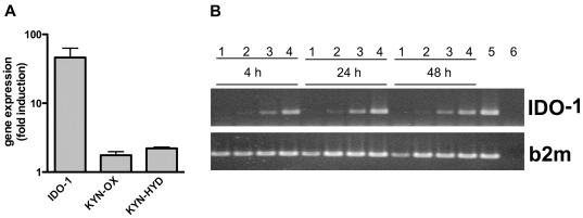
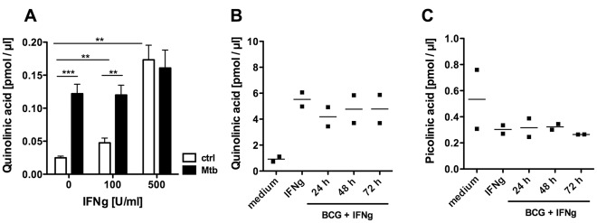
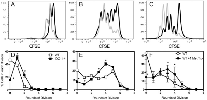
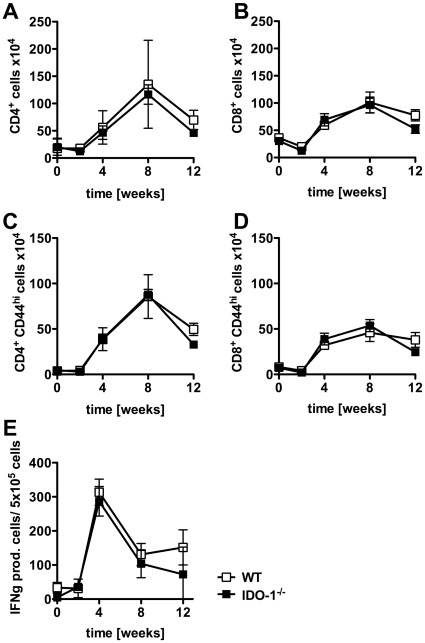
Indoleamine 2,3-dioxygenesae-1 (IDO-1) catalyses the initial, rate-limiting step in tryptophan metabolism, thereby regulating tryptophan availability and the formation of downstream metabolites, including picolinic and quinolinic acid. We found that Mycobacterium tuberculosis infection induced marked upregulation of IDO-1 expression in both human and murine macrophages in vitro and in the lungs of mice following aerosol challenge with M. tuberculosis. The absence of IDO-1 in dendritic cells enhanced the activation of mycobacteria-specific T cells in vitro. Interestingly, IDO-1-deficiency during M. tuberculosis infection in mice was not associated with altered mycobacteria-specific T cell responses in vivo. The bacterial burden of infected organs, pulmonary inflammatory responses, and survival were also comparable in M. tuberculosis-infected IDO-1 deficient and wild type animals. Tryptophan is metabolised into either picolinic acid or quinolinic acid, but only picolinic acid inhibited the growth of M. tuberculosis in vitro. By contrast macrophages infected with pathogenic mycobacteria, produced quinolinic, rather than picolinic acid, which did not reduce M. tuberculosis growth in vitro. Therefore, although M. tuberculosis induces robust expression of IDO-1 and activation of tryptophan metabolism, IDO-1-deficiency fails to impact on the immune control and the outcome of the infection in the mouse model of tuberculosis.
Overview
The claims and statistics of this paper haven’t been checked yet.
Run the check — free, no account needed. It reads the open-access text and checks the sources it cites.
Integrity signals
Registry
Cites
Statistics
Figures
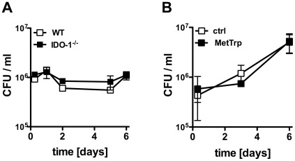
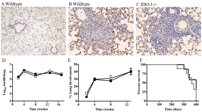
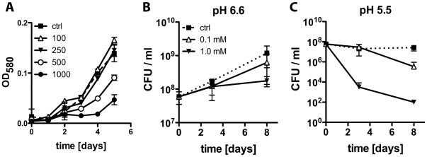
Verification
Claims the paper makes
The claims and statistics of this paper haven’t been checked yet.
Run the check — free, no account needed. It reads the open-access text and checks the sources it cites.
Claims other papers make about this one
Each is checked against this paper’s own text: does it say what the citing paper says it does?
In the citing paper
“However, in the setting of M. tuberculosis infection, IDO activation appears to be ineffective in controlling the infection.”
From Macrophage defense mechanisms against intracellular bacteria (2015) · cites this paper as [96]
In the citing paper
“IDO deficient mice were able to control M. tuberculosis infection as good as wildtype animals and lack of IDO did not affect mycobacterial specific T-cell responses (96).”
From Macrophage defense mechanisms against intracellular bacteria (2015) · cites this paper as [96]
In the citing paper
“Tryptophan is essential for M. tuberculosis growth, however, IDO deficient mice were able to control M. tuberculosis infection as good as wildtype animals and lack of IDO did not affect mycobacterial specific T-cell responses (96).”
From Macrophage defense mechanisms against intracellular bacteria (2015) · cites this paper as [96]
Self-check
Percentages, sums and reported differences re-computed from the paper’s own numbers.
The self-check needs the paper’s full text.
Citations
Cites
Prevention of Allogeneic Fetal Rejection by Tryptophan Catabolism
Munn, D. H., Zhou, M., Attwood, J. T., Bondarev, I. E., Conway, S. J., Marshall, B., Brown, C. C., & Mellor, A. L. (1998) · Science · doi:10.1126/science.281.5380.1191 · Cited by 2,589
An essential role for interferon gamma in resistance to Mycobacterium tuberculosis infection.
Flynn, J. L., Chan, J., Triebold, K. J., Dalton, D. K., Stewart, T. A., & Bloom, B. R. (1993) · The Journal of Experimental Medicine · doi:10.1084/jem.178.6.2249 · Cited by 2,468
Disseminated tuberculosis in interferon gamma gene-disrupted mice.
Cooper, A. M., Dalton, D. K., Stewart, T. A., Griffin, J. P., Russell, D. G., & Orme, I. M. (1993) · The Journal of Experimental Medicine · doi:10.1084/jem.178.6.2243 · Cited by 1,987
Inhibition of T Cell Proliferation by Macrophage Tryptophan Catabolism
Munn, D. H., Shafizadeh, E., Attwood, J. T., Bondarev, I. E., Pashine, A., & Mellor, A. L. (1999) · The Journal of Experimental Medicine · doi:10.1084/jem.189.9.1363 · Cited by 1,640
Frumento, G., Rotondo, R., Tonetti, M., Damonte, G., Benatti, U., & Ferrara, G. B. (2002) · The Journal of Experimental Medicine · doi:10.1084/jem.20020121 · Cited by 1,133
Potential Regulatory Function of Human Dendritic Cells Expressing Indoleamine 2,3-Dioxygenase
Munn, D. H., Sharma, M. D., Lee, J. R., Jhaver, K. G., Johnson, T. S., Keskin, D. B., Marshall, B., Chandler, P., Antonia, S. J., Burgess, R. E., et al. (2002) · Science · doi:10.1126/science.1073514 · Cited by 1,028
Genetic Dissection of Immunity to Mycobacteria: The Human Model
Casanova, J., & Abel, L. (2002) · Annual Review of Immunology · doi:10.1146/annurev.immunol.20.081501.125851 · Cited by 1,006
Pfefferkorn, E. (1984) · Proceedings of the National Academy of Sciences of the United States of America · doi:10.1073/pnas.81.3.908 · Cited by 870
Monitoring tryptophan metabolism in chronic immune activation
Schröcksnadel, K., Wirleitner, B., Winkler, C., & Fuchs, D. (2006) · Clinica Chimica Acta · doi:10.1016/j.cca.2005.06.013 · Cited by 551 · cited 1×
“IDO-2 remained unchanged during Plasmodium berghei ANKA infection [46] and TDO activity has also not been associated with immunerelated tryptophan metabolism [47].”
BackgroundFavre, D., Mold, J. E., Hunt, P. W., Kanwar, B., Loke, P., Seu, L., Barbour, J. D., Lowe, M. M., Jayawardene, A., Aweeka, F. T., et al. (2010) · Science Translational Medicine · doi:10.1126/scitranslmed.3000632 · Cited by 545
Cited by
Sorted by citations
Macrophage defense mechanisms against intracellular bacteria
Weiss, G., & Schaible, U. E. (2015) · Immunological Reviews · doi:10.1111/imr.12266 · Cited by 989
Tryptophan and indole metabolism in immune regulation
Fiore, A., & Murray, P. J. (2021) · Current Opinion in Immunology · doi:10.1016/j.coi.2020.12.001 · Cited by 269
Role of indoleamine 2,3-dioxygenase in health and disease
Yeung, A. W. S., Terentis, A. C., King, N. J. C., & Thomas, S. R. (2015) · Clinical Science · doi:10.1042/cs20140392 · Cited by 244
Addressing the tuberculosis–depression syndemic to end the tuberculosis epidemic
Sweetland, A. C., Kritski, A., Oquendo, M. A., Sublette, M. E., Norcini Pala, A., Silva, L. R. B., Karpati, A., Silva, E. C., Moraes, M. O., Silva, J. R. L. E., et al. (2017) · The International Journal of Tuberculosis and Lung Disease · doi:10.5588/ijtld.16.0584 · Cited by 177
New Insights into IDO Biology in Bacterial and Viral Infections
Schmidt, S. V., & Schultze, J. L. (2014) · Frontiers in Immunology · doi:10.3389/fimmu.2014.00384 · Cited by 176
Feast or famine: the host-pathogen battle over amino acids
Zhang, Y. J., & Rubin, E. J. (2013) · Cellular Microbiology · doi:10.1111/cmi.12140 · Cited by 117
Immunometabolism within the tuberculosis granuloma: amino acids, hypoxia, and cellular respiration
Qualls, J. E., & Murray, P. J. (2015) · Seminars in Immunopathology · doi:10.1007/s00281-015-0534-0 · Cited by 151
Mehra, S., Alvarez, X. A., Didier, P. J., Doyle, L. A., Blanchard, J., Lackner, A. A., & Kaushal, D. (2012) · The Journal of Infectious Diseases · doi:10.1093/infdis/jis778 · Cited by 106
Tryptophan catabolism reflects disease activity in human tuberculosis
Collins, J. M., Siddiqa, A., Jones, D. P., Liu, K., Kempker, R. R., Nizam, A., Shah, N. S., Ismail, N. A., Ouma, S. G., Tukvadze, N., et al. (2020) · JCI Insight · doi:10.1172/jci.insight.137131 · Cited by 103
The Tuberculous Granuloma and Preexisting Immunity
Cohen, S. B., Gern, B. H., & Urdahl, K. B. (2022) · Annual Review of Immunology · doi:10.1146/annurev-immunol-093019-125148 · Cited by 105
Shon, W.-J., Lee, Y.-K., Shin, J. H., Choi, E. Y., & Shin, D.-M. (2015) · Scientific Reports · doi:10.1038/srep17305 · Cited by 98
Ying, X., Han, X., Guo, S., Xu, B., Liao, Y., Wu, Y., & Zhang, G. (2018) · Cell Death and Disease · doi:10.1038/s41419-018-0389-0 · Cited by 93
Immune mechanisms of granuloma formation in sarcoidosis and tuberculosis
Weeratunga, P. N., Möller, D. R., & Ho, L. (2024) · Journal of Clinical Investigation · doi:10.1172/jci175264 · Cited by 78
Ryndak, M. B., Singh, K. K., Peng, Z., & Laal, S. (2015) · PLoS ONE · doi:10.1371/journal.pone.0123745 · Cited by 69
Kynurenines in the Pathogenesis of Multiple Sclerosis: Therapeutic Perspectives
Biernacki, T., Sandi, D., Bencsik, K., & Vécsei, L. (2020) · Cells · doi:10.3390/cells9061564 · Cited by 68
Feng, S., Du, Y., Li, Z., Zhang, L., Feng, R.-R., & Liu, S.-Y. (2015) · Chinese Medical Journal · doi:10.4103/0366-6999.149188 · Cited by 61
Immunomodulatory Effects of Genetic Alterations Affecting the Kynurenine Pathway
Boros, F. A., & Vécsei, L. (2019) · Frontiers in Immunology · doi:10.3389/fimmu.2019.02570 · Cited by 58
Lavalett, L., Rodríguez-Cabal, H. A., Ortega, H., Sadée, W., Schlesinger, L. S., & Barrera, L. F. (2017) · Tuberculosis · doi:10.1016/j.tube.2017.08.012 · Cited by 58
Digestive physiology of spiny lobsters: implications for formulated diet development
Perera, E., & Simon, C. J. (2014) · Reviews in Aquaculture · doi:10.1111/raq.12066 · Cited by 54
Niño‐Castro, A., Abdullah, Z., Popov, A., Thabet, Y., Beyer, M., Knolle, P. A., Domann, E., Chakraborty, T., Schmidt, S. V., & Schultze, J. L. (2013) · Innate Immunity · doi:10.1177/1753425913496442 · Cited by 55
Combustion Behavior of Algal Biomass: Carbon Release, Nitrogen Release, and Char Reactivity
Lane, D. J., Ashman, P. J., Zevenhoven, M., Hupa, M. M., van Eyk, P. J., de Nys, R., Karlström, O., & Lewis, D. M. (2013) · Energy & Fuels · doi:10.1021/ef4014983 · Cited by 48
Blohmke, C. J., Darton, T. C., Jones, C., Suárez, N. M., Waddington, C. S., Angus, B., Zhou, L., Hill, J. W., Clare, S., Kane, L., et al. (2016) · The Journal of Experimental Medicine · doi:10.1084/jem.20151025 · Cited by 43
Koebele, S. V., & Bimonte‐Nelson, H. A. (2016) · Experimental Gerontology · doi:10.1016/j.exger.2016.12.011 · Cited by 49
Cell-Autonomous Effector Mechanisms against Mycobacterium tuberculosisPreprint
MacMicking, J. D. (2014) · Cold Spring Harbor Perspectives in Medicine · doi:10.1101/cshperspect.a018507 · Cited by 42
Phycoremediation of Heavy Metals Coupled with Generation of Bioenergy
Chabukdhara, M., Gupta, S. K., & Gogoi, M. (2017) · Springer eBooks · doi:10.1007/978-3-319-51010-1_9 · Cited by 41
Kaylor, M. J., & Warren, D. R. (2017) · Ecosphere · doi:10.1002/ecs2.1845 · Cited by 41
Prakapenka, A. V., Hiroi, R., Quihuis, A. M., Carson, C., Patel, S., Berns-Leone, C., Fox, C., Sirianni, R. W., & Bimonte‐Nelson, H. A. (2017) · Neurobiology of Aging · doi:10.1016/j.neurobiolaging.2017.11.015 · Cited by 46
New insight into arginine and tryptophan metabolism in macrophage activation during tuberculosis
Zhang, K., Mishra, A., & Jagannath, C. (2024) · Frontiers in Immunology · doi:10.3389/fimmu.2024.1363938 · Cited by 40
Crowther, R. R., & Qualls, J. E. (2021) · Frontiers in Immunology · doi:10.3389/fimmu.2020.628432 · Cited by 36
Singh, B., Moodley, C., Singh, D. K., Escobedo, R. A., Sharan, R., Arora, G., Ganatra, S., Shivanna, V., González, O., Hall-Ursone, S., et al. (2023) · JCI Insight · doi:10.1172/jci.insight.163101 · Cited by 34
Modulating Iron for Metabolic Support of TB Host Defense
Phelan, J. J., Basdeo, S. A., Tazoll, S. C., McGivern, S., Saborido, J. R., & Keane, J. (2018) · Frontiers in Immunology · doi:10.3389/fimmu.2018.02296 · Cited by 37
Host Immune-Metabolic Adaptations Upon Mycobacterial Infections and Associated Co-Morbidities
Llibre, A., Dedicoat, M. J., Burel, J. G., Demangel, C., O’Shea, M. K., & Mauro, C. (2021) · Frontiers in Immunology · doi:10.3389/fimmu.2021.747387 · Cited by 37
Tientcheu, L. D., Maertzdorf, J., Weiner, J. M., Adetifa, I., Mollenkopf, H., Sutherland, J. S., Donkor, S. A., Kampmann, B., Kaufmann, S. H. E., Dockrell, H. M., et al. (2015) · Genes and Immunity · doi:10.1038/gene.2015.21 · Cited by 35
Mason, S., van Furth, A. M., Solomons, R., Wevers, R. A., van Reenen, M., & Reinecke, C. J. (2016) · Metabolomics · doi:10.1007/s11306-016-1053-2 · Cited by 34
Adu-Gyamfi, C. G., Savulescu, D. M., George, J. A., & Suchard, M. S. (2019) · Frontiers in Cellular and Infection Microbiology · doi:10.3389/fcimb.2019.00372 · Cited by 33
Indoleamine-2,3-Dioxygenase as a Perioperative Marker of the Immune System
Bello, C., Heinisch, P. P., Mihalj, M., Carrel, T. P., & Luedi, M. M. (2021) · Frontiers in Physiology · doi:10.3389/fphys.2021.766511 · Cited by 32
Tryptophan as a Central Hub for Host/Microbial Symbiosis
Borghi, M., Puccetti, M., Pariano, M., Renga, G., Stincardini, C., Ricci, M., Giovagnoli, S., Costantini, C., & Romani, L. R. (2020) · International Journal of Tryptophan Research · doi:10.1177/1178646920919755 · Cited by 30
Kim, D. J., Oh, J. Y., Rhee, C. K., Park, S. J., Shim, J. J., & Cho, J. (2021) · Frontiers in Medicine · doi:10.3389/fmed.2021.619077 · Cited by 30
du Plessis, N., Jacobs, R., Gutschmidt, A., Fang, Z., van Helden, P. D., Lutz, M. B., Hesseling, A. C., & Walzl, G. (2016) · European Journal of Immunology · doi:10.1002/eji.201646658 · Cited by 29
Adu-Gyamfi, C. G., Snyman, T., Makhathini, L., Otwombe, K. N., Darboe, F., Penn‐Nicholson, A., Fisher, M., Savulescu, D. M., Hoffmann, C. J., Chaisson, R. E., et al. (2020) · International Journal of Infectious Diseases · doi:10.1016/j.ijid.2020.08.028 · Cited by 29
Nora, R. L. D., Walburg, K. V., van Hagen, P. M., Swagemakers, S. M. A., van der Spek, P. J., Quinten, E., van Velthoven, M. E. J., Ottenhoff, T. H. M., Dik, W. A., & Haks, M. C. (2018) · Investigative Ophthalmology & Visual Science · doi:10.1167/iovs.17-23246 · Cited by 28
Kynurenine Pathway in Respiratory Diseases
Pamart, G., Gosset, P., Rouzic, O. L., Pichavant, M., & Poulain‐Godefroy, O. (2024) · International Journal of Tryptophan Research · doi:10.1177/11786469241232871 · Cited by 27
Comparing the Metabolic Capabilities of Bacteria in the Mycobacterium tuberculosis Complex
Fieweger, R. A., Wilburn, K. M., & VanderVen, B. C. (2019) · Microorganisms · doi:10.3390/microorganisms7060177 · Cited by 28
Ufermann, C.-M., Domröse, A., Babel, T., Tersteegen, A., Cengiz, S. C., Eller, S. K., Spekker, K., Sorg, U. R., Förster, I., & Däubener, W. (2019) · Frontiers in Cellular and Infection Microbiology · doi:10.3389/fcimb.2019.00184 · Cited by 23
Rodríguez-Viera, L., Perera, E., Martos-Sitcha, J. A., Perdomo-Morales, R., Casuso, A., Montero-Alejo, V., García-Galano, T., Martínez-Rodríguez, G., & Mancera, J. M. (2016) · PLoS ONE · doi:10.1371/journal.pone.0158919 · Cited by 25
Trypsin isozymes in the lobster Panulirus argus (Latreille, 1804): from molecules to physiology
Perera, E., Rodríguez-Viera, L., Perdomo-Morales, R., Montero-Alejo, V., Moyano, F. J., Martı́nez-Rodrı́guez, G., & Mancera, J. M. (2014) · Journal of Comparative Physiology B · doi:10.1007/s00360-014-0851-y · Cited by 25
Translational Potential of Therapeutics Targeting Regulatory Myeloid Cells in Tuberculosis
du Plessis, N., Kotzé, L. A., Leukes, V., & Walzl, G. (2018) · Frontiers in Cellular and Infection Microbiology · doi:10.3389/fcimb.2018.00332 · Cited by 23
Friends and foes of tuberculosis: modulation of protective immunity
Brighenti, S., & Joosten, S. A. (2018) · Journal of Internal Medicine · doi:10.1111/joim.12778 · Cited by 20
Assessment of gender divide in scientific communities
Nicola, A. D., & D’Agostino, G. (2021) · Scientometrics · doi:10.1007/s11192-021-03885-3 · Cited by 21
McCaffrey, E., Delmastro, A. C., Fitzhugh, I., Ranek, J. S., Douglas, S., Peters, J. M., Fullaway, C. C., Bosse, M. M., Liu, C. C., Gillen, C., et al. (2026) · Nature Immunology · doi:10.1038/s41590-026-02431-8 · Cited by 37
Jayashankar, L., & Hafner, R. (2016) · Frontiers in Immunology · doi:10.3389/fimmu.2016.00577 · Cited by 20
Animal models for COVID-19 and tuberculosis
Corleis, B., Bastian, M., Hoffmann, D., Beer, M., & Dorhoi, A. (2023) · Frontiers in Immunology · doi:10.3389/fimmu.2023.1223260 · Cited by 21
Development and function of the filter-press in spiny lobster, Sagmariasus verreauxi, phyllosoma
Simon, C. J., Carter, C. G., & Battaglene, S. (2012) · Aquaculture · doi:10.1016/j.aquaculture.2012.10.003 · Cited by 19
The Kynurenine Pathway of Tryptophan Catabolism and AIDS-Associated Kaposi Sarcoma in Africa
Byakwaga, H., Hunt, P. W., Laker‐Oketta, M., Glidden, D. V., Huang, Y., Bwana, B. M., Mocello, A. R., Bennett, J., Walusansa, V., Dollard, S. C., et al. (2015) · JAIDS Journal of Acquired Immune Deficiency Syndromes · doi:10.1097/qai.0000000000000747 · Cited by 19
Koebele, S. V., Mennenga, S. E., Poisson, M. L., Hewitt, L. T., Patel, S., Mayer, L. P., Dyer, C. A., & Bimonte‐Nelson, H. A. (2020) · Hormones and Behavior · doi:10.1016/j.yhbeh.2020.104854 · Cited by 19
Bateman, D. S., Gresswell, R. E., Warren, D. R., Hockman-Wert, D. P., Leer, D. W., Light, J. T., & Stednick, J. D. (2018) · Forest Ecology and Management · doi:10.1016/j.foreco.2018.01.030 · Cited by 19
“Are you sure it’s about ‘age’?”
Rusínová, K., & Guidet, B. (2013) · Intensive Care Medicine · doi:10.1007/s00134-013-3147-x · Cited by 19
Isaiah, S., Loots, D. T., Solomons, R., van der Kuip, M., van Furth, A. M., & Mason, S. (2020) · Frontiers in Neuroscience · doi:10.3389/fnins.2020.00296 · Cited by 20
Bioanalytical equivalents and relative potencies for predicting the biological effects of mixtures
Zhou, S., Peng, S., Brack, W., Doering, J. A., Seiler, T., & Hollert, H. (2020) · The Science of The Total Environment · doi:10.1016/j.scitotenv.2020.143030 · Cited by 17
Innate Lymphoid Cells in Response to Intracellular Pathogens: Protection Versus Immunopathology
Korchagina, A. A., Koroleva, E. P., & Tumanov, A. V. (2021) · Frontiers in Cellular and Infection Microbiology · doi:10.3389/fcimb.2021.775554 · Cited by 19
Mycobacterial tryptophan biosynthesis: A promising target for tuberculosis drug development?
Consalvi, S., Scarpecci, C., Biava, M., & Poce, G. (2019) · Bioorganic & Medicinal Chemistry Letters · doi:10.1016/j.bmcl.2019.126731 · Cited by 18
Wawrocki, S., Kielnierowski, G., Rudnicka, W., Seweryn, M. T., & Druszczyńska, M. (2020) · Pathogens · doi:10.3390/pathogens9060451 · Cited by 17
Tryptophan Catabolites and Their Impact on Multiple Sclerosis Progression
Watzlawik, J. O., Wootla, B., & Rodriguez, M. (2015) · Current Pharmaceutical Design · doi:10.2174/1381612822666151215095940 · Cited by 18
Fadhilah, F., Indrati, A. R., Dewi, S., & Santoso, P. (2023) · International Journal of General Medicine · doi:10.2147/ijgm.s438364 · Cited by 16
Host Directed Therapies for Tuberculous Meningitis
Davis, A., Donovan, J., Bremer, M., van Toorn, R., Schoeman, J. F., Dadabhoy, A., Lai, R., Cresswell, F. V., Boulware, D. R., Wilkinson, R. J., et al. (2021) · Wellcome Open Research · doi:10.12688/wellcomeopenres.16474.2 · Cited by 16
Virok, D. P., Raffai, T., Kókai, D., Paróczai, D., Bogdanov, A., Veres, G., Vécsei, L., Póliska, S., Tiszlavicz, L., Somogyvári, F., et al. (2019) · Frontiers in Cellular and Infection Microbiology · doi:10.3389/fcimb.2019.00192 · Cited by 15
Jortzik, E., Zocher, K., Isernhagen, A., Mailu, B. M., Rahlfs, S., Viola, G., Wittlin, S., Hunt, N. H., Ihmels, H., & Becker, K. (2015) · Antimicrobial Agents and Chemotherapy · doi:10.1128/aac.01066-15 · Cited by 15
Host Directed Therapies for Tuberculous Meningitis
Davis, A., Donovan, J., Bremer, M., van Toorn, R., Schoeman, J. F., Dadabhoy, A., Lai, R., Cresswell, F. V., Boulware, D. R., Wilkinson, R. J., et al. (2020) · Wellcome Open Research · doi:10.12688/wellcomeopenres.16474.1 · Cited by 14
Comparative modelling studies of fruit bromelain using molecular dynamics simulation
Pang, W. C., Ramli, A. N. M., & Hamid, A. A. A. (2020) · Journal of Molecular Modeling · doi:10.1007/s00894-020-04398-1 · Cited by 14
McCaffrey, E., Delmastro, A. C., Fitzhugh, I., Ranek, J. S., Douglas, S., Peters, J. M., Fullaway, C. C., Bosse, M. M., Liu, C. C., Gillen, C., et al. (2025) · bioRxiv (Cold Spring Harbor Laboratory) · doi:10.1101/2025.02.18.638923 · Cited by 14
Mycobacterial glycolipid Di-O-acyl trehalose promotes a tolerogenic profile in dendritic cells
Magallanes-Puebla, A., Espinosa-Cueto, P., Lopez-Marin, L. M., & Mancilla, R. (2018) · PLoS ONE · doi:10.1371/journal.pone.0207202 · Cited by 13
Kaushal, D., Singh, D. K., & Mehra, S. (2023) · Pathogens · doi:10.3390/pathogens12091120 · Cited by 12
Mycobacterium tuberculosis and host interactions in the manifestation of tuberculosis
Abbasnia, S., Asnaashari, A. M. H., Sharebiani, H., Soleimanpour, S., Mosavat, A., & Rezaee, S. A. (2024) · Journal of Clinical Tuberculosis and Other Mycobacterial Diseases · doi:10.1016/j.jctube.2024.100458 · Cited by 14
Metabolic insights into HIV/TB co-infection: an untargeted urinary metabolomics approach
Olivier, C., & Luies, L. (2024) · Metabolomics · doi:10.1007/s11306-024-02148-5 · Cited by 12
Sharebiani, H., Hajimiri, S., Abbasnia, S., Soleimanpour, S., Asnaashari, A. M. H., Valizadeh, N., Derakhshan, M., Pilpa, R., Firouzeh, A., Ghazvini, K., et al. (2021) · PubMed Central · doi:10.22038/ijbms.2021.55471.12410 · Cited by 12
Suchard, M. S., Adu-Gyamfi, C. G., Cumming, B. M., & Savulescu, D. M. (2020) · BioEssays · doi:10.1002/bies.201900220 · Cited by 12
Simper, J., Perez, E., Schlesinger, L. S., & Azad, A. K. (2022) · Pathogens · doi:10.3390/pathogens11101153 · Cited by 11
Oliveira, J. P., Gandini, M., Sales, J., Fujimori, S. K., Barbosa, M. G. M., Frutuoso, V., Moraes, M., Sarno, E., Pessolani, M., & Pinheiro, R. (2020) · Journal of Leukocyte Biology · doi:10.1002/jlb.4a0320-188r · Cited by 10
Tiwari, P., Gosain, T. P., Chugh, S., Singh, M., Sankhe, G. D., Arora, G., Kidwai, S., Agarwal, S., Saini, D. K., & Singh, R. (2022) · Microbial Pathogenesis · doi:10.1016/j.micpath.2022.105885 · Cited by 8
Russell, D. G. (2013) · Cell · doi:10.1016/j.cell.2013.11.015 · Cited by 9
Interleukin-1 receptor antagonist is a conserved factor for exacerbating tuberculosis susceptibilityPreprint
Lee, O. V., Pathak, E., Hernández-Bazán, S., Ji, D. X., Rosa, B. A., Jaye, D. L., Suliman, S., Mitreva, M., Gabay, C., Vance, R. E., et al. (2023) · bioRxiv (Cold Spring Harbor Laboratory) · doi:10.1101/2023.10.27.564420 · Cited by 9
Cao, T., Dai, G., Chu, H., Kong, C., Duan, H., Tian, N., & Sun, Z. (2022) · Hereditas · doi:10.1186/s41065-022-00219-y · Cited by 10
Fernandes, C. S. M., Pina, A. S., Dias, A. M. G. C., Branco, R. J. F., & Roque, A. C. A. (2014) · Journal of Biotechnology · doi:10.1016/j.jbiotec.2014.06.016 · Cited by 8
Zhang, X., Mardinoğlu, A., Joosten, L. A. B., Kuivenhoven, J. A., Li, Y., Netea, M. G., & Groen, A. K. (2018) · Frontiers in Physiology · doi:10.3389/fphys.2018.00139 · Cited by 7
Lee, J., Jang, J., Cha, S.-R., Lee, S. B., Hong, S., Bae, H.-S., Lee, Y. J., & Yang, S. (2023) · Immune Network · doi:10.4110/in.2023.23.e48 · Cited by 7
van Reenen, M., Westerhuis, J. A., Reinecke, C. J., & Venter, J. H. (2017) · BMC Bioinformatics · doi:10.1186/s12859-017-1480-8 · Cited by 6
Ferreiro‐Posse, A., Boada-Pérez, M., Hatfull, G. F., Revilla‐López, E., Sáez, B., López‐Meseguer, M., Gavaldà, J., Parra, A. P., Roch-Santed, M., Tórtola, T., et al. (2025) · American Journal of Transplantation · doi:10.1016/j.ajt.2025.11.008 · Cited by 3
Tryptophan and Its Derived Metabolites as Biomarkers for Tuberculosis Disease: A Systematic Review
Maulina, N., Hayati, Z., Hasballah, K., & Zulkarnain, Z. (2024) · ̒Ulūm va funūn-i bastah/bandī · doi:10.61186/ibj.4174 · Cited by 3
Consalvi, S., Venditti, G., Zhu, J., Boshoff, H. I., Arora, K., Logu, A. D., Ioerger, T. R., Rubin, E. J., Biava, M., & Poce, G. (2021) · European Journal of Medicinal Chemistry · doi:10.1016/j.ejmech.2021.113843 · Cited by 3
Targeting Inhibitory Cells Such as Tregs and MDSCs in the Tuberculous Granuloma
Parveen, S., Murphy, J. R., & Bishai, W. R. (2020) · Springer eBooks · doi:10.1007/978-3-030-56905-1_11 · Cited by 2
Sharebiani, H., Hajimiri, S., Abbasnia, S., Soleimanpour, S., Asnaashari, H., Valizadeh, N., Derakhshan, M., Pilpa, R., Firouzeh, A., Ghazvini, K., et al. (2019) · bioRxiv (Cold Spring Harbor Laboratory) · doi:10.1101/2019.12.26.888602 · Cited by 2
Yang, B., Zhai, F., An, H., Jiang, J., Cao, Z., Liu, Y., Su, J., Wang, R., & Cheng, X. (2022) · Current Medical Science · doi:10.1007/s11596-022-2518-3 · Cited by 1
Nagalingam, G., Vinuesa, C. G., Britton, W. J., & Saunders, B. M. (2017) · The Journal of Immunology · doi:10.4049/jimmunol.1602069 · Cited by 1
Self-citationMetabolic Host Response to Intracellular Infections
Ferreira, C. M., Barbosa, A. M., Pereira, I. M., & Torrado, E. (2018) · Experientia supplementum · doi:10.1007/978-3-319-74932-7_8 · Cited by 1
Jose, S., Jerome, R., Krishnan, A., Jagan, O., Li, D.-M., & Menon, V. P. (2023) · Viral immunology · doi:10.1089/vim.2022.0160 · Cited by 1
Role of Kynurenine Pathway in the Respiratory System
Lutter, R. (2015) · Springer eBooks · doi:10.1007/978-3-319-11870-3_11
O'Connell, M. L., & Morrison, J. A. (2018) · Innovative Higher Education · doi:10.1007/s10755-018-9441-3
Metabolic biological markers for diagnosing and monitoring the course of tuberculosis
Korotetskaya, M. V., & Rubakova, E. I. (2022) · Russian Journal of Infection and Immunity · doi:10.15789/2220-7619-mbm-1947
Análisis in silico de genes diana de miRNAs posiblemente inducidos por la infección con tuberculosis
Rodríguez-Hernández, E., Granados, L. I. Q., Suazo, F. M., & Escalera, A. M. A. (2024) · Revista Mexicana de Ciencias Pecuarias · doi:10.22319/rmcp.v15i1.6463
Metadata
- Authors
- Blumenthal, A., Nagalingam, G., Huch, J. H., Walker, L., Guillemin, G. J., Smythe, G. A., Ehrt, S., Britton, W. J., & Saunders, B. M.
- Affiliations
- The University of Queensland (AU)
- Centenary Institute (AU)
- UNSW Sydney (AU)
- Cornell University (US)
- The University of Sydney (AU)
- Corresponding author
- Blumenthal, Antje
- Saunders, Bernadette M.
- ORCID
- Venue
- PLoS ONE
- Publisher
- Public Library of Science (PLoS)
- Published
- 2012-05-23
- Volume
- 7
- Issue
- 5
- Pages
- e37314
- ISSN
- 1932-6203 (ISSN-L)
- PMC
- PMC3359358
- OpenAlex
- W2025969665
- Cited by
- 105
- References
- 55
- Citations by year
- 2026: 4 · 2025: 3 · 2024: 7 · 2023: 7 · 2022: 6 · 2021: 10 · 2020: 14 · 2019: 7 · 2018: 10 · 2017: 7 · 2016: 6 · 2015: 11 · 2014: 6 · 2013: 5 · 2012: 2
- FWCI
- 5.30
- Type
- journal article
- Language
- en
- Open access
- gold
- doi.org
- Locations
- PLoS ONE (publishedVersion, cc-by)
- PubMed Central (publishedVersion, cc-by)
- PubMed (publishedVersion)
- DOAJ (DOAJ: Directory of Open Access Journals) (submittedVersion, cc-by-sa)
- Figshare (submittedVersion, cc-by)
- UTS ePRESS (University of Technology Sydney) (submittedVersion)
- Funders
- National Institutes of Health
- Medical Research Council
- National Health and Medical Research Council
- National Heart, Lung, and Blood Institute (R01HL068525, HL68525)
- Topics
- Health Sciences › Medicine › Infectious Diseases › Tuberculosis Research and Epidemiology
- Life Sciences › Immunology and Microbiology › Immunology › Immune responses and vaccinations
- Health Sciences › Medicine › Infectious Diseases › Infectious Diseases and Tuberculosis
- Keywords
- Mycobacterium tuberculosis, IDO1, tryptophan metabolism, tryptophan catabolism, picolinic acid, quinolinic acid, macrophages, dendritic cells, tuberculosis immunology, bacterial growth inhibition
- MeSH terms
- Animals, Female, Humans, Immunohistochemistry, Lung, Macrophages, Mice, Inbred C57BL, Mycobacterium tuberculosis, Picolinic Acids, T-Lymphocytes, Tryptophan, Tuberculosis, Blotting, Western, DNA Primers, Mice, Knockout, Reverse Transcriptase Polymerase Chain Reaction, Gene Expression Profiling, Microarray Analysis, Indoleamine-Pyrrole 2,3,-Dioxygenase, Mice, Kaplan-Meier Estimate, Real-Time Polymerase Chain Reaction
About these checks
Registry, retraction, Cites and statistics results are computed, not judged, with one exception: a printed reference that no index lists is read once by a model before it counts as a citation error. Claims are extracted by a model and kept only when their quote appears verbatim in the text; each cited source is then checked, with a model’s help, for whether it says what the claim relies on. Those verdicts can be wrong — read the claim’s quote and the cited source before relying on one.
Statistics and arithmetic are recomputed, not judged.
Confidence bands are shown only for fields calibrated on our annotated test set. None is calibrated yet, so no band is shown.
What counts as a claim ›How we measure confidence ›How we check this ›
Are you an author, or do you see a problem with this page? Request a review
