

The DOI matches its Crossref record; 1 of the 1 references it cites were found in a registry or an index.
Transcriptional Repression of E-Cadherin by Human Papillomavirus Type 16 E6
Overview
The claims and statistics of this paper haven’t been checked yet.
Run the check — free, no account needed. It reads the open-access text and checks the sources it cites.
Integrity signals
Registry
Cites
Statistics
Figures
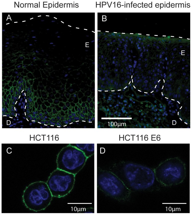
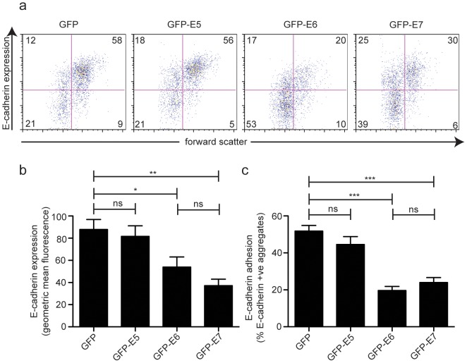
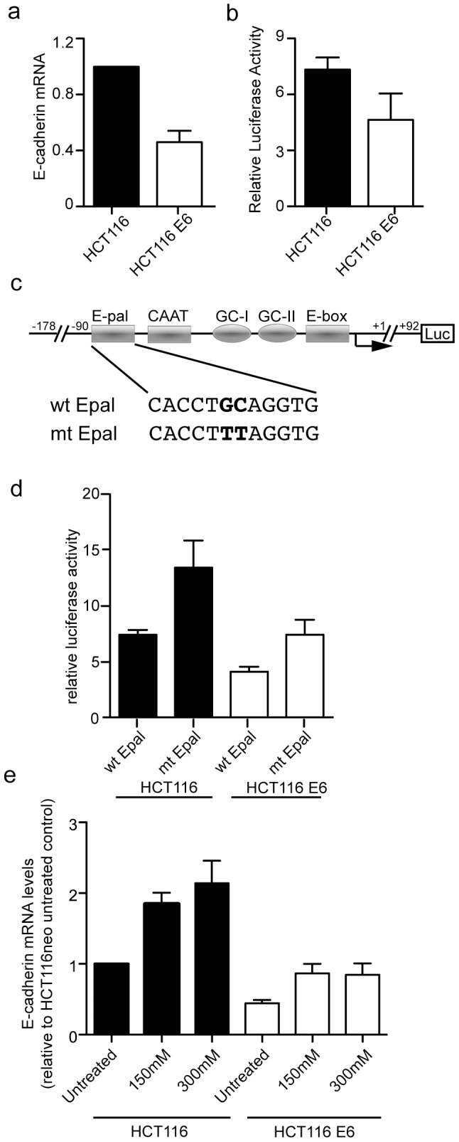
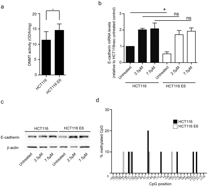
Verification
Claims the paper makes
The claims and statistics of this paper haven’t been checked yet.
Run the check — free, no account needed. It reads the open-access text and checks the sources it cites.
Claims other papers make about this one
Each is checked against this paper’s own text: does it say what the citing paper says it does?
In the citing paper
“HPV oncoproteins E6 and E7 have been shown to activate EMT-inducing transcriptional factors—Slug, Twizt, ZEB1 and ZEB2—resulting in an increase in mesenchymal markers such as N-cadherin, fibronectin and vimentin and a decrease in epithelial cell markers like E-cadherin [257,258]”
From The Not-So-Good, the Bad and the Ugly: HPV E5, E6 and E7 Oncoproteins in the Orchestration of Carcinogenesis (2021) · cites this paper as [257]
Self-check
Percentages, sums and reported differences re-computed from the paper’s own numbers.
The self-check needs the paper’s full text.
Citations
Cites
Snail, Zeb and bHLH factors in tumour progression: An alliance against the epithelial phenotype?
Peinado, H., Olmeda, D., & Cano, A. (2007) · Nature reviews. Cancer · doi:10.1038/nrc2131 · Cited by 3,121
Research works printed but not linked: 53
- CB Woodman, 2007, The natural history of cervical HPV infection: unresolved issues, Nat Rev Cancer, 7, 11, 10.1038/nrc2050 · DOI
- J Doorbar, 2005, The papillomavirus life cycle, J Clin Virol, 32 Suppl 1, S7, 10.1016/j.jcv.2004.12.006 · DOI
- SS Tungteakkhun, 2008, Cellular binding partners of the human papillomavirus E6 protein, Arch Virol, 153, 397, 10.1007/s00705-007-0022-5 · DOI
- K Zanier, 2012, Solution structure analysis of the HPV16 E6 oncoprotein reveals a self-association mechanism required for E6-mediated degradation of p53, Structure, 20, 604, 10.1016/j.str.2012.02.001 · DOI · Paper page
- M Scheffner, 1990, The E6 oncoprotein encoded by human papillomavirus types 16 and 18 promotes the degradation of p53, Cell, 63, 1129, 10.1016/0092-8674(90)90409-8 · DOI · Paper page
- AJ Klingelhutz, 1996, Telomerase activation by the E6 gene product of human papillomavirus type 16, Nature, 380, 79, 10.1038/380079a0 · DOI · Paper page
- VK Subbaiah, 2011, PDZ domains: the building blocks regulating tumorigenesis, Biochem J, 439, 195, 10.1042/BJ20110903 · DOI · Paper page
- BM Gumbiner, 2005, Regulation of cadherin-mediated adhesion in morphogenesis, Nat Rev Mol Cell Biol, 6, 622, 10.1038/nrm1699 · DOI
- H Wakita, 2003, Aberrant suprabasal P-cadherin expression in acanthotic but not psoriatic thickened epidermis, Arch Dermatol Res, 295 Suppl 1, S71, 10.1007/s00403-002-0377-0 · DOI
- L Larue, 2005, Epithelial-mesenchymal transition in development and cancer: role of phosphatidylinositol 3′ kinase/AKT pathways, Oncogene, 24, 7443, 10.1038/sj.onc.1209091 · DOI
- A Naito, 2001, Clinical significance of E-cadherin expression in thyroid neoplasms, J Surg Oncol, 76, 176, 10.1002/jso.1031 · DOI
- E Carico, 2001, E-cadherin and alpha-catenin expression during tumor progression of cervical carcinoma, Gynecol Oncol, 80, 156, 10.1006/gyno.2000.6035 · DOI
- Y Yuecheng, 2006, Clinical evaluation of E-cadherin expression and its regulation mechanism in epithelial ovarian cancer, Clin Exp Metastasis, 23, 65, 10.1007/s10585-006-9020-3 · DOI
- K Matthews, 2003, Depletion of Langerhans cells in human papillomavirus type 16-infected skin is associated with E6-mediated down regulation of E-cadherin, J Virol, 77, 8378, 10.1128/JVI.77.15.8378-8385.2003 · DOI · Paper page
- CJ Vessey, 1995, Altered expression and function of E-cadherin in cervical intraepithelial neoplasia and invasive squamous cell carcinoma, J Pathol, 176, 151, 10.1002/path.1711760208 · DOI
- JH Caberg, 2008, Silencing of E7 oncogene restores functional E-cadherin expression in human papillomavirus 16-transformed keratinocytes, Carcinogenesis, 29, 1441, 10.1093/carcin/bgn145 · DOI
- J Laurson, 2010, Epigenetic repression of E-cadherin by human papillomavirus 16 E7 protein, Carcinogenesis, 31, 918, 10.1093/carcin/bgq027 · DOI
- ZJ D'Costa, 2012, Screening of drugs to counteract human papillomavirus 16 E6 repression of E-cadherin expression, Invest New Drugs
- M Saitoh, 2009, Regulation of the stability of cell surface E-cadherin by the proteasome, Biochem Biophys Res Commun, 381, 560, 10.1016/j.bbrc.2009.02.098 · DOI
- L Roger, 2010, Gain of oncogenic function of p53 mutants regulates E-cadherin expression uncoupled from cell invasion in colon cancer cells, J Cell Sci, 123, 1295, 10.1242/jcs.061002 · DOI
- SM Park, 2008, The miR-200 family determines the epithelial phenotype of cancer cells by targeting the E-cadherin repressors ZEB1 and ZEB2, Genes Dev, 22, 894, 10.1101/gad.1640608 · DOI · Paper page
- WC Reinhold, 2007, Detailed DNA methylation profiles of the E-cadherin promoter in the NCI-60 cancer cells, Mol Cancer Ther, 6, 391, 10.1158/1535-7163.MCT-06-0609 · DOI
- J Behrens, 1991, The E-cadherin promoter: functional analysis of a G.C-rich region and an epithelial cell-specific palindromic regulatory element, Proc Natl Acad Sci U S A, 88, 11495, 10.1073/pnas.88.24.11495 · DOI
- A Cano, 2000, The transcription factor snail controls epithelial-mesenchymal transitions by repressing E-cadherin expression, Nat Cell Biol, 2, 76, 10.1038/35000025 · DOI
- H Peinado, 2004, Snail mediates E-cadherin repression by the recruitment of the Sin3A/histone deacetylase 1 (HDAC1)/HDAC2 complex, Mol Cell Biol, 24, 306, 10.1128/MCB.24.1.306-319.2004 · DOI
- RL Momparler, 2000, Evaluation of an inhibitor of DNA methylation, 5-aza-2′-deoxycytidine, for the treatment of lung cancer and the future role of gene therapy, Adv Exp Med Biol, 465, 433, 10.1007/0-306-46817-4_38 · DOI
- YN Liu, 2005, Regulatory mechanisms controlling human E-cadherin gene expression, Oncogene, 24, 8277, 10.1038/sj.onc.1208991 · DOI
- CL Au Yeung, 2010, HPV-16 E6 upregulation of DNMT1 through repression of tumor suppressor p53, Oncol Rep, 24, 1599
- RL Momparler, 2000, DNA methylation and cancer, J Cell Physiol, 183, 145, 10.1002/(SICI)1097-4652(200005)183:2<145::AID-JCP1>3.0.CO;2-V · DOI
- Q Lu, 2004, Methods for Analyzing the Role of DNA Methylation and Chromatin Structure in Regulating T Lymphocyte Gene Expression, Biol procedures online, 6, 189, 10.1251/bpo89 · DOI
- MA Whiteside, 2008, Human papillomavirus and molecular considerations for cancer risk, Cancer, 113, 2981, 10.1002/cncr.23750 · DOI
- Y Kondo, 2008, Gene silencing in cancer by histone H3 lysine 27 trimethylation independent of promoter DNA methylation, Nat Genet, 40, 741, 10.1038/ng.159 · DOI
- Y Kondo, 2003, Critical role of histone methylation in tumor suppressor gene silencing in colorectal cancer, Mol Cell Biol, 23, 206, 10.1128/MCB.23.1.206-215.2003 · DOI
- S Fujii, 2008, Enhancer of zeste homolog 2 downregulates E-cadherin by mediating histone H3 methylation in gastric cancer cells, Cancer science, 99, 738, 10.1111/j.1349-7006.2008.00743.x · DOI
- K Kokura, 2010, Methyl-H3K9-binding protein MPP8 mediates E-cadherin gene silencing and promotes tumour cell motility and invasion, EMBO J, 29, 3673, 10.1038/emboj.2010.239 · DOI
- WG Zhu, 2004, 5-aza-2′-deoxycytidine activates the p53/p21Waf1/Cip1 pathway to inhibit cell proliferation, J Biol Chem, 279, 15161, 10.1074/jbc.M311703200 · DOI
- S Milutinovic, 2000, DNA methyltransferase inhibition induces the transcription of the tumor suppressor p21(WAF1/CIP1/sdi1), J Biol Chem, 275, 6353, 10.1074/jbc.275.9.6353 · DOI
- S Milutinovic, 2004, DNA methyltransferase 1 knock down induces gene expression by a mechanism independent of DNA methylation and histone deacetylation, J Biol Chem, 279, 27915, 10.1074/jbc.M312823200 · DOI
- KM Hajra, 2002, The SLUG zinc-finger protein represses E-cadherin in breast cancer, Cancer Res, 62, 1613
- J Comijn, 2001, The two-handed E box binding zinc finger protein SIP1 downregulates E-cadherin and induces invasion, Mol Cell, 7, 1267, 10.1016/S1097-2765(01)00260-X · DOI
- MA Perez-Moreno, 2001, A new role for E12/E47 in the repression of E-cadherin expression and epithelial-mesenchymal transitions, J Biol Chem, 276, 27424, 10.1074/jbc.M100827200 · DOI
- F Vesuna, 2008, Twist is a transcriptional repressor of E-cadherin gene expression in breast cancer, Biochem Biophys Res Commun, 367, 235, 10.1016/j.bbrc.2007.11.151 · DOI
- CN Tsai, 2002, The Epstein-Barr virus oncogene product, latent membrane protein 1, induces the downregulation of E-cadherin gene expression via activation of DNA methyltransferases, Proc Natl Acad Sci U S A, 99, 10084, 10.1073/pnas.152059399 · DOI
- S Leonard, 2011, Epigenetic and transcriptional changes which follow Epstein-Barr virus infection of germinal center B cells and their relevance to the pathogenesis of Hodgkin's lymphoma, J Virol, 85, 9568, 10.1128/JVI.00468-11 · DOI
- WA Burgers, 2007, Viral oncoproteins target the DNA methyltransferases, Oncogene, 26, 1650, 10.1038/sj.onc.1209950 · DOI · Paper page
- H Gowher, 2005, Mechanism of stimulation of catalytic activity of Dnmt3A and Dnmt3B DNA-(cytosine-C5)-methyltransferases by Dnmt3L, J Biol Chem, 280, 13341, 10.1074/jbc.M413412200 · DOI
- P Arora, 2008, Hepatitis C virus core protein downregulates E-cadherin expression via activation of DNA methyltransferase 1 and 3b, Cancer Lett, 261, 244, 10.1016/j.canlet.2007.11.033 · DOI
- J Liu, 2006, Downregulation of E-cadherin by hepatitis B virus X antigen in hepatocellullar carcinoma, Oncogene, 25, 1008, 10.1038/sj.onc.1209138 · DOI
- SM Krishna, 2005, Down regulation of adhesion protein E-cadherin in Epstein-Barr virus infected nasopharyngeal carcinomas, Cancer biomarkers : section A of Disease markers, 1, 271, 10.3233/CBM-2005-1602 · DOI
- CM Leong, 2010, Loss of epidermal Langerhans cells occurs in human papillomavirus alpha, gamma, and mu but not beta genus infections, J Invest Dermatol, 130, 472, 10.1038/jid.2009.266 · DOI · Paper page
- A Tang, 1993, Adhesion of epidermal Langerhans cells to keratinocytes mediated by E-cadherin, Nature, 361, 82, 10.1038/361082a0 · DOI
- P Hubert, 2005, E-cadherin-dependent adhesion of dendritic and Langerhans cells to keratinocytes is defective in cervical human papillomavirus-associated (pre)neoplastic lesions, J Pathol, 206, 346, 10.1002/path.1771 · DOI
- R Ravizza, 2004, Role of the p53/p21 system in the response of human colon carcinoma cells to Doxorubicin, BMC cancer, 4, 92, 10.1186/1471-2407-4-92 · DOI
Cited by
Carcinogenic human papillomavirus infection
Schiffman, M., Doorbar, J., Wentzensen, N., de Sanjosé, S., Fakhry, C., Monk, B. J., Stanley, M. A., & Franceschi, S. (2016) · Nature Reviews Disease Primers · doi:10.1038/nrdp.2016.86 · Cited by 999
Basukala, O., & Banks, L. (2021) · Viruses · doi:10.3390/v13101892 · Cited by 137
Deegan, D. B., Zimmerman, C., Skardal, A., Atala, A., & Shupe, T. D. (2016) · Journal of the Mechanical Behavior of Biomedical Materials · doi:10.1016/j.jmbbm.2015.10.016 · Cited by 79
Metadata
- Authors
- D'Costa, Z. J., Jolly, C., Androphy, E. J., Mercer, A., Matthews, C. M., & Hibma, M. H.
- Venue
- PLoS ONE
- Publisher
- Public Library of Science (PLoS)
- Published
- 2012-11-26
- Volume
- 7
- Issue
- 11
- Pages
- e48954
- ISSN
- 1932-6203
- PMC
- PMC3506579
- Cited by
- 83
- References
- 1
- Language
- en
About these checks
Registry, retraction, Cites and statistics results are computed, not judged, with one exception: a printed reference that no index lists is read once by a model before it counts as a citation error. Claims are extracted by a model and kept only when their quote appears verbatim in the text; each cited source is then checked, with a model’s help, for whether it says what the claim relies on. Those verdicts can be wrong — read the claim’s quote and the cited source before relying on one.
Statistics and arithmetic are recomputed, not judged.
Confidence bands are shown only for fields calibrated on our annotated test set. None is calibrated yet, so no band is shown.
What counts as a claim ›How we measure confidence ›How we check this ›
Are you an author, or do you see a problem with this page? Request a review
