

Relationships between Mitochondrial Function and Metabolic Flexibility in Type 2 Diabetes Mellitus
Open accessThe DOI matches its Crossref record; 41 of the 41 references it cites were found in a registry or an index.
Abstract
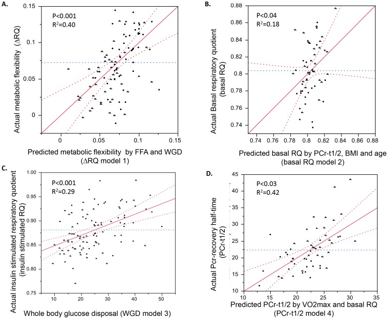
INTRODUCTION: Mitochondrial dysfunction, lipid accumulation, insulin resistance and metabolic inflexibility have been implicated in the etiology of type 2 diabetes (T2D), yet their interrelationship remains speculative. We investigated these interrelationships in a group of T2D and obese normoglycemic control subjects. METHODS: 49 non-insulin dependent male T2D patients and 54 male control subjects were enrolled, and a hyperinsulinemic-euglycemic clamp and indirect calorimetry were performed. A muscle biopsy was taken and intramyocellular lipid (IMCL) was measured. In vivo mitochondrial function was measured by PCr recovery in 30 T2D patients and 31 control subjects. RESULTS: Fasting NEFA levels were significantly elevated in T2D patients compared with controls, but IMCL was not different. Mitochondrial function in T2D patients was compromised by 12.5% (p<0.01). Whole body glucose disposal (WGD) was higher at baseline and lower after insulin stimulation. Metabolic flexibility (ΔRER) was lower in the type 2 diabetic patients (0.050±0.033 vs. 0.093±0.050, p<0.01). Mitochondrial function was the sole predictor of basal respiratory exchange ratio (RER) (R(2) = 0.18, p<0.05); whereas WGD predicted both insulin-stimulated RER (R(2) = 0.29, p<0.001) and metabolic flexibility (R(2) = 0.40, p<0.001). CONCLUSIONS: These results indicate that defects in skeletal muscle in vivo mitochondrial function in type 2 diabetic patients are only reflected in basal substrate oxidation and highlight the importance of glucose disposal rate as a determinant of substrate utilization in response to insulin.
Overview
The claims and statistics of this paper haven’t been checked yet.
Free: it reads the open-access text and checks the first 5 claims that cite a source we can check. Create a free account to check every claim.
Integrity signals
Registry
Cites
Statistics
Figures
Verification
Claims the paper makes
The claims and statistics of this paper haven’t been checked yet.
Free: it reads the open-access text and checks the first 5 claims that cite a source we can check. Create a free account to check every claim.
Claims other papers make about this one
Each is checked against this paper’s own text: does it say what the citing paper says it does?
The cited source doesn’t support the claim as written.
In the citing paper
“Many studies support the idea that deregulation of mitochondrial function underlies the onset of metabolic inflexibility [reviewed in Muoio (4)], although a causal link between the two still remains to be fully established (100, 101).”
From Metabolic Flexibility as an Adaptation to Energy Resources and Requirements in Health and Disease (2018) · cites this paper as [101]
Checked by a reasoning model — may be wrong.
Experimental — accuracy not yet measuredThe source backs part of the claim, not all of it — or backs it for a different population, condition or comparison.
In the citing paper
“However, studies have found that metabolic inflexibility of glucose is largely due to defective glucose transport,201,202 which suggests that skeletal muscle is unable to promote sufficient insulin secretion from type 2 diabetic islet-beta cells via any signalling mechanisms, in which it likely attempts to achieve given its need for increased glucose uptake.3,197”
From A review of current trends with type 2 diabetes epidemiology, aetiology, pathogenesis, treatments and future perspectives (2021) · cites this paper as [201]
Experimental — accuracy not yet measuredThe cited source says what the claim attributes to it.
In the citing paper
“Obese normoglycemic individuals were shown to have better metabolic flexibility than weight-matched T2D patients.201”
From A review of current trends with type 2 diabetes epidemiology, aetiology, pathogenesis, treatments and future perspectives (2021) · cites this paper as [201]
Self-check
Percentages, sums and reported differences re-computed from the paper’s own numbers.
The self-check needs the paper’s full text.
Citations
Cites
Glucose clamp technique: a method for quantifying insulin secretion and resistance.
DeFronzo, R. A., Tobin, J. D., & Andres, R. (1979) · American Journal of Physiology-Endocrinology and Metabolism · doi:10.1152/ajpendo.1979.237.3.e214 · Cited by 7,425 · cited 2×
“Hyperinsulinemic-euglycemic clamp In all subjects a 3-h hyperinsulinemic-euglycemic (40 mU /m(2)/ min) clamp was performed according to DeFronzo [20].”
MethodEckel, R. H., Alberti, K., Grundy, S. M., & Zimmet, P. Z. (2010) · The Lancet · doi:10.1016/s0140-6736(09)61794-3 · Cited by 6,295
Eckel, R. H., Grundy, S. M., & Zimmet, P. Z. (2005) · The Lancet · doi:10.1016/s0140-6736(05)66378-7 · Cited by 5,550
Randle, P. J., Garland, P. B., Hales, C. N., & Newsholme, E. A. (1963) · The Lancet · doi:10.1016/s0140-6736(63)91500-9 · Cited by 5,093 · cited 2×
“signaling pathways and a subsequent decline in glucose uptake by lipotoxic intermediates [26,27,28,29,30,31].”
BackgroundMechanisms of fatty acid-induced inhibition of glucose uptake.
Boden, G., Chen, X., Ruiz, J., White, J. V., & Rossetti, L. (1994) · Journal of Clinical Investigation · doi:10.1172/jci117252 · Cited by 3,705 · cited 1×
“signaling pathways and a subsequent decline in glucose uptake by lipotoxic intermediates [26,27,28,29,30,31].”
BackgroundCalculation of substrate oxidation rates in vivo from gaseous exchange.
Frayn, K. N. (1983) · Journal of Applied Physiology · doi:10.1152/jappl.1983.55.2.628 · Cited by 2,365 · cited 1×
“From oxygen consumption and carbondioxide production levels, whole body glucose oxidation and fat oxidation rates could be calculated using stoichiometric equations according to Frayn [24] with the assumption that protein oxidation was negligible.”
MethodThe Gross Composition of the Body
SIRI, W. E. (1956) · Advances in Biological and Medical Physics · doi:10.1016/b978-1-4832-3110-5.50011-x · Cited by 1,765
Diet, muscle glycogen and physical performance.
Bergström, J., Hermansen, L., Hultman, E., & Saltin, B. (1967) · Acta Physiologica Scandinavica · doi:10.1111/j.1748-1716.1967.tb03720.x · Cited by 1,740 · cited 1×
“Muscle biopsies were performed prior to the clamp under local anesthesia (2% lidocaine) according to the Bergstrom technique [22].”
MethodINFLUENCES OF GLUCOSE LOADING AND OF INJECTED INSULIN ON HEPATIC GLUCOSE OUTPUT*
Steele, R. (1959) · Annals of the New York Academy of Sciences · doi:10.1111/j.1749-6632.1959.tb44923.x · Cited by 1,670 · cited 11×
“Calculations Steele’s single-pool non–steady-state equations were used to calculate glucose endogenous glucose production (EGP) and whole body glucose disposal (WGD) [25].”
MethodInfluentialDresner, A., Laurent, D., Marcucci, M., Griffin, M. E., Dufour, S., Cline, G. W., Slezak, L. A., Andersen, D. K., Hundal, R. S., Rothman, D. L., et al. (1999) · Journal of Clinical Investigation · doi:10.1172/jci5001 · Cited by 1,311 · cited 1×
“These factors have been suggested to interfere with muscle insulin sensitivity but may also affect mitochondrial function or vice versa [7,8,9,10,11,12].”
Background
Cited by
Metabolic Flexibility as an Adaptation to Energy Resources and Requirements in Health and Disease
Smith, R. L., Soeters, M. R., Wüst, R. C. I., & Houtkooper, R. H. (2018) · Endocrine Reviews · doi:10.1210/er.2017-00211 · Cited by 787
Reed, J., Bain, S., & Kanamarlapudi, V. (2021) · Diabetes, Metabolic Syndrome and Obesity: Targets and Therapy · doi:10.2147/dmso.s319895 · Cited by 441
Virmani, M. A., & Cirulli, M. (2022) · International Journal of Molecular Sciences · doi:10.3390/ijms23052717 · Cited by 276
Insulin Resistance and Mitochondrial Dysfunction
González-Franquesa, A., & Patti, M. (2017) · Advances in experimental medicine and biology · doi:10.1007/978-3-319-55330-6_25 · Cited by 173
Trimethylamine N-oxide impairs pyruvate and fatty acid oxidation in cardiac mitochondria
Makrecka‐Kuka, M., Volska, K., Antone, U., Vilšķe̅rsts, R., Grīnberga, S., Bandere, D., Liepinsh, E., & Dambrova, M. (2017) · Toxicology Letters · doi:10.1016/j.toxlet.2016.12.017 · Cited by 150
Papaetis, G. S., Papakyriakou, P., & Panagiotou, T. N. (2015) · Archives of Medical Science · doi:10.5114/aoms.2015.52350 · Cited by 130
Andriessen, C., Fealy, C. E., Veelen, A., van Beek, S. M. M., Roumans, K. H. M., Connell, N. J., Mevenkamp, J., Moonen‐Kornips, E., Havekes, B., Schrauwen‐Hinderling, V. B., et al. (2022) · Diabetologia · doi:10.1007/s00125-022-05752-z · Cited by 140
Self-citationMost, J., Timmers, S., Warnke, I., Jocken, J. W. E., Boekschoten, M. V., de Groot, P., Bendik, I., Schrauwen, P., Goossens, G. H., & Blaak, E. E. (2016) · American Journal of Clinical Nutrition · doi:10.3945/ajcn.115.122937 · Cited by 127
Self-citationPathophysiological role of metabolic flexibility on metabolic health
Galgani, J. E., & Fernández‐Verdejo, R. (2021) · Obesity Reviews · doi:10.1111/obr.13131 · Cited by 90
Cavaliere, G., Cimmino, F., Trinchese, G., Catapano, A., Petrella, L., D’Angelo, M., Lucchin, L., & Mollica, M. P. (2023) · Antioxidants · doi:10.3390/antiox12061172 · Cited by 118
“Indeed, many studies support the idea that the deregulation of mitochondrial function underlies the onset of metabolic inflexibility [73].”
BackgroundMalin, S. K., Haus, J. M., Solomon, T. P. J., Blaszczak, A., Kashyap, S. R., & Kirwan, J. P. (2013) · American Journal of Physiology-Endocrinology and Metabolism · doi:10.1152/ajpendo.00441.2013 · Cited by 89
“Skeletal muscle biopsies are needed to determine whether the presence of IFG alters in vivo mitochondrial oxidative capacity in adults with IGT (20, 29, 42).”
BackgroundVanweert, F., Neinast, M. D., Erazo‐Tapia, E., van de Weijer, T., Hoeks, J., Schrauwen‐Hinderling, V. B., Blair, M. C., Bornstein, M. R., Hesselink, M. K. C., Schrauwen, P., et al. (2022) · Nature Communications · doi:10.1038/s41467-022-31249-9 · Cited by 96
Self-citationAmin, M. M., Asaad, G. F., Abdel Salam, R. M., El-Abhar, H. S., & Arbid, M. S. (2014) · PLoS ONE · doi:10.1371/journal.pone.0089169 · Cited by 82
Most, J., Goossens, G. H., Jocken, J. W. E., & Blaak, E. E. (2013) · International Journal of Obesity · doi:10.1038/ijo.2013.231 · Cited by 89
Metabolic Flexibility and Its Impact on Health Outcomes
Palmer, B. F., & Clegg, D. J. (2022) · Mayo Clinic Proceedings · doi:10.1016/j.mayocp.2022.01.012 · Cited by 93
“Insulin resistance may also play a primary role in creating this abnormality.(8,9)”
BackgroundDiMenna, F. J., & Arad, A. D. (2018) · BMC Sports Science, Medicine and Rehabilitation · doi:10.1186/s13102-018-0110-8 · Cited by 68
Jonscher, K. R., Stewart, M. S., Garcia, A. A., DeFelice, B. C., Wang, X. X., Luo, Y., Levi, M., Heerwagen, M. J. R., Janssen, R. C., de la Houssaye, B. A., et al. (2016) · The FASEB Journal · doi:10.1096/fj.201600906r · Cited by 72
“Insulin resistance andalteredglucosedisposal rate, as well as dysfunction in fatty acid oxidation in mitochondria, impair metabolic flexibility in patients with type 2 diabetes (65).”
BackgroundPhielix, E., Jeleník, T., Nowotny, P., Szendroedi, J., & Roden, M. (2013) · Diabetologia · doi:10.1007/s00125-013-3127-2 · Cited by 67
“This is partly in line with results of a recent paper in which in vivo mitochondrial function assessed from post-exercise phosphocreatine recovery rates predicted RQ under baseline conditions [32].”
ResultMitochondrial dynamics in skeletal muscle insulin resistance and type 2 diabetes
Fealy, C. E., Mulya, A., Axelrod, C. L., & Kirwan, J. P. (2018) · Translational Research · doi:10.1016/j.trsl.2018.07.011 · Cited by 63
Exercise counteracts lipotoxicity by improving lipid turnover and lipid droplet quality
Zacharewicz, E., Hesselink, M. K. C., & Schrauwen, P. (2018) · Journal of Internal Medicine · doi:10.1111/joim.12729 · Cited by 63
Self-citationTsilingiris, D., Tzeravini, E., Koliaki, C. C., Dalamaga, Μ., & Kokkinos, A. D. (2021) · Current Obesity Reports · doi:10.1007/s13679-021-00434-0 · Cited by 63
“In the same direction, patients with T2DM displayed lower increases in RQ after insulin infusion and longer PCr recovery half-time than healthy controls in a study using both (31)P-MRS and indirect calorimetry to assess mitochondrial plasticity [91].”
BackgroundLong-chain acylcarnitine content determines the pattern of energy metabolism in cardiac mitochondria
Makrecka‐Kuka, M., Kuka, J., Volska, K., Antone, U., Sevostjanovs, E., Cirule, H., Grīnberga, S., Pugovičs, O., Dambrova, M., & Liepinsh, E. (2014) · Molecular and Cellular Biochemistry · doi:10.1007/s11010-014-2106-3 · Cited by 66
Vanweert, F., de Ligt, M., Hoeks, J., Hesselink, M. K. C., Schrauwen, P., & Phielix, E. (2020) · The Journal of Clinical Endocrinology & Metabolism · doi:10.1210/clinem/dgaa751 · Cited by 65
Self-citationWelch, A. A., Hayhoe, R. P. G., & Cameron, D. (2020) · Proceedings of the Nutrition Society · doi:10.1017/s0029665119001150 · Cited by 51
Tarpey, M. D., Davy, K. P., McMillan, R. P., Bowser, S. M., Halliday, T. M., Boutagy, N. E., Davy, B. M., Frisard, M. I., & Hulver, M. W. (2017) · Molecular Metabolism · doi:10.1016/j.molmet.2017.10.006 · Cited by 48
“[13] reported that in-vivo mitochondrial function, assessed via PCr recovery, was the major determinant of basal respiratory exchange ratio (RER) but was not a major contributor under insulin-stimulated conditions.”
BackgroundTyagi, A., Cohen, M. M., Reece, J., Telles, S., & Jones, L. (2016) · Applied Psychophysiology and Biofeedback · doi:10.1007/s10484-016-9340-2 · Cited by 62
“This may lead to the development of conditions such as metabolic syndrome (MetS), accelerated aging and cardiovascular disease (Thayer et al. 2012; van de Weijer et al. 2013).”
BackgroundAmin, M. M., & Arbid, M. S. (2016) · Applied Physiology Nutrition and Metabolism · doi:10.1139/apnm-2016-0429 · Cited by 47
Aw, W. C., Garvin, M. R., Melvin, R. G., & Ballard, J. W. O. (2017) · PLoS ONE · doi:10.1371/journal.pone.0187554 · Cited by 43
Malin, S. K., Liu, Z., Barrett, E. J., & WELTMAN, A. L. (2016) · Reviews in Endocrine and Metabolic Disorders · doi:10.1007/s11154-016-9352-5 · Cited by 39
Frankó, A., Neschen, S., Rozman, J., Rathkolb, B., Aichler, M., Feuchtinger, A., Brachthäuser, L., Neff, F., Kovářová, M., Wolf, E., et al. (2017) · Molecular Metabolism · doi:10.1016/j.molmet.2016.12.007 · Cited by 34
Altered intramuscular network of lipid droplets and mitochondria in type 2 diabetes
de Almeida, M. E., Nielsen, J., Petersen, M. H., Wentorf, E. K., Pedersen, N. B., Jensen, K., Højlund, K., & Ørtenblad, N. (2022) · American Journal of Physiology-Cell Physiology · doi:10.1152/ajpcell.00470.2022 · Cited by 38
Tyagi, A., Cohen, M. M., Reece, J., & Telles, S. (2014) · BMC Complementary and Alternative Medicine · doi:10.1186/1472-6882-14-445 · Cited by 37
Remchak, M. E., Heiston, E. M., Ballantyne, A. C., Dotson, B. L., Stewart, N. R., Spaeth, A. M., & Malin, S. K. (2022) · The Journal of Clinical Endocrinology & Metabolism · doi:10.1210/clinem/dgac233 · Cited by 36
Metformin May Contribute to Inter-individual Variability for Glycemic Responses to Exercise
Malin, S. K., & Stewart, N. R. (2020) · Frontiers in Endocrinology · doi:10.3389/fendo.2020.00519 · Cited by 34
“The reason FFAs contribute to this multi-tissue insulin resistance is beyond the scope of this review, but likely relates to elevated plasma FFA concentrations being linked with reduced mitochondrial function and metabolic flexibility (58), Therefore, it would be reasonable to expect aerobic exercise interventions des…”
Backgroundfor the GDS group, Apostolopoulou, M., Straßburger, K., Herder, C., Knebel, B., Kotzka, J., Szendroedi, J., & Roden, M. (2016) · Diabetologia · doi:10.1007/s00125-016-4038-9 · Cited by 29
“If impaired metabolic flexibility is the result of impaired mitochondrial oxidation,ΔRQ should correlate with V : O2max, as demonstrated for muscle ATP synthase flux and phosphocreatine recovery [4].”
BackgroundMethodVaitheesvaran, B., Hartil, K., Navare, A., Zheng, C., Broin, P. Ó., Golden, A., Guha, C., Lee, W. N., Kurland, I. J., & Bruce, J. E. (2014) · Metabolomics · doi:10.1007/s11306-014-0639-9 · Cited by 28
Parry, H. A., & Glancy, B. C. (2021) · Current Opinion in Physiology · doi:10.1016/j.cophys.2022.100487 · Cited by 25
Whytock, K. L., Pino, M. F., Sun, Y., Yu, G., de Carvalho, F. G., Yeo, R. X., Vega, R. B., Parmar, G., Divoux, A., Kapoor, N., et al. (2023) · American Journal of Physiology-Endocrinology and Metabolism · doi:10.1152/ajpendo.00143.2023 · Cited by 29
Self-citationDubé, J. J., Collyer, M. L., Trant, S., Toledo, F. G. S., Goodpaster, B. H., Kershaw, E. E., & DeLany, J. P. (2019) · The Journal of Clinical Endocrinology & Metabolism · doi:10.1210/clinem/dgz272 · Cited by 25
Brunetta, H. S., de Paula, G. C., de Oliveira, J., Martins, E. L., dos Santos, G. J., Galina, A., Rafacho, A., Bem, A. F. D., & Nunes, E. A. (2018) · Experimental Physiology · doi:10.1113/ep087317 · Cited by 21
Characteristics of Glucose Metabolism in Nordic and South Asian Subjects with Type 2 Diabetes
Wium, C., Gulseth, H. L., Eriksen, E. F., & Birkeland, K. I. (2013) · PLoS ONE · doi:10.1371/journal.pone.0083983 · Cited by 22
Stimulatory Effects of Balanced Deep Sea Water on Mitochondrial Biogenesis and Function
Ha, B. G., Park, J., Hyun-Jung, C., & Shon, Y. H. (2015) · PLoS ONE · doi:10.1371/journal.pone.0129972 · Cited by 21
“Emerging evidence suggests that impaired mitochondrial metabolism is accompanied by reduced transcription factor activity regulating mitochondrial biogenesis in patients with insulin resistance, type 2 diabetes and obesity [2, 3].”
Chen, H., Zhang, J., Wang, T., Zhang, S., Lai, Q., Huang, S., & Zeng, P. (2021) · The Journal of Clinical Endocrinology & Metabolism · doi:10.1210/clinem/dgab465 · Cited by 17
The Mitochondrial Proteomic Signatures of Human Skeletal Muscle Linked to Insulin Resistance
Kruse, R., Sahebekhtiari, N., & Højlund, K. (2020) · International Journal of Molecular Sciences · doi:10.3390/ijms21155374 · Cited by 19
Liu, F., Wanigatunga, A. A., Zampino, M., Knuth, N. D., Simonsick, E. M., Schrack, J. A., & Ferrucci, L. (2020) · The Journals of Gerontology Series A · doi:10.1093/gerona/glaa201 · Cited by 21
“Another study found that muscle oxidative capacity was the sole predictor of basal RER among patients with type 2 diabetes (42).”
BackgroundMetabolic flexibility across the spectrum of glycemic regulation in youth
Bacha, F. F., Bartz, S. K., Puyau, M. R., Adolph, A. L., & Sharma, S. (2021) · JCI Insight · doi:10.1172/jci.insight.146000 · Cited by 20
“Similarly, in adults with type 2 diabetes compared with obese controls, insulin-stimulated glucose oxidation during the hyperinsulinemic clamp was determined by insulin-stimulated glucose uptake (25), suggesting that the primary defect is in substrate availability secondary to limitation in substrate transport.”
InfluentialMalin, S. K., Viskochil, R. H., Oliver, C., & Braun, B. (2013) · Journal of Applied Physiology · doi:10.1152/japplphysiol.00084.2013 · Cited by 17
“Since elevated plasma NEFA concentrations have been linked with mitochondrial dysfunction and metabolic inflexibility (22, 38), and we observed elevated NEFAs during exercise, our findings highlight the need for future work to use lipid tracers and examine skeletal muscle biopsies in adults with IFG IGT to elucidate m…”
BackgroundCarnero, E. Á., Bock, C. P., Distéfano, G., Corbin, K. D., Stephens, N. A., Pratley, R. E., Smith, S. R., Goodpaster, B. H., & Sparks, L. M. (2021) · Diabetologia · doi:10.1007/s00125-021-05535-y · Cited by 18
Self-citationGarthwaite, T., Sjöros, T., Laine, S., Koivumäki, M., Vähä‐Ypyä, H., Verho, T., Norha, J., Kallio, P., Saarenhovi, M., Löyttyniemi, E., et al. (2024) · American Journal of Physiology-Endocrinology and Metabolism · doi:10.1152/ajpendo.00338.2023 · Cited by 23
Bakker, L. E. H., Guigas, B., van Schinkel, L. D., van der Zon, G. C. M., Streefland, T. C. M., van Klinken, J. B., Jonker, J. T., Lamb, H. J., Smit, J. W. A., Pijl, H., et al. (2014) · Diabetologia · doi:10.1007/s00125-014-3408-4 · Cited by 17
“Indeed, fatty acid oxidation rates, which are positively associated with plasma fatty acid levels, were reported to be an important determinant of metabolic flexibility [31, 32].”
BackgroundFernández‐Verdejo, R., Malo‐Vintimilla, L., Gutiérrez‐Pino, J., López‐Fuenzalida, A., Olmos, P. R., Pablo Irarrázaval, M., & Galgani, J. E. (2021) · Frontiers in Nutrition · doi:10.3389/fnut.2021.745907 · Cited by 12
Rodriguez, J. C., Peterman, J. E., Fleenor, B. S., WHALEY, M. H., Kaminsky, L. A., & Harber, M. P. (2022) · Metabolic Syndrome and Related Disorders · doi:10.1089/met.2021.0130 · Cited by 8
Shi, W., Hu, S., Wang, W., Zhou, X., & Qiu, W. (2013) · Journal of Diabetes Research · doi:10.1155/2013/163062 · Cited by 8
RetractedCan we alter dietary macronutrient compositions and alleviate mitochondrial disease?
Ballard, J. W. O. (2016) · Journal of Rare Diseases Research & Treatment · doi:10.29245/2572-9411/2016/3.1043 · Cited by 9
von Rauchhaupt, E., Rodemer, C., Kliemank, E., Bulkescher, R., Campos, M., Kopf, S., Fleming, T., Herzig, S., Nawroth, P. P., Szendroedi, J., et al. (2024) · Diabetes Care · doi:10.2337/dc24-0209 · Cited by 11
Bartlett, M. F., Fitzgerald, L. F., Nagarajan, R., & Kent, J. A. (2023) · Applied Physiology, Nutrition, and Metabolism · doi:10.1139/apnm-2023-0315 · Cited by 8
Chaos, mitochondria and type 2 diabetes; does type 2 diabetes arise from a metabolic dysrhythmia?
Baskerville, R. (2019) · Medical Hypotheses · doi:10.1016/j.mehy.2019.03.032 · Cited by 7
“However the precise relationship between metabolic flexibility, insulin resistance and mitochondrial dysfunction is unclear as they are mutually interdependent [24].”
BackgroundRagland, T. J., & Malin, S. K. (2024) · Physiological Reports · doi:10.14814/phy2.15987 · Cited by 8
Fonkoua, M., Boris, N. S., Kengne, S. R., Chia, T. E., Guy, T. G., Boris, A. K., Ngondi, J. L., & Gouado, I. (2017) · Biology and Medicine · doi:10.4172/0974-8369.1000407 · Cited by 7
“Oxidative stress aggravates type 2 DM [1].”
Exogenous Factors May Differentially Influence the Selective Costs of mtDNA Mutations
Aw, W. C., Garvin, M. R., & Ballard, J. W. O. (2018) · Advances in anatomy, embryology and cell biology · doi:10.1007/102_2018_2 · Cited by 5
Are Individuals With Type 2 Diabetes Metabolically Inflexible? A Systematic Review and Meta‐Analysis
Hansen, M., Lange, K. K., Stausholm, M. B., & Dela, F. (2025) · Endocrinology Diabetes & Metabolism · doi:10.1002/edm2.70044 · Cited by 10
“Furthermore, van de Weijer et al. found that in overweight individuals with and without T2D, the relationship between mitochondrial capacity and metabolic flexibility disappeared when correcting for whole-body glucose disposal [50].”
Rahbar, S., Naimi, S. S., Soltani, A. R., Rahimi, A., Baghban, A. A., & Khorami, N. (2018) · PubMed Central · Cited by 4
Liu, J., Fan, J., & Zhou, B. (2026) · Physiology International · doi:10.1556/2060.2026.00780 · Cited by 3
Hayes, A., Swackhamer, C., Quezada‐Calvillo, R., Butte, N. F., Sterchi, E. E., Nichols, B. L., & Hamaker, B. R. (2025) · European Journal of Nutrition · doi:10.1007/s00394-025-03585-1 · Cited by 3
“Metabolic flexibility has been studied in the context of disease states [5, 10, 11], exercise [6, 12], and fasting [5, 13], yet evidence causally linking metabolic flexibility with metabolic health remains inconclusive [14, 15].”
Wilson, T. D., Philips, E. A., Alharithi, Y. J., Broberg, C., Vanderpool, J., Rakshe, S. K., Fei, S. S., Davis, B. A., Koch, S. E., Zhu, Z., et al. (2025) · bioRxiv (Cold Spring Harbor Laboratory) · doi:10.1101/2025.04.15.648971 · Cited by 2
de Kam, S. S., van der Velde, J. H. P. M., Veelen, A., Jörgensen, J. A., Hoeks, J., & Schrauwen, P. (2026) · Diabetologia · doi:10.1007/s00125-026-06736-z · Cited by 1
Self-citationTrexler, E. T., Smith‐Ryan, A. E., Wingfield, H. L., Blue, M. N. M., Roelofs, E. J., & Hirsch, K. R. (2017) · The Journal of Sports Medicine and Physical Fitness · doi:10.23736/s0022-4707.16.06284-8 · Cited by 2
Fetal Origins of Adult Disease
Friedman, J., & Baker, P. R. (2016) · Elsevier eBooks · doi:10.1016/b978-0-323-35214-7.00015-9 · Cited by 1
Room Calorimetry as a Method of Measuring Metabolic Flexibility
Leger, J. (2015) · University Libraries (University of Maryland) · doi:10.13016/m24065
A genetikai diabeteskockázat hatása a csontanyagcsere–energia-háztartás kapcsolatokra
Fék, A., Buday, B., Kovács, G., Vitai, M., Vecsei, Z., Pauer, J., Literáti-Nagy, B., Bezzegh, K., Péterfai, É., & Koranyi, L. I. (2015) · Orvosi Hetilap · doi:10.1556/650.2015.30185
Skeletal muscle autophagy and mitophagy in response to high-fat feeding and endurance training
Tarpey, M. D. (2016) · VTechWorks (Virginia Tech)
The influence of age and nutrients on insulin sensitivity
Chee, C. (2016) · Nottingham ePrints (University of Nottingham) · Cited by 2
Dietary polyphenols: modulators of energy and substrate metabolism in obese humans
Most, J. (2016) · Research Publications (Maastricht University) · doi:10.26481/dis.20160115jm
Warnke (2018) · Research Publications (Maastricht University) · doi:10.26481/dis.20180316iw
Loehr, S. (2013) · ERA: Education and Research Archive (University of Alberta) · doi:10.7939/r3t43jb3c
The Immediate and Extended Effect of Diet and Exercise on Metabolic Flexibility
Allerton, T. D. (2017) · Louisiana State University · doi:10.31390/gradschool_dissertations.4493
Vanweert, F., Neinast, M. D., Erazo‐Tapia, E., van de Weijer, T., Joris Hoeks, J., Schrauwen‐Hinderling, V. B., Blair, M. C., Bornstein, M. R., Hesselink, M. K. C., Schrauwen, P., et al. (2021) · SSRN Electronic Journal · doi:10.2139/ssrn.3944597
Self-citationExercise and training in NAFLD and insulin resistance: psychological approach in humans
Brouwers, B. (2016) · Research Publications (Maastricht University) · doi:10.26481/dis.20160624bb
Improving flexibility in substrate metabolism: a pharmacological and lifestyle approach
Veelen, A. (2022) · Research Publications (Maastricht University) · doi:10.26481/dis.20230125av
Moderating carbohydrate digestion rate promotes metabolic flexibility in micePreprint
Hayes, A., Swackhamer, C., Quezada‐Calvillo, R., Butte, N. F., Sterchi, E. E., Nichols, B. L., & Hamaker, B. R. (2023) · bioRxiv (Cold Spring Harbor Laboratory) · doi:10.1101/2023.01.08.523060
A metabolically healthy lifestyle: A matter of timing?
Andriessen, C. (2023) · Research Publications (Maastricht University) · doi:10.26481/dis.20230706ca
Vanweert, F. (2023) · Research Publications (Maastricht University) · doi:10.26481/dis.20231117fv · Cited by 1
Philips, E. A., Alharithi, Y. J., Wilson, T. D., Broberg, C., Davis, B. A., Koch, S. E., Carbone, L., Rubinstein, J. H., Gurley, S. B., Kumar, S., et al. (2025) · SSRN Electronic Journal · doi:10.2139/ssrn.5228904 · Cited by 2
Mendonca, G. V., Cidrais, M., & Silva, A. M. (2026) · Annals of Nutrition and Metabolism · doi:10.1159/anm/abmag007
Hansen, M., Møller, S., Blom, I., Lange, K. K., Ziegler, A. K., Farlov, J. E., Borch, J., Hesselager, S. A., Hansen, J. K., Liebmann, A., et al. (2026) · Scandinavian Journal of Medicine and Science in Sports · doi:10.1111/sms.70383
“Studies that account for glucose disposal have reported that the apparent impairment in metabolic flexibility in people with T2D can be eliminated when an appropriate control group and correction are applied [27, 28].”
Influential
Metadata
- Authors
- van de Weijer, T., Sparks, L. M., Phielix, E., Meex, R. C., van Herpen, N. A., Hesselink, M. K. C., Schrauwen, P., & Schrauwen-Hinderling, V. B.
- Affiliations
- Maastricht University Medical Centre (NL)
- Venue
- PLoS ONE
- Publisher
- Public Library of Science (PLoS)
- Published
- 2013-02-13
- Volume
- 8
- Issue
- 2
- Pages
- e51648
- ISSN
- 1932-6203 (ISSN-L)
- PMC
- PMC3572106
- OpenAlex
- W2079395532
- Cited by
- 87
- References
- 41
- Citations by year
- 2026: 4 · 2025: 4 · 2024: 3 · 2023: 6 · 2022: 9 · 2021: 8 · 2020: 5 · 2019: 4 · 2018: 8 · 2017: 8 · 2016: 12 · 2015: 4 · 2014: 5 · 2013: 7
- FWCI
- 6.48
- Type
- journal article
- Language
- en
- Open access
- gold
- doi.org
- Locations
- PLoS ONE (publishedVersion, cc-by)
- Europe PMC (PubMed Central) (publishedVersion, cc-by)
- PubMed Central (publishedVersion, cc-by)
- PubMed (publishedVersion)
- Research Publications (Maastricht University) (publishedVersion, cc-by)
- DOAJ (DOAJ: Directory of Open Access Journals) (submittedVersion, cc-by-sa)
- Research Publications (Maastricht University) (submittedVersion, other-oa)
- Funders
- Nierstichting
- Diabetes Fonds
- Nederlandse Organisatie voor Wetenschappelijk Onderzoek (918.96.618)
- Center for Translational Molecular Medicine
- Topics
- Health Sciences › Medicine › Physiology (medical) › Adipose Tissue and Metabolism
- Life Sciences › Biochemistry, Genetics and Molecular Biology › Molecular Biology › Metabolism, Diabetes, and Cancer
- Life Sciences › Biochemistry, Genetics and Molecular Biology › Molecular Biology › Mitochondrial Function and Pathology
- Keywords
- type 2 diabetes, mitochondrial function, metabolic flexibility, skeletal muscle, intramyocellular lipid, respiratory exchange ratio, glucose disposal, insulin resistance, fatty acid oxidation, phosphocreatine recovery, free fatty acids, substrate oxidation
- MeSH terms
- Aged, Blood Glucose, Diabetes Mellitus, Type 2, Fatty Acids, Nonesterified, Humans, Insulin, Insulin Resistance, Male, Middle Aged, Mitochondria, Obesity, Glucose Clamp Technique, Muscle, Skeletal, Lipid Metabolism
About these checks
Registry, retraction, Cites and statistics results are computed, not judged, with one exception: a printed reference that no index lists is read once by a model before it counts as a citation error. Claims are extracted by a model and kept only when their quote appears verbatim in the text; each cited source is then checked, with a model’s help, for whether it says what the claim relies on. Those verdicts can be wrong — read the claim’s quote and the cited source before relying on one.
Statistics and arithmetic are recomputed, not judged.
Confidence bands are shown only for fields calibrated on our annotated test set. None is calibrated yet, so no band is shown.
What counts as a claim ›How we measure confidence ›How we check this ›
Are you an author, or do you see a problem with this page? Request a review
Data sources
Bibliographic data: Semantic Scholar, OpenAlex, Crossref.
Contains information from the Semantic Scholar Academic Graph Datasets, made available under the ODC Attribution License (ODC-BY 1.0).
Semantic Scholar’s data is described in: Kinney, R., et al. (2023). The Semantic Scholar Open Data Platform. arXiv:2301.10140.
