

Computer Assisted Assembly of Connectomes from Electron Micrographs: Application to Caenorhabditis elegans
Open accessThe DOI matches its Crossref record; 21 of the 21 references it cites were found in a registry or an index.
Abstract
A rate-limiting step in determining a connectome, the set of all synaptic connections in a nervous system, is extraction of the relevant information from serial electron micrographs. Here we introduce a software application, Elegance, that speeds acquisition of the minimal dataset necessary, allowing the discovery of new connectomes. We have used Elegance to obtain new connectivity data in the nematode worm Caenorhabditis elegans. We analyze the accuracy that can be obtained, which is limited by unresolvable ambiguities at some locations in electron microscopic images. Elegance is useful for reconstructing connectivity in any region of neuropil of sufficiently small size.
Overview
The claims and statistics of this paper haven’t been checked yet.
Free: it reads the open-access text and checks the first 5 claims that cite a source we can check. Create a free account to check every claim.
Integrity signals
Registry
Cites
Statistics
Figures

Verification
Claims the paper makes
The claims and statistics of this paper haven’t been checked yet.
Free: it reads the open-access text and checks the first 5 claims that cite a source we can check. Create a free account to check every claim.
Claims other papers make about this one
Each is checked against this paper’s own text: does it say what the citing paper says it does?
The source backs part of the claim, not all of it — or backs it for a different population, condition or comparison.
In the citing paper
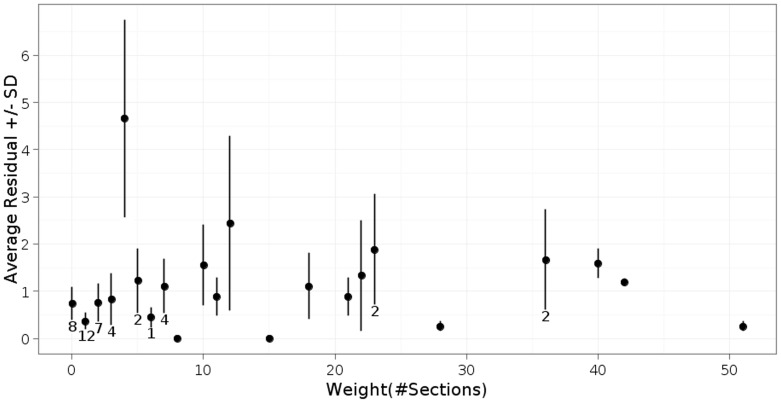
“the percentage uncertainty (s.d.) in the number of sections scored ranged for chemical synapses between 10% for edge weights of 10 sections or less to 2% for edge weights of 40 sections or more.”
From Whole-animal connectomes of both Caenorhabditis elegans sexes (2019) · cites this paper as [62]
Experimental — accuracy not yet measuredThe cited source says what the claim attributes to it.
In the citing paper
“Reproducibility of the annotation has been assessed by comparing the results of independent annotators62.”
From Whole-animal connectomes of both Caenorhabditis elegans sexes (2019) · cites this paper as [62]
Self-check
Percentages, sums and reported differences re-computed from the paper’s own numbers.
The self-check needs the paper’s full text.
Citations
Cites
THE GENETICS OF CAENORHABDITIS ELEGANS
Brenner, S. (1974) · Genetics · doi:10.1093/genetics/77.1.71 · Cited by 15,081
The structure of the nervous system of the nematode Caenorhabditis elegans
White, J. G., Southgate, E., Thomson, J. N., & Brenner, S. (1986) · Philosophical Transactions of the Royal Society of London. B, Biological Sciences · doi:10.1098/rstb.1986.0056 · Cited by 5,589 · cited 2×
“elegans hermaphrodite from existing electron micrographs (JSE series, see [2]) in 131 hr.”
BackgroundOdorant-selective genes and neurons mediate olfaction in C. elegans
Bargmann, C. I., Hartwieg, E., & Horvitz, H. R. (1993) · Cell · doi:10.1016/0092-8674(93)80053-h · Cited by 1,225
The neural circuit for touch sensitivity in Caenorhabditis elegans
Chalfie, M., Sulston, J., White, J., Southgate, E., Thomson, J., & Brenner, S. (1985) · The Journal of Neuroscience · doi:10.1523/jneurosci.05-04-00956.1985 · Cited by 1,093 · cited 1×
“[4–8]).”
Quantitative ultrastructural analysis of hippocampal excitatory synapses
Schikorski, T., & Stevens, C. F. (1997) · The Journal of Neuroscience · doi:10.1523/jneurosci.17-15-05858.1997 · Cited by 997
Network anatomy and in vivo physiology of visual cortical neurons
Bock, D. D., Lee, W.-C. A., Kerlin, A. M., Andermann, M. L., Hood, G., Wetzel, A. W., Yurgenson, S., Soucy, E. R., Kim, H. S., & Reid, R. C. (2011) · Nature · doi:10.1038/nature09802 · Cited by 870
Structural Properties of the Caenorhabditis elegans Neuronal Network
Varshney, L. R., Chen, B. L., Paniagua, E., Hall, D. H., & Chklovskii, D. B. (2011) · PLoS Computational Biology · doi:10.1371/journal.pcbi.1001066 · Cited by 867 · cited 1×
“elegans adult hermaphrodite [2,3].”
Self-citationA circuit for navigation in Caenorhabditis elegans
Gray, J. M., Hill, J. J., & Bargmann, C. I. (2005) · Proceedings of the National Academy of Sciences · doi:10.1073/pnas.0409009101 · Cited by 811
Neural regulation of thermotaxis in Caenorhabditis elegans
Mori, I., & Ohshima, Y. (1995) · Nature · doi:10.1038/376344a0 · Cited by 594
The Caenorhabditis elegans male: Postembryonic development of nongonadal structures
Sulston, J. E., Albertson, D. G., & Thomson, J. N. (1980) · Developmental Biology · doi:10.1016/0012-1606(80)90352-8 · Cited by 582
The Connectome of a Decision-Making Neural Network
Jarrell, T. A., Wang, Y., Bloniarz, A. E., Brittin, C. A., Xu, M., Thomson, J. N., Albertson, D. G., Hall, D. H., & Emmons, S. W. (2012) · Science · doi:10.1126/science.1221762 · Cited by 474 · cited 7×
“PVX is connected to 55 neurons via 420 chemical synapses [9].”
BackgroundMethodSelf-citationCardona, A., Saalfeld, S., Preibisch, S., Schmid, B., Cheng, A., Pulokas, J., Tomancak, P., & Hartenstein, V. (2010) · PLoS Biology · doi:10.1371/journal.pbio.1000502 · Cited by 372
High-accuracy neurite reconstruction for high-throughput neuroanatomy
Helmstaedter, M., Briggman, K. L., & Denk, W. (2011) · Nature Neuroscience · doi:10.1038/nn.2868 · Cited by 270
Hall, D., & Russell, R. (1991) · The Journal of Neuroscience · doi:10.1523/jneurosci.11-01-00001.1991 · Cited by 268
Self-citationHa, H.-I., Hendricks, M., Shen, Y., Gabel, C. V., Fang-Yen, C., Qin, Y., Colón-Ramos, D., Shen, K., Samuel, A. D. T., & Zhang, Y. (2010) · Neuron · doi:10.1016/j.neuron.2010.11.025 · Cited by 208
Semi-automated reconstruction of neural circuits using electron microscopy
Chklovskii, D. B., Vitaladevuni, S., & Scheffer, L. K. (2010) · Current Opinion in Neurobiology · doi:10.1016/j.conb.2010.08.002 · Cited by 171
Machines that learn to segment images: a crucial technology for connectomics
Jain, V., Seung, H. S., & Turaga, S. C. (2010) · Current Opinion in Neurobiology · doi:10.1016/j.conb.2010.07.004 · Cited by 155
Mishchenko, Y. (2009) · Journal of Neuroscience Methods · doi:10.1016/j.jneumeth.2008.09.006 · Cited by 73
Scalable and Interactive Segmentation and Visualization of Neural Processes in EM Datasets
Jeong, W., Beyer, J., Hadwiger, M., Vazquez, A., Pfister, H., & Whitaker, R. (2009) · IEEE Transactions on Visualization and Computer Graphics · doi:10.1109/tvcg.2009.178 · Cited by 72
Jeong, W., Beyer, J., Hadwiger, M., Blue, R., Law, C., Vázquez-Reina, A., Reid, R. C., Lichtman, J. W., & Pfister, H. (2010) · IEEE Computer Graphics and Applications · doi:10.1109/mcg.2010.56 · Cited by 70
Contour-propagation algorithms for semi-automated reconstruction of neural processes
Macke, J. H., Maack, N., Gupta, R., Denk, W. T., Schölkopf, B., & Borst, A. (2007) · Journal of Neuroscience Methods · doi:10.1016/j.jneumeth.2007.07.021 · Cited by 66
Cited by
Whole-animal connectomes of both Caenorhabditis elegans sexes
Cook, S. J., Jarrell, T. A., Brittin, C. A., Wang, Y., Bloniarz, A. E., Yakovlev, M. A., Nguyen, K. C. Q., Tang, L. T.-H., Bayer, E. A., Duerr, J. S., et al. (2019) · Nature · doi:10.1038/s41586-019-1352-7 · Cited by 921
Self-citationCellular-resolution connectomics: challenges of dense neural circuit reconstruction
Helmstaedter, M. (2013) · Nature Methods · doi:10.1038/nmeth.2476 · Cited by 313
Glia-derived neurons are required for sex-specific learning in C. elegans
Sammut, M., Cook, S. J., Nguyen, K. C. Q., Felton, T., Hall, D. H., Emmons, S. W., Poole, R. J., & Barrios, A. (2015) · Nature · doi:10.1038/nature15700 · Cited by 136
Self-citationSex-specific pruning of neuronal synapses in Caenorhabditis elegans
Oren-Suissa, M., Bayer, E. A., & Hobert, O. (2016) · Nature · doi:10.1038/nature17977 · Cited by 121
A multi-scale brain map derived from whole-brain volumetric reconstructions
Brittin, C. A., Cook, S. J., Hall, D. H., Emmons, S. W., & Cohen, N. (2021) · Nature · doi:10.1038/s41586-021-03284-x · Cited by 116
Self-citation
Metadata
- Authors
- Xu, M., Jarrell, T. A., Wang, Y., Cook, S. J., Hall, D. H., & Emmons, S. W.
- Affiliations
- Albert Einstein College of Medicine (US)
- Corresponding author
- Emmons, Scott W.
- Venue
- PLoS ONE
- Publisher
- Public Library of Science (PLoS)
- Published
- 2013-01-16
- Volume
- 8
- Issue
- 1
- Pages
- e54050
- ISSN
- 1932-6203 (ISSN-L)
- PMC
- PMC3546938
- OpenAlex
- W2060731407
- Cited by
- 55
- References
- 21
- Citations by year
- 2026: 1 · 2024: 4 · 2023: 1 · 2022: 6 · 2021: 7 · 2020: 4 · 2019: 4 · 2018: 4 · 2017: 5 · 2016: 3 · 2015: 6 · 2014: 8 · 2013: 2
- FWCI
- 3.84
- Type
- journal article
- Language
- en
- Open access
- gold
- doi.org
- Locations
- PLoS ONE (publishedVersion, cc-by)
- PubMed Central (publishedVersion, cc-by)
- PubMed (publishedVersion)
- DOAJ (DOAJ: Directory of Open Access Journals) (submittedVersion, cc-by-sa)
- Funders
- National Institutes of Health (5r21mh063223-02, R24OD010943)
- National Institute of Mental Health (R21MH63223)
- National Institute of General Medical Sciences (T32GM007491)
- Topics
- Life Sciences › Biochemistry, Genetics and Molecular Biology › Biophysics › Cell Image Analysis Techniques
- Life Sciences › Neuroscience › Cellular and Molecular Neuroscience › Neurobiology and Insect Physiology Research
- Life Sciences › Biochemistry, Genetics and Molecular Biology › Structural Biology › Advanced Electron Microscopy Techniques and Applications
- Keywords
- connectome reconstruction, Caenorhabditis elegans, electron microscopy, serial section imaging, synaptic connectivity, image analysis, neuropil
- MeSH terms
- Animals, Microscopy, Electron, Software, Synapses, Caenorhabditis elegans, Connectome
About these checks
Registry, retraction, Cites and statistics results are computed, not judged, with one exception: a printed reference that no index lists is read once by a model before it counts as a citation error. Claims are extracted by a model and kept only when their quote appears verbatim in the text; each cited source is then checked, with a model’s help, for whether it says what the claim relies on. Those verdicts can be wrong — read the claim’s quote and the cited source before relying on one.
Statistics and arithmetic are recomputed, not judged.
Confidence bands are shown only for fields calibrated on our annotated test set. None is calibrated yet, so no band is shown.
What counts as a claim ›How we measure confidence ›How we check this ›
Are you an author, or do you see a problem with this page? Request a review
Data sources
Bibliographic data: Semantic Scholar, OpenAlex, Crossref.
Contains information from the Semantic Scholar Academic Graph Datasets, made available under the ODC Attribution License (ODC-BY 1.0).
Semantic Scholar’s data is described in: Kinney, R., et al. (2023). The Semantic Scholar Open Data Platform. arXiv:2301.10140.
