

The DOI matches its Crossref record; 32 of the 32 references it cites were found in a registry or an index.
Impact of the 3D Microenvironment on Phenotype, Gene Expression, and EGFR Inhibition of Colorectal Cancer Cell Lines
Open accessAbstract
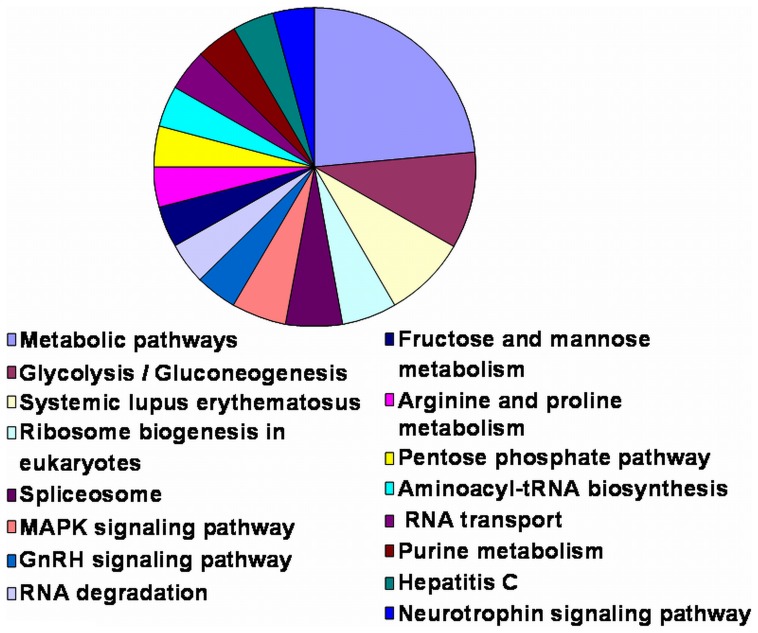
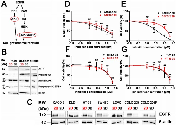
Three-dimensional (3D) tumor cell cultures grown in laminin-rich-extracellular matrix (lrECM) are considered to reflect human tumors more realistic as compared to cells grown as monolayer on plastic. Here, we systematically investigated the impact of ECM on phenotype, gene expression, EGFR signaling pathway, and on EGFR inhibition in commonly used colorectal cancer (CRC) cell lines. LrECM on-top (3D) culture assays were performed with the CRC cell lines SW-480, HT-29, DLD-1, LOVO, CACO-2, COLO-205 and COLO-206F. Morphology of lrECM cultivated CRC cell lines was determined by phase contrast and confocal laser scanning fluorescence microscopy. Proliferation of cells was examined by MTT assay, invasive capacity of the cell lines was assayed using Matrigel-coated Boyden chambers, and migratory activity was determined employing the Fence assay. Differential gene expression was analyzed at the transcriptional level by the Agilent array platform. EGFR was inhibited by using the specific small molecule inhibitor AG1478. A specific spheroid growth pattern was observed for all investigated CRC cell lines. DLD-1, HT-29 and SW-480 and CACO-2 exhibited a clear solid tumor cell formation, while LOVO, COLO-205 and COLO-206F were characterized by forming grape-like structures. Although the occurrence of a spheroid morphology did not correlate with an altered migratory, invasive, or proliferative capacity of CRC cell lines, gene expression was clearly altered in cells grown on lrECM as compared to 2D cultures. Interestingly, in KRAS wild-type cell lines, inhibition of EGFR was less effective in lrECM (3D) cultures as compared to 2D cell cultures. Thus, comparing both 2D and 3D cell culture models, our data support the influence of the ECM on cancer growth. Compared to conventional 2D cell culture, the lrECM (3D) cell culture model offers the opportunity to investigate permanent CRC cell lines under more physiological conditions, i.e. in the context of molecular therapeutic targets and their pharmacological inhibition.
Overview
The claims and statistics of this paper haven’t been checked yet.
Run the check — free, no account needed. It reads the open-access text and checks the sources it cites.
Integrity signals
Registry
Cites
Statistics
Figures
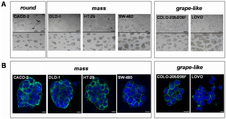
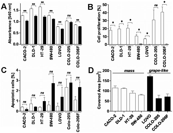
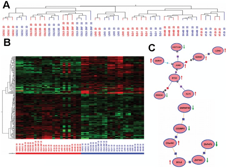
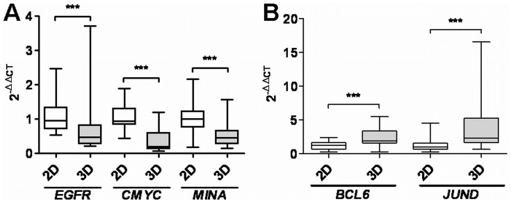
Verification
Claims the paper makes
The claims and statistics of this paper haven’t been checked yet.
Run the check — free, no account needed. It reads the open-access text and checks the sources it cites.
Claims other papers make about this one
Each is checked against this paper’s own text: does it say what the citing paper says it does?
The source backs part of the claim, not all of it — or backs it for a different population, condition or comparison.
In the citing paper
“The proliferation rates of cells cultured in 3D and 2D are usually different, and are cell line and matrix dependent.”
From Three-dimensional cell culture systems and their applications in drug discovery and cell-based biosensors (2014) · cites this paper as [29]
Experimental — accuracy not yet measuredThe cited source says what the claim attributes to it.
In the citing paper
“Overall, the morphological appearance of 3D spheroids is primarily cell line dependent. 29,35,37”
From Three-dimensional cell culture systems and their applications in drug discovery and cell-based biosensors (2014) · cites this paper as [29]
The cited source says what the claim attributes to it.
In the citing paper
“A variety of cell lines showed reduced proliferation rate in 3D cultures compared to those cultured in 2D. 17,27-31”
From Three-dimensional cell culture systems and their applications in drug discovery and cell-based biosensors (2014) · cites this paper as [29]
The check wasn’t confident either way, so it gives no verdict — never disputed.
In the citing paper
“A similar trend was observed in hepatocellular carcinoma models, where monolayers showed enrichment in structural gene expression, whereas spheroids upregulated genes related to metabolism and biosynthesis [196,197,198].”
From Modeling Tumor Microenvironment Complexity In Vitro: Spheroids as Physiologically Relevant Tumor Models and Strategies for Their Analysis (2025) · cites this paper as [197]
Experimental — accuracy not yet measured
Self-check
Percentages, sums and reported differences re-computed from the paper’s own numbers.
The self-check needs the paper’s full text.
Citations
Cites
Analysis of Relative Gene Expression Data Using Real-Time Quantitative PCR and the 2−ΔΔCT Method
Livak, K. J., & Schmittgen, T. D. (2001) · Methods · doi:10.1006/meth.2001.1262 · Cited by 180,860
Integrins: Versatility, modulation, and signaling in cell adhesion
Hynes, R. O. (1992) · Cell · doi:10.1016/0092-8674(92)90115-s · Cited by 9,857
Wild-Type BRAF Is Required for Response to Panitumumab or Cetuximab in Metastatic Colorectal Cancer
Di Nicolantonio, F., Martini, M., Molinari, F., Sartore-Bianchi, A., Arena, S., Saletti, P., De Dosso, S., Mazzucchelli, L., Frattini, M., Siena, S., et al. (2008) · Journal of Clinical Oncology · doi:10.1200/jco.2008.18.0786 · Cited by 1,591 · cited 1×
“KRAS and BRAF mutational status of patients is usually assessed before EGFR treatment on the basis that only patients exhibiting BRAF and KRAS wild type profit from treatments with anti-EGFR therapies [29,30].”
BackgroundBasement membrane complexes with biological activity
Kleinman, H. K., McGarvey, M. L., Hassell, J. R., Star, V. L., Cannon, F. B., Laurie, G. W., & Martin, G. R. (1986) · Biochemistry · doi:10.1021/bi00350a005 · Cited by 1,502
Three-dimensional culture models of normal and malignant breast epithelial cells
Lee, G. Y., Kenny, P. A., Lee, E. H., & Bissell, M. J. (2007) · Nature Methods · doi:10.1038/nmeth1015 · Cited by 1,336
Kleinman, H. K., McGarvey, M. L., Liotta, L. A., Robey, P. G., Tryggvason, K., & Martin, G. R. (1982) · Biochemistry · doi:10.1021/bi00267a025 · Cited by 1,231
PETERSEN, O. W., Rønnov‐Jessen, L., Howlett, A. R., & Bissell, M. J. (1992) · Proceedings of the National Academy of Sciences · doi:10.1073/pnas.89.19.9064 · Cited by 1,096
Kenny, P. A., Lee, G. Y., Myers, C. A., Neve, R. M., Semeiks, J. R., Spellman, P. T., Lorenz, K., Lee, E. H., Barcellos‐Hoff, M. H., PETERSEN, O. W., et al. (2008) · eScholarship (California Digital Library) · Cited by 1,039
Benvenuti, S., Sartore-Bianchi, A., Di Nicolantonio, F., Zanon, C., Moroni, M., Veronese, S., Siena, S., & Bardelli, A. (2007) · Cancer Research · doi:10.1158/0008-5472.can-06-4158 · Cited by 830
Weaver, V. M., Lelièvre, S. A., Lakins, J. N., Chrenek, M. A., Jones, J., Giancotti, F. G., Werb, Z., & Bissell, M. J. (2002) · Cancer Cell · doi:10.1016/s1535-6108(02)00125-3 · Cited by 794
Research works printed but not linked: 1
- PD Yurchenco, 1992, Laminin forms an independent network in basement membranes, J Cell Biol, 117, 1119
Cited by
Sorted by citations
Edmondson, R., Broglie, J. J., Adcock, A. F., & Yang, L. (2014) · ASSAY and Drug Development Technologies · doi:10.1089/adt.2014.573 · Cited by 2,515
3D Cell Culture Systems: Advantages and Applications
Ravi, M., Paramesh, V., Kaviya, S. R., Anuradha, E., & Solomon, F. D. P. (2015) · Journal of Cellular Physiology · doi:10.1002/jcp.24683 · Cited by 1,206
3D tumor spheroids: an overview on the tools and techniques used for their analysis
Costa, E. C., Moreira, A. F., de Melo-Diogo, D., Gaspar, V. M., Carvalho, M. P., & Correia, I. J. (2016) · Biotechnology Advances · doi:10.1016/j.biotechadv.2016.11.002 · Cited by 929
In Vitro Tumor Models: Advantages, Disadvantages, Variables, and Selecting the Right Platform
Katt, M. E., Placone, A. L., Wong, A. D., Xu, Z. S., & Searson, P. C. (2016) · Frontiers in Bioengineering and Biotechnology · doi:10.3389/fbioe.2016.00012 · Cited by 754
3D In Vitro Model (R)evolution: Unveiling Tumor–Stroma Interactions
Rodrigues, J., Heinrich, M. A., Teixeira, L. M., & Prakash, J. (2021) · Trends in Cancer · doi:10.1016/j.trecan.2020.10.009 · Cited by 405
“Gene expression Cell adhesion-, proliferation-, and survival-related genes are usually modified Accurate representation of gene expression patterns [16,85,86]”
Background
Metadata
- Authors
- Luca, A. C., Mersch, S., Deenen, R., Schmidt, S., Messner, I., Schäfer, K.-L., Baldus, S. E., Huckenbeck, W., Piekorz, R. P., Knoefel, W. T., Krieg, A., & Stoecklein, N. H.
- Affiliations
- Düsseldorf University Hospital (DE)
- Heinrich Heine University Düsseldorf (DE)
- Corresponding author
- Krieg, Andreas
- Stoecklein, Nikolas H.
- ORCID
- Venue
- PLoS ONE
- Publisher
- Public Library of Science (PLoS)
- Published
- 2013-03-26
- Volume
- 8
- Issue
- 3
- Pages
- e59689
- ISSN
- 1932-6203 (ISSN-L)
- PMC
- PMC3608563
- OpenAlex
- W1983051794
- Cited by
- 341
- References
- 31
- Citations by year
- 2026: 11 · 2025: 14 · 2024: 18 · 2023: 29 · 2022: 27 · 2021: 39 · 2020: 40 · 2019: 29 · 2018: 25 · 2017: 33 · 2016: 29 · 2015: 18 · 2014: 25 · 2013: 4
- FWCI
- 13.56
- Type
- journal article
- Language
- en
- Open access
- gold
- doi.org
- Locations
- PLoS ONE (publishedVersion, cc-by)
- Europe PMC (PubMed Central) (publishedVersion, other-oa)
- PubMed Central (publishedVersion, cc-by)
- PubMed (publishedVersion)
- DOAJ (DOAJ: Directory of Open Access Journals) (submittedVersion, cc-by-sa)
- Univ. Duesseldorf: Duesseldorfer Dokumenten- und Publikationsserver (submittedVersion)
- Figshare (submittedVersion, cc-by)
- Funders
- Deutsche Forschungsgemeinschaft (SFB728)
- Topics
- Health Sciences › Medicine › Oncology › Cancer Cells and Metastasis
- Life Sciences › Biochemistry, Genetics and Molecular Biology › Cell Biology › Cellular Mechanics and Interactions
- Physical Sciences › Engineering › Biomedical Engineering › 3D Printing in Biomedical Research
- Keywords
- colorectal cancer, 3D cell culture, tumor microenvironment, EGFR signaling, EGFR inhibition, AG1478, gene expression, cell morphology, spheroid formation, cell invasion, cell migration, KRAS wild-type, 2D vs 3D culture comparison, colorectal cancer cell lines
- MeSH terms
- Extracellular Matrix, Humans, Laminin, Phenotype, Colorectal Neoplasms, Gene Expression Regulation, Neoplastic, Spheroids, Cellular, Cell Culture Techniques, Cell Line, Tumor, Protein Kinase Inhibitors, Cell Proliferation, Tumor Microenvironment, ErbB Receptors
About these checks
Registry, retraction, Cites and statistics results are computed, not judged, with one exception: a printed reference that no index lists is read once by a model before it counts as a citation error. Claims are extracted by a model and kept only when their quote appears verbatim in the text; each cited source is then checked, with a model’s help, for whether it says what the claim relies on. Those verdicts can be wrong — read the claim’s quote and the cited source before relying on one.
Statistics and arithmetic are recomputed, not judged.
Confidence bands are shown only for fields calibrated on our annotated test set. None is calibrated yet, so no band is shown.
What counts as a claim ›How we measure confidence ›How we check this ›
Are you an author, or do you see a problem with this page? Request a review
