

The DOI matches its Crossref record; 38 of the 38 references it cites were found in a registry or an index.
Rapamycin attenuates the progression of tau pathology in P301S tau transgenic mice.
Open accessAbstract
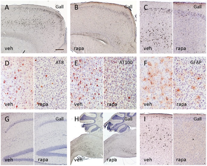
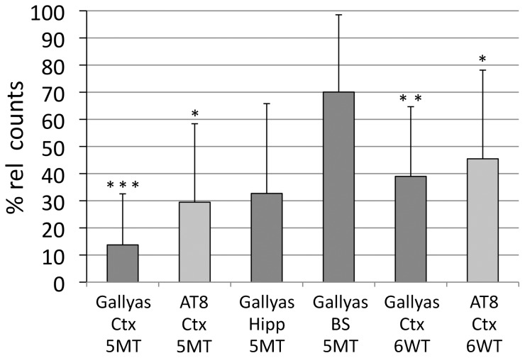
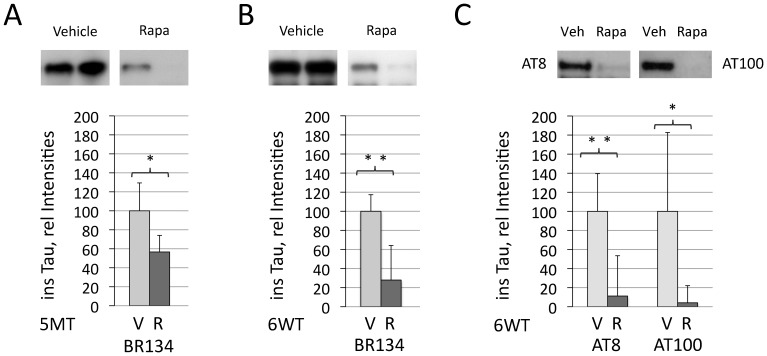
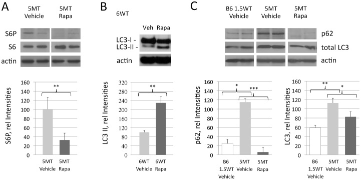
Altered autophagy contributes to the pathogenesis of Alzheimer's disease and other tauopathies, for which curative treatment options are still lacking. We have recently shown that trehalose reduces tau pathology in a tauopathy mouse model by stimulation of autophagy. Here, we studied the effect of the autophagy inducing drug rapamycin on the progression of tau pathology in P301S mutant tau transgenic mice. Rapamycin treatment resulted in a significant reduction in cortical tau tangles, less tau hyperphosphorylation, and lowered levels of insoluble tau in the forebrain. The favourable effect of rapamycin on tau pathology was paralleled by a qualitative reduction in astrogliosis. These effects were visible with early preventive or late treatment. We further noted an accumulation of the autophagy associated proteins p62 and LC3 in aged tangle bearing P301S mice that was lowered upon rapamycin treatment. Thus, rapamycin treatment defers the progression of tau pathology in a tauopathy animal model and autophagy stimulation may constitute a therapeutic approach for patients suffering from tauopathies.
Overview
The claims and statistics of this paper haven’t been checked yet.
Run the check — free, no account needed. It reads the open-access text and checks the sources it cites.
Integrity signals
Registry
Cites
Statistics
Figures
Verification
Claims the paper makes
The claims and statistics of this paper haven’t been checked yet.
Run the check — free, no account needed. It reads the open-access text and checks the sources it cites.
Claims other papers make about this one
Each is checked against this paper’s own text: does it say what the citing paper says it does?
The cited source says what the claim attributes to it.
In the citing paper
“Rapamycin treatment inhibits both tau hyperphosphorylation (Liu et al., 2013; Ozcelik et al., 2013; Tramutola et al., 2017) and the development of status epilepticus and chronic epilepsy in models of pharmacological seizure induction (Zeng et al., 2008; Huang et al., 2010), TLE (Drion et al., 2016), and PTE (Guo et al., 2013; Butler et al., 2015).”
From Tauopathy and Epilepsy Comorbidities and Underlying Mechanisms (2022) · cites this paper as Ozcelik et al., 2013
Self-check
Percentages, sums and reported differences re-computed from the paper’s own numbers.
The self-check needs the paper’s full text.
Citations
Cites
Repertoires of Autophagy in the Pathogenesis of Ocular Diseases
Li, Y., Jiang, Q. L., Cao, G.-F., Yao, J., & Yan, B. (2015) · Cellular Physiology and Biochemistry · doi:10.1159/000373980 · Cited by 16,351
Autophagy fights disease through cellular self-digestion
Mizushima, N., Levine, B., Cuervo, A. M., & Klionsky, D. J. (2008) · Nature · doi:10.1038/nature06639 · Cited by 6,452
West, M. J., Slomianka, L., & Gundersen, H. J. G. (1991) · The Anatomical Record · doi:10.1002/ar.1092310411 · Cited by 2,994
Goedert, M., Spillantini, M. G., Jakes, R., Rutherford, D., & Crowther, R. A. (1989) · Neuron · doi:10.1016/0896-6273(89)90210-9 · Cited by 2,492
Self-citationRavikumar, B., Vacher, C., Berger, Z., Davies, J. E., Luo, S., Oroz, L. G., Scaravilli, F., Easton, D. F., Duden, R., O'Kane, C. J., et al. (2004) · Nature Genetics · doi:10.1038/ng1362 · Cited by 2,347
Synapse Loss and Microglial Activation Precede Tangles in a P301S Tauopathy Mouse Model
Yoshiyama, Y., Higuchi, M., Zhang, B., Huang, S.-M., Iwata, N., Saido, T. C., Maeda, J., Suhara, T., Trojanowski, J. Q., & Lee, V. M.-Y. (2007) · Neuron · doi:10.1016/j.neuron.2007.01.010 · Cited by 2,208
A Century of Alzheimer's Disease
Goedert, M., & Spillantini, M. G. (2006) · Science · doi:10.1126/science.1132814 · Cited by 2,150
Self-citationThe roles of intracellular protein-degradation pathways in neurodegeneration
Rubinsztein, D. C. (2006) · Nature · doi:10.1038/nature05291 · Cited by 1,625
Boland, B., Kumar, A., Lee, S., Platt, F. M., Wegiel, J., Yu, W. H., & Nixon, R. A. (2008) · Journal of Neuroscience · doi:10.1523/jneurosci.0800-08.2008 · Cited by 1,119
Alzheimer's disease: strategies for disease modification
Citron, M. (2010) · Nature Reviews Drug Discovery · doi:10.1038/nrd2896 · Cited by 1,075
Cited by
Sorted by citations
Tau in physiology and pathology
Wang, Y., & Mandelkow, E. (2016) · Nature Reviews Neuroscience · doi:10.1038/nrn.2015.1 · Cited by 2,251
Autophagy and Neurodegeneration: Pathogenic Mechanisms and Therapeutic Opportunities
Menzies, F. M., Fleming, A., Caricasole, A., Bento, C. F., Andrews, S. P., Ashkenazi, A., Füllgrabe, J., Jackson, A., Jimenez Sanchez, M., Karabiyik, C., et al. (2017) · Neuron · doi:10.1016/j.neuron.2017.01.022 · Cited by 1,134
The mechanistic target of rapamycin: the grand conducTOR of metabolism and aging
Kennedy, B. K., & Lamming, D. W. (2016) · Cell Metabolism · doi:10.1016/j.cmet.2016.05.009 · Cited by 544
The quest to slow ageing through drug discovery
Partridge, L., Fuentealba, M., & Kennedy, B. K. (2020) · Nature Reviews Drug Discovery · doi:10.1038/s41573-020-0067-7 · Cited by 468
The Autophagy–Lysosomal Pathway in Neurodegeneration: A TFEB Perspective
Martini-Stoica, H., Xu, Y., Ballabio, A., & Zheng, H. (2016) · Trends in Neurosciences · doi:10.1016/j.tins.2016.02.002 · Cited by 436
Metadata
- Authors
- Ozcelik, S., Fraser, G., Castets, P., Schaeffer, V., Skachokova, Z., Breu, K., Clavaguera, F., Sinnreich, M., Kappos, L., Goedert, M., Tolnay, M., & Winkler, D. T.
- Affiliations
- MRC Laboratory of Molecular Biology (GB)
- University Hospital of Basel (CH)
- Corresponding author
- Winkler, David Theo
- ORCID
- Venue
- PLoS ONE
- Publisher
- Public Library of Science (PLoS)
- Published
- 2013-05-07
- Volume
- 8
- Issue
- 5
- Pages
- e62459
- ISSN
- 1932-6203 (ISSN-L)
- PMC
- PMC3646815
- OpenAlex
- W1989723790
- Cited by
- 226
- References
- 38
- Citations by year
- 2026: 7 · 2025: 16 · 2024: 9 · 2023: 15 · 2022: 27 · 2021: 26 · 2020: 31 · 2019: 17 · 2018: 17 · 2017: 17 · 2016: 18 · 2015: 17 · 2014: 7 · 2013: 2
- FWCI
- 10.42
- Type
- journal article
- Language
- en
- Open access
- gold
- doi.org
- Locations
- PLoS ONE (publishedVersion, cc-by)
- Europe PMC (PubMed Central) (publishedVersion, cc-by)
- PubMed Central (publishedVersion, cc-by)
- PubMed (publishedVersion)
- Archive ouverte UNIGE (University of Geneva) (submittedVersion, cc-by)
- DOAJ (DOAJ: Directory of Open Access Journals) (submittedVersion)
- Figshare (submittedVersion, cc-by)
- Funders
- National Science Foundation
- University of Pennsylvania
- Velux Stiftung
- Parkinson's UK (K-1012)
- Schweizerischer Nationalfonds zur Förderung der Wissenschaftlichen Forschung (135214, 123812, 310030)
- Topics
- Life Sciences › Neuroscience › Cellular and Molecular Neuroscience › Alzheimer's disease research and treatments
- Life Sciences › Biochemistry, Genetics and Molecular Biology › Cell Biology › Autophagy in Disease and Therapy
- Life Sciences › Neuroscience › Neurology › Neuroinflammation and Neurodegeneration Mechanisms
- Keywords
- rapamycin, tau pathology, P301S tau transgenic mice, tauopathy, Alzheimer's disease, autophagy, tau hyperphosphorylation, tau tangles, astrocyte activation, p62, LC3, trehalose, neuroprotection
- MeSH terms
- Animals, Astrocytes, Humans, Mice, Transgenic, Phosphorylation, Solubility, Time Factors, tau Proteins, Disease Progression, Sirolimus, Tauopathies, Mice
About these checks
Registry, retraction, Cites and statistics results are computed, not judged, with one exception: a printed reference that no index lists is read once by a model before it counts as a citation error. Claims are extracted by a model and kept only when their quote appears verbatim in the text; each cited source is then checked, with a model’s help, for whether it says what the claim relies on. Those verdicts can be wrong — read the claim’s quote and the cited source before relying on one.
Statistics and arithmetic are recomputed, not judged.
Confidence bands are shown only for fields calibrated on our annotated test set. None is calibrated yet, so no band is shown.
What counts as a claim ›How we measure confidence ›How we check this ›
Are you an author, or do you see a problem with this page? Request a review
