Cited by 321Cell death mechanisms and regulationUbiquitin and proteasome pathwaysNF-κB Signaling Pathways

The DOI matches its Crossref record; 48 of the 48 references it cites were found in a registry or an index.

CYLD Deubiquitinates RIP1 in the TNFα-Induced Necrosome to Facilitate Kinase Activation and Programmed Necrosis

Open access

Abstract

BACKGROUND: Necroptosis/programmed necrosis is initiated by a macro-molecular protein complex termed the necrosome. Receptor interacting protein kinase 1 (RIPK1/RIP1) and RIP3 are key components of the necrosome. TNFα is a prototypic inducer of necrosome activation, and it is widely believed that deubiquitination of RIP1 at the TNFR-1 signaling complex precedes transition of RIP1 into the cytosol where it forms the RIP1-RIP3 necrosome. Cylindromatosis (CYLD) is believed to promote programmed necrosis by facilitating RIP1 deubiquitination at this membrane receptor complex. METHODOLOGY/PRINCIPAL FINDINGS: We demonstrate that RIP1 is indeed the primary target of CYLD in TNFα-induced programmed necrosis. We observed that CYLD does not regulate RIP1 ubiquitination at the TNF receptor. TNF and zVAD-induced programmed necrosis was highly attenuated in CYLD(-/-) cells. However, in the presence of cycloheximide or SMAC mimetics, programmed necrosis was only moderately reduced in CYLD(-/-) cells. Under the latter conditions, RIP1-RIP3 necrosome formation is only delayed, but not abolished in CYLD(-/-) cells. We further demonstrate that RIP1 within the NP-40 insoluble necrosome is ubiquitinated and that CYLD regulates RIP1 ubiquitination in this compartment. Hence, RIP1 ubiquitination in this late-forming complex is greatly increased in CYLD(-/-) cells. Increased RIP1 ubiquitination impairs RIP1 and RIP3 phosphorylation, a signature of kinase activation. CONCLUSIONS/SIGNIFICANCE: Our results show that CYLD regulates RIP1 ubiquitination in the TNFα-induced necrosome, but not in the TNFR-1 signaling complex. In cells sensitized to programmed necrosis with SMAC mimetics, CYLD is not essential for necrosome assembly. Since SMAC mimetics induces the loss of the E3 ligases cIAP1 and cIAP2, reduced RIP1 ubiquitination could lead to reduced requirement for CYLD to remove ubiquitin chains from RIP1 in the TNFR-1 complex. As increased RIP1 ubiquitination in the necrosome correlates with impaired RIP1 and RIP3 phosphorylation and function, these results suggest that CYLD controls RIP1 kinase activity during necrosome assembly.

Overview

The claims and statistics of this paper haven’t been checked yet.

Run the check — free, no account needed. It reads the open-access text and checks the sources it cites.

A few free checks a day · papers already verified open instantly · results are public

Integrity signals

Registry

Crossref · 10.1371/journal.pone.0076841

Cites

48 of 48 references verified0 citation errors · 0 not checkable

Statistics

Figures

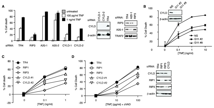
Figure 1 Partial protection against TNF-induced necrosis by CYLD siRNAs. (A) FADD-deficient Jurkat cells were transiently transfected with the indicated siRNAs. Forty-eight hours post-transfection, the cells were treated with TNF to induce necrosis. The percentage cell death was determined by propidium iodide staining and flow cytometry. Western Blots on the right show the efficiency of gene silencing. Representative of three experiments. (B) HT-29 cells were stably transfected with either non-specific shRNA (control) or CYLD targeting shRNA (clones: G11-2 and G11-8). Cells were treated with TNF, LBW242 and zVAD-fmk. Cell viability was determined by MTS assay (Promega). Western blot shows that expression of CYLD was inhibited in the two selected clones, G11 #2 and G11 #8. Representative of three experiments. (C-D) L929 cells were transfected with the indicated siRNAs. Necrosis was induced with (C) TNF or (D) TNF and zVAD-fmk. Cell death was determined by MTS assay. The panel on the right shows the reduction in protein expression of the siRNA transfected cells. Representative of two experiments.
Figure 1 Partial protection against TNF-induced necrosis by CYLD siRNAs. (A) FADD-deficient Jurkat cells were transiently transfected with the indicated siRNAs. Forty-eight hours post-transfection, the cells were treated with TNF to induce necrosis. The percentage cell death was determined by propidium iodide staining and flow cytometry. Western Blots on the right show the efficiency of gene silencing. Representative of three experiments. (B) HT-29 cells were stably transfected with either non-specific shRNA (control) or CYLD targeting shRNA (clones: G11-2 and G11-8). Cells were treated with TNF, LBW242 and zVAD-fmk. Cell viability was determined by MTS assay (Promega). Western blot shows that expression of CYLD was inhibited in the two selected clones, G11 #2 and G11 #8. Representative of three experiments. (C-D) L929 cells were transfected with the indicated siRNAs. Necrosis was induced with (C) TNF or (D) TNF and zVAD-fmk. Cell death was determined by MTS assay. The panel on the right shows the reduction in protein expression of the siRNA transfected cells. Representative of two experiments.CC BY · PubMed Central
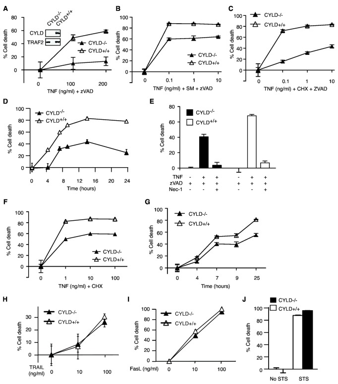
Figure 2 RIP1-dependent necrosis occurs in CYLD-/- MEFs. Wild type (CYLD+/+) and CYLD-/- MEFs were treated with (A) TNF and zVAD, (B) TNF, Smac mimetic and zVAD, or (C) TNF, cycloheximide (CHX) and zVAD-fmk for 12 hours. Cell death was determined by staining with propidium iodide (PI) and analyzed via flow cytometry. The inset shows loss of CYLD expression in CYLD-/- MEFs using a C-terminal specific CYLD antibody. In (D), cells were treated with 10 ng/ml TNF, CHX and zVAD-fmk and cell death was measured at the indicated times following treatment. Representative of three experiments. (E) Nec-1 inhibited TNF-induced necrosis in CYLD-/- MEFs. Representative of two experiments. (F) CYLD+/+ and CYLD-/- MEFs were treated with TNF and CHX for 12 hours. Cell death was determined by PI staining and flow cytometry. Representative of three experiments. (G) MEFs were treated with 10 ng/ml TNF and CHX for the indicated times. Representative of two experiments. (H-J) MEFs were treated with (H) TRAIL, (I) FasL or (J) staurosporine (STS) for 12 hours. Cell death was determined by PI exclusion and flow cytometry.
Figure 2 RIP1-dependent necrosis occurs in CYLD-/- MEFs. Wild type (CYLD+/+) and CYLD-/- MEFs were treated with (A) TNF and zVAD, (B) TNF, Smac mimetic and zVAD, or (C) TNF, cycloheximide (CHX) and zVAD-fmk for 12 hours. Cell death was determined by staining with propidium iodide (PI) and analyzed via flow cytometry. The inset shows loss of CYLD expression in CYLD-/- MEFs using a C-terminal specific CYLD antibody. In (D), cells were treated with 10 ng/ml TNF, CHX and zVAD-fmk and cell death was measured at the indicated times following treatment. Representative of three experiments. (E) Nec-1 inhibited TNF-induced necrosis in CYLD-/- MEFs. Representative of two experiments. (F) CYLD+/+ and CYLD-/- MEFs were treated with TNF and CHX for 12 hours. Cell death was determined by PI staining and flow cytometry. Representative of three experiments. (G) MEFs were treated with 10 ng/ml TNF and CHX for the indicated times. Representative of two experiments. (H-J) MEFs were treated with (H) TRAIL, (I) FasL or (J) staurosporine (STS) for 12 hours. Cell death was determined by PI exclusion and flow cytometry.CC BY · PubMed Central
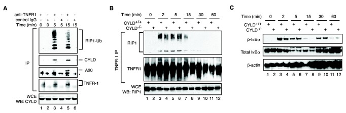
Figure 3 CYLD does not regulate RIP1 ubiquitination within the TNFR1 complex. (A) Recruitment of CYLD and A20 to the TNFR-1 complex. TNFR1 complex was purified from TNF, zVAD and CHX treated cells by immunoprecipitation (IP). The recruitment of RIP1, CYLD and A20 was assessed via Western Blot. Control IPs with isotype-matched IgG were included to show the specificity of binding to TNFR-1. (B) CYLD+/+ and CYLD-/- MEFs were treated with TNF for the indicated times. Recruitment of polyubiquitinated RIP1 to TNFR-1 was determined by Western Blot. (C) IκBα phosphorylation and degradation was normal in CYLD-/- MEFs.
Figure 3 CYLD does not regulate RIP1 ubiquitination within the TNFR1 complex. (A) Recruitment of CYLD and A20 to the TNFR-1 complex. TNFR1 complex was purified from TNF, zVAD and CHX treated cells by immunoprecipitation (IP). The recruitment of RIP1, CYLD and A20 was assessed via Western Blot. Control IPs with isotype-matched IgG were included to show the specificity of binding to TNFR-1. (B) CYLD+/+ and CYLD-/- MEFs were treated with TNF for the indicated times. Recruitment of polyubiquitinated RIP1 to TNFR-1 was determined by Western Blot. (C) IκBα phosphorylation and degradation was normal in CYLD-/- MEFs.CC BY · PubMed Central
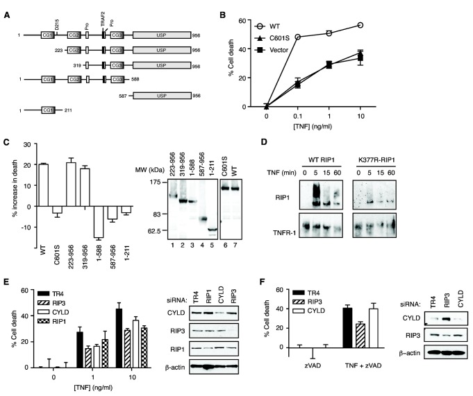
Figure 4 Poly-ubiquitinated RIP1 is the major substrate for CYLD in TNF-induced necrosis. (A) Schematic diagram of wild-type CYLD and deletion mutants used in the experiments. (B-C) CYLD-/- MEFs were transiently transfected with the indicated GFP-tagged CYLD. Necrosis was induced with TNF, CHX and zVAD-fmk. Cell death was determined in the GFP+ population by PI staining and flow cytometry. The panel on the right of (C) shows that the GFP-tagged CYLD mutants were of the correct sizes. (D) RIP1-deficient Jurkat cells reconstituted with wild type RIP1 or RIP1-K377R were stimulated with TNF. The recruitment of RIP1 to TNFR-1 was determined by Western blot. (E) RIP1-deficient Jurkat cells stably expressing either WT RIP1-GFP or (F) RIP1-K377R-GFP mutant were transfected with the indicated siRNAs. Necrosis was induced with TNF and zVAD-fmk. Cell death was determined as described before [48]. The Western blots provide validation of siRNA knock-down efficiency.
Figure 4 Poly-ubiquitinated RIP1 is the major substrate for CYLD in TNF-induced necrosis. (A) Schematic diagram of wild-type CYLD and deletion mutants used in the experiments. (B-C) CYLD-/- MEFs were transiently transfected with the indicated GFP-tagged CYLD. Necrosis was induced with TNF, CHX and zVAD-fmk. Cell death was determined in the GFP+ population by PI staining and flow cytometry. The panel on the right of (C) shows that the GFP-tagged CYLD mutants were of the correct sizes. (D) RIP1-deficient Jurkat cells reconstituted with wild type RIP1 or RIP1-K377R were stimulated with TNF. The recruitment of RIP1 to TNFR-1 was determined by Western blot. (E) RIP1-deficient Jurkat cells stably expressing either WT RIP1-GFP or (F) RIP1-K377R-GFP mutant were transfected with the indicated siRNAs. Necrosis was induced with TNF and zVAD-fmk. Cell death was determined as described before [48]. The Western blots provide validation of siRNA knock-down efficiency.CC BY · PubMed Central
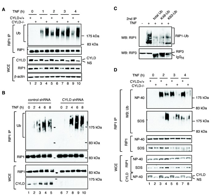
Figure 5 CYLD regulates RIP1 ubiquitination in the necrosome. (A-B) Increased RIP1 ubiquitination in the absence of CYLD. (A) CYLD+/+ and CYLD-/- MEFs or (B) control and CYLD knock-down HT29 cells were treated with TNF, the Smac mimetic LBW242 [35] and zVAD-fmk for the indicated times. Total lysis in 1% SDS was performed as described in the methods. The level of RIP1 ubiquitination was examined by Western Blot. NS: non-specific band. (C) Necrosome-associated RIP1 contains ubiquitin chains of different linkage types. Cells were treated with TNF and zVAD-fmk for 3 hours or left untreated. RIP3 immune complexes were denatured in urea, followed by immunoprecipitation with the indicated antibodies against ubiquitin. (D) Selective accumulation of poly-ubiquitinated proteins in RIP1 complexes in the NP-40 insoluble compartment. WT MEFs or CYLD-/- MEFs were treated to undergo necrosis with TNF, zVAD-fmk, and CHX for the indicated times. Cells were lysed in NP-40 lysis buffer and insoluble material was solubilized with SDS. RIP1 was immunoprecipitated from both fractions followed by Western blot with the indicated antibodies.
Figure 5 CYLD regulates RIP1 ubiquitination in the necrosome. (A-B) Increased RIP1 ubiquitination in the absence of CYLD. (A) CYLD+/+ and CYLD-/- MEFs or (B) control and CYLD knock-down HT29 cells were treated with TNF, the Smac mimetic LBW242 [35] and zVAD-fmk for the indicated times. Total lysis in 1% SDS was performed as described in the methods. The level of RIP1 ubiquitination was examined by Western Blot. NS: non-specific band. (C) Necrosome-associated RIP1 contains ubiquitin chains of different linkage types. Cells were treated with TNF and zVAD-fmk for 3 hours or left untreated. RIP3 immune complexes were denatured in urea, followed by immunoprecipitation with the indicated antibodies against ubiquitin. (D) Selective accumulation of poly-ubiquitinated proteins in RIP1 complexes in the NP-40 insoluble compartment. WT MEFs or CYLD-/- MEFs were treated to undergo necrosis with TNF, zVAD-fmk, and CHX for the indicated times. Cells were lysed in NP-40 lysis buffer and insoluble material was solubilized with SDS. RIP1 was immunoprecipitated from both fractions followed by Western blot with the indicated antibodies.CC BY · PubMed Central
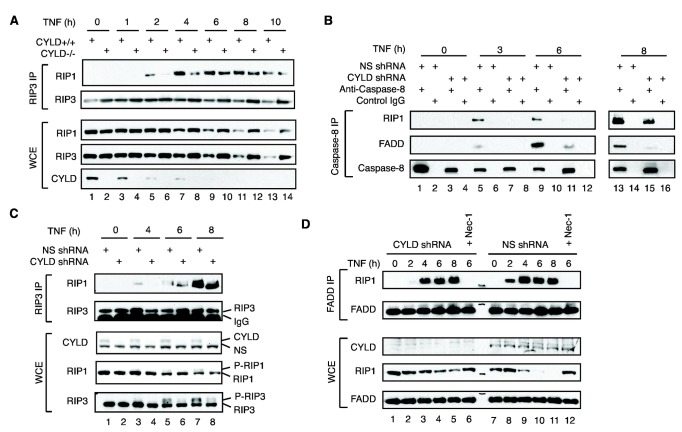
Figure 6 CYLD regulates the kinetics of RIP1-RIP3 necrosome assembly. (A) WT or CYLD-/- MEFs were treated with TNF, LBW242 and zVAD-fmk for the indicated times. Recruitment of RIP1 to RIP3 was determined by Western Blot. (B) HT-29 cells expressing non-specific shRNA (NS) or CYLD shRNA were treated with TNF, LBW242 and zVAD-fmk. RIP3 complexes were immunoprecipitated and RIP1 association was determined by Western Blot. (C) HT-29 cells were treated with TNF, the SM LBW242 and zVAD-fmk. Caspase-8 complexes were immunoprecipitated, and recruitment of RIP1 and FADD was determined by Western blot. (D) HT-29 cells were treated with TNF, Smac mimetic and zVAD-fmk. FADD complexes were immunoprecipitated, and recruitment of RIP1 was determined by Western blot.
Figure 6 CYLD regulates the kinetics of RIP1-RIP3 necrosome assembly. (A) WT or CYLD-/- MEFs were treated with TNF, LBW242 and zVAD-fmk for the indicated times. Recruitment of RIP1 to RIP3 was determined by Western Blot. (B) HT-29 cells expressing non-specific shRNA (NS) or CYLD shRNA were treated with TNF, LBW242 and zVAD-fmk. RIP3 complexes were immunoprecipitated and RIP1 association was determined by Western Blot. (C) HT-29 cells were treated with TNF, the SM LBW242 and zVAD-fmk. Caspase-8 complexes were immunoprecipitated, and recruitment of RIP1 and FADD was determined by Western blot. (D) HT-29 cells were treated with TNF, Smac mimetic and zVAD-fmk. FADD complexes were immunoprecipitated, and recruitment of RIP1 was determined by Western blot.CC BY · PubMed Central
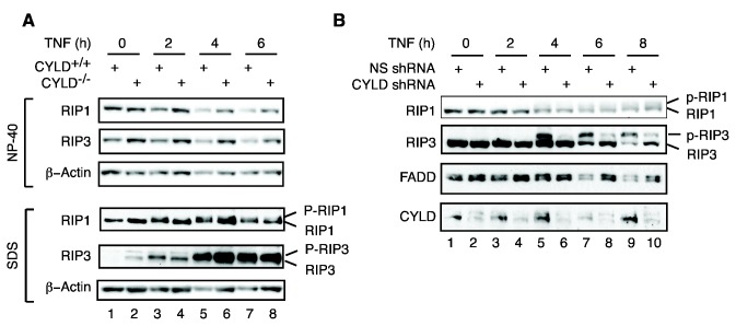
Figure 7 Impaired RIP1 and RIP3 phosphorylation in the absence of CYLD. (A) MEFs were treated with TNF, zVAD-fmk and the LBW242. Cell lysates were extracted by sequential detergent lysis in NP-40 and SDS as described in methods. RIP1 and RIP3 in each fraction were examined by Western blot. Note that phosphorylated RIP1 and RIP3 (p-RIP1 and p-RIP3) were exclusively detected in the SDS fractions. (B) HT-29 cells stably expressing non-specific (NS) shRNA or CYLD shRNA were treated with TNF, LBW242 and zVAD-fmk for the indicated times. Unmodified and phospho-RIP1 and phospho-RIP3 were analyzed by Western Blot.
Figure 7 Impaired RIP1 and RIP3 phosphorylation in the absence of CYLD. (A) MEFs were treated with TNF, zVAD-fmk and the LBW242. Cell lysates were extracted by sequential detergent lysis in NP-40 and SDS as described in methods. RIP1 and RIP3 in each fraction were examined by Western blot. Note that phosphorylated RIP1 and RIP3 (p-RIP1 and p-RIP3) were exclusively detected in the SDS fractions. (B) HT-29 cells stably expressing non-specific (NS) shRNA or CYLD shRNA were treated with TNF, LBW242 and zVAD-fmk for the indicated times. Unmodified and phospho-RIP1 and phospho-RIP3 were analyzed by Western Blot.CC BY · PubMed Central
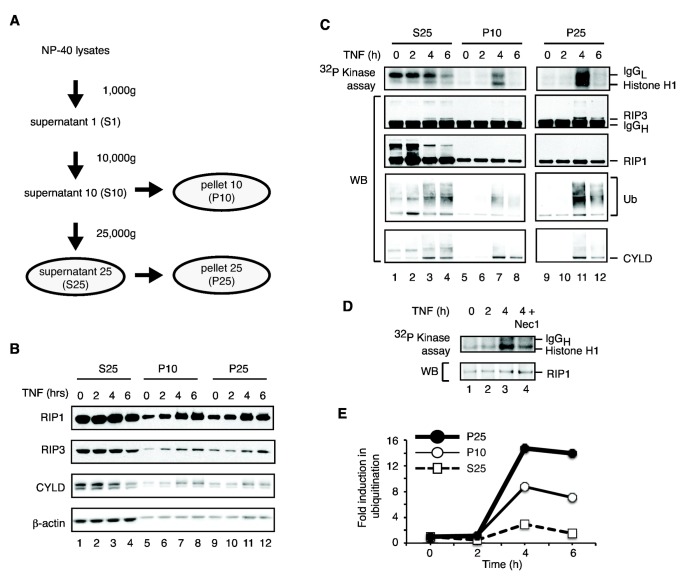
Figure 8 Induction of RIP1 associated kinase activity in the NP-40 insoluble compartment. (A) Schematic flowchart showing the procedures by which the different NP-40 soluble and insoluble fractions were obtained. (B) Accumulation of necrosis signaling adaptors in the NP-40 insoluble fractions. The indicated fractions were analyzed for levels of RIP1, RIP3 and CYLD in response to necrosis induction by TNF. β-actin was used as internal control. (C) The kinase activity of the necrosome is selectively activated in the NP-40 insoluble fractions. Cell lysates were subjected to differential centrifugation as described in (A). The RIP1 immune complexes were subjected to in vitro kinase assays using histone H1 as substrate. RIP1 ubiquitination and recruitment of RIP3 and CYLD were determined by Western blot (lower panels). (D) Induction of kinase activity in the NP-40 insoluble fraction is partially dependent on RIP1 kinase activity. The P10 fractions were prepared and RIP1 immune complexes were isolated. Where indicated, 30 µM of Nec-1 was added to the in vitro kinase assay. (E) Densitometry quantification of RIP1 ubiquitination in the different fractions in (C).
Figure 8 Induction of RIP1 associated kinase activity in the NP-40 insoluble compartment. (A) Schematic flowchart showing the procedures by which the different NP-40 soluble and insoluble fractions were obtained. (B) Accumulation of necrosis signaling adaptors in the NP-40 insoluble fractions. The indicated fractions were analyzed for levels of RIP1, RIP3 and CYLD in response to necrosis induction by TNF. β-actin was used as internal control. (C) The kinase activity of the necrosome is selectively activated in the NP-40 insoluble fractions. Cell lysates were subjected to differential centrifugation as described in (A). The RIP1 immune complexes were subjected to in vitro kinase assays using histone H1 as substrate. RIP1 ubiquitination and recruitment of RIP3 and CYLD were determined by Western blot (lower panels). (D) Induction of kinase activity in the NP-40 insoluble fraction is partially dependent on RIP1 kinase activity. The P10 fractions were prepared and RIP1 immune complexes were isolated. Where indicated, 30 µM of Nec-1 was added to the in vitro kinase assay. (E) Densitometry quantification of RIP1 ubiquitination in the different fractions in (C).CC BY · PubMed Central
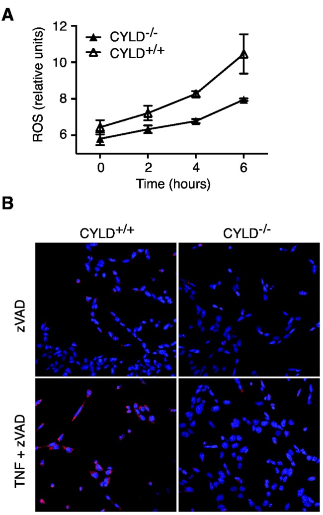
Figure 9 ROS production is reduced in CYLD-/- MEFs. (A) CYLD-/- or CYLD+/+ MEFs were stimulated with TNF and zVAD for the indicated times. ROS generation was determined by staining with CM-H 2DCFDA as per manufacturer’s instructions. Results shown are average +/- SEM of triplicates. (B) CYLD-/- or CYLD+/+ MEFs were stimulated with TNF and zVAD. ROS production was determined by staining with MitoSox as per manufacturer’s instructions.
Figure 9 ROS production is reduced in CYLD-/- MEFs. (A) CYLD-/- or CYLD+/+ MEFs were stimulated with TNF and zVAD for the indicated times. ROS generation was determined by staining with CM-H 2DCFDA as per manufacturer’s instructions. Results shown are average +/- SEM of triplicates. (B) CYLD-/- or CYLD+/+ MEFs were stimulated with TNF and zVAD. ROS production was determined by staining with MitoSox as per manufacturer’s instructions.CC BY · PubMed Central
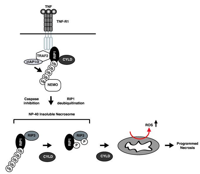
Figure 10 Schematic diagram of the proposed mechanism of CYLD-mediated necrosis. Although CYLD is recruited to the TNFR-1 complex, it dose not deubiquitinate RIP1 within the this compartment. Upon transition to the cytosol, CYLD deubiquitinates RIP1 within the NP-40 insoluble fraction to actively promote necrosome phosphorylation and activation. In addition to regulating RIP1 ubiquitination in the necrosome, CYLD also controls downstream events of necrosis such as ROS production.
Figure 10 Schematic diagram of the proposed mechanism of CYLD-mediated necrosis. Although CYLD is recruited to the TNFR-1 complex, it dose not deubiquitinate RIP1 within the this compartment. Upon transition to the cytosol, CYLD deubiquitinates RIP1 within the NP-40 insoluble fraction to actively promote necrosome phosphorylation and activation. In addition to regulating RIP1 ubiquitination in the necrosome, CYLD also controls downstream events of necrosis such as ROS production.CC BY · PubMed Central

Verification

Claims the paper makes

The claims and statistics of this paper haven’t been checked yet.

Run the check — free, no account needed. It reads the open-access text and checks the sources it cites.

A few free checks a day · papers already verified open instantly · results are public

Claims other papers make about this one

Each is checked against this paper’s own text: does it say what the citing paper says it does?

Self-check

Percentages, sums and reported differences re-computed from the paper’s own numbers.

The self-check needs the paper’s full text.

Citations

Cites

Show all 48 references

Cited by

Sorted by citations

See all 321

Metadata

Authors
Moquin, D. M., McQuade, T., & Chan, F. K.-M.
Affiliations
University of Massachusetts Chan Medical School (US)
Corresponding author
Chan, Francis Ka-Ming
Venue
PLoS ONE
Published
2013-10-02
Volume
8
Issue
10
Pages
e76841
ISSN
1932-6203 (ISSN-L)
OpenAlex
W2060803227
Cited by
320
References
48
Citations by year
2026: 14 · 2025: 22 · 2024: 18 · 2023: 29 · 2022: 21 · 2021: 33 · 2020: 18 · 2019: 26 · 2018: 30 · 2017: 25 · 2016: 33 · 2015: 19 · 2014: 31 · 2013: 1
FWCI
12.55
Type
journal article
Language
en
Open access
Locations
  • PLoS ONE (publishedVersion, cc-by)
  • Europe PMC (PubMed Central) (publishedVersion, cc-by)
  • PubMed Central (publishedVersion, cc-by)
  • PubMed (publishedVersion)
  • DOAJ (DOAJ: Directory of Open Access Journals) (submittedVersion)
Funders
  • National Institutes of Health (NIH T32, AI083497)
  • National Institute of Allergy and Infectious Diseases (T32 AI07349, R21AI088502, AI083497, AI088502, R01AI083497, T32AI007349)
Topics
  • Life Sciences › Biochemistry, Genetics and Molecular Biology › Molecular Biology › Cell death mechanisms and regulation
  • Life Sciences › Biochemistry, Genetics and Molecular Biology › Molecular Biology › Ubiquitin and proteasome pathways
  • Life Sciences › Biochemistry, Genetics and Molecular Biology › Cancer Research › NF-κB Signaling Pathways
Keywords
CYLD, RIPK1, RIPK3, necrosome, necroptosis, TNF signaling, deubiquitination, ubiquitination, IAP antagonists, cIAP1, cIAP2, E3 ubiquitin ligase, zVAD
MeSH terms
Deubiquitinating Enzyme CYLD, Animals, Cycloheximide, Cysteine Endopeptidases, Gene Expression Regulation, Humans, Necrosis, Oligopeptides, Phosphorylation, Tumor Necrosis Factor-alpha, Signal Transduction, GTPase-Activating Proteins, Cell Line, Tumor, Receptors, Tumor Necrosis Factor, Type I, Mice, Receptor-Interacting Protein Serine-Threonine Kinases, Ubiquitination, Gene Knockdown Techniques

About these checks

Registry, retraction, Cites and statistics results are computed, not judged, with one exception: a printed reference that no index lists is read once by a model before it counts as a citation error. Claims are extracted by a model and kept only when their quote appears verbatim in the text; each cited source is then checked, with a model’s help, for whether it says what the claim relies on. Those verdicts can be wrong — read the claim’s quote and the cited source before relying on one.

Statistics and arithmetic are recomputed, not judged.

Confidence bands are shown only for fields calibrated on our annotated test set. None is calibrated yet, so no band is shown.

What counts as a claim ›How we measure confidence ›How we check this ›

Are you an author, or do you see a problem with this page? Request a review