

The DOI matches its Crossref record; 76 of the 76 references it cites were found in a registry or an index.
Identify Changes of Brain Regional Homogeneity in Bipolar Disorder and Unipolar Depression Using Resting-State fMRI
Open accessAbstract
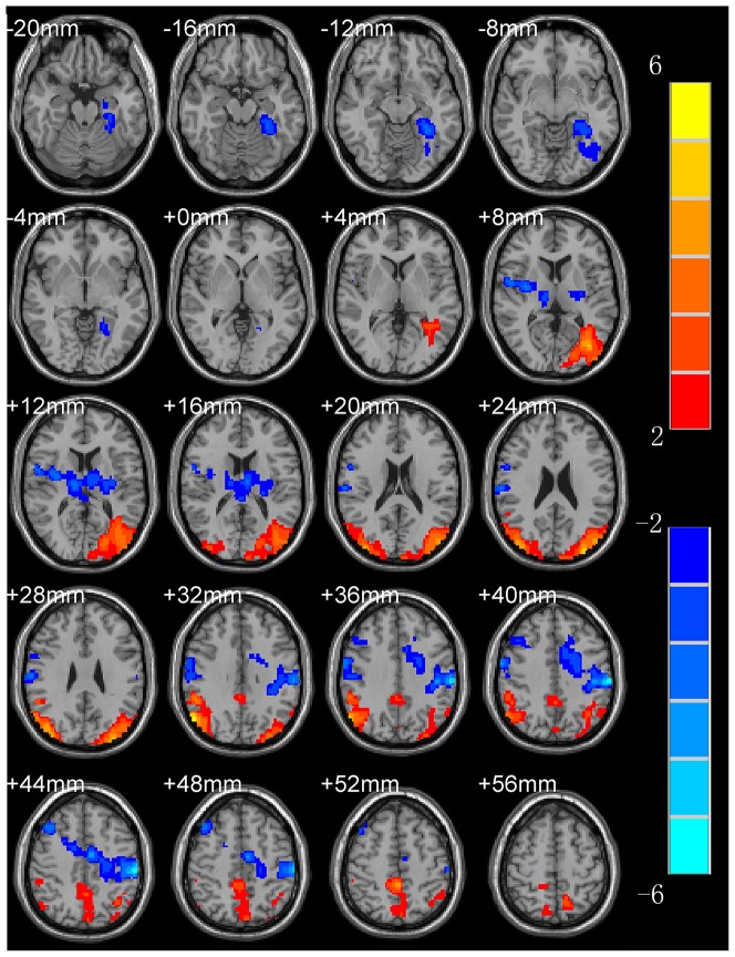
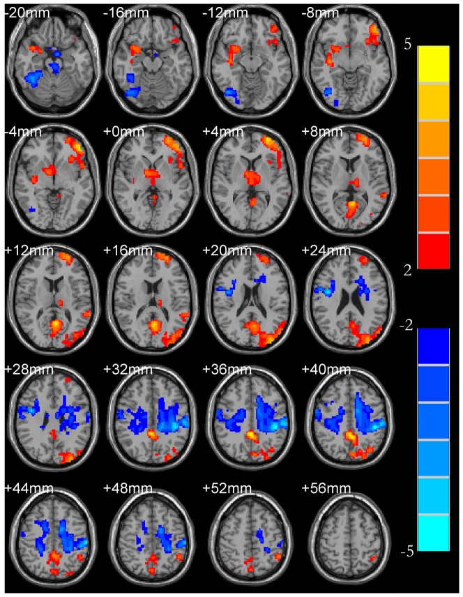
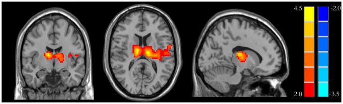
BACKGROUND: To identify changes in brain activation patterns in bipolar disorder (BD) and unipolar depression (UD) patients. METHODOLOGY/PRINCIPAL FINDINGS: Resting-state fMRI scans of 16 healthy controls, 17 BD and 16 UD patients were obtained. T-test of normalized regional homogeneity (ReHo) was performed in a voxel-by-voxel manner. A combined threshold of á = 0.05, minimum cluster volume of V = 10503 mm(3) (389 voxels) were used to determine ReHo differences between groups. In UD group, fMRI revealed ReHo increases in the left middle occipital lobe, right inferior parietal lobule, right precuneus and left convolution; and ReHo decreases in the left parahippocampalgyrus, right precentralgyrus, left postcentralgyrus, left precentralgyrus and left cingulated. In BD group, ReHo increases in the right insular cortex, left middle frontal gyrus, left precuneus, left occipital lobe, left parietal, left superior frontal gyrus and left thalamus; and ReHo decreases in the right anterior lobe of cerebellum, pons, right precentralgyrus, left postcentralgyrus, left inferior frontal gyrus, and right cingulate. There were some overlaps in ReHo profiles between UD and BD groups, but a marked difference was seen in the thalamus of BD. CONCLUSIONS/SIGNIFICANCE: The resting-state fMRI and ReHo mapping are a promising tool to assist the detection of functional deficits and distinguish clinical and pathophysiological signs of BD and UD.
Overview
The claims and statistics of this paper haven’t been checked yet.
Run the check — free, no account needed. It reads the open-access text and checks the sources it cites.
Integrity signals
Registry
Cites
Statistics
Figures
Verification
Claims the paper makes
The claims and statistics of this paper haven’t been checked yet.
Run the check — free, no account needed. It reads the open-access text and checks the sources it cites.
Claims other papers make about this one
Each is checked against this paper’s own text: does it say what the citing paper says it does?
The source backs part of the claim, not all of it — or backs it for a different population, condition or comparison.
In the citing paper
“In bipolar disorder, subjects show reduced cerebellar volumes (Baldacara et al., 2011; DelBello et al., 1999; Mills et al., 2005), abnormal cerebellar functional connectivity (Liang et al., 2013), and abnormal cerebellar metabolism (Johnson et al., 2018; Magnotta et al., 2022) compared to neuronormative populations.”
From A double-blind, randomized, sham-controlled clinical trial of cerebellar intermittent theta burst stimulation for bipolar disorder: Safety, tolerability, mood and cognitive effects (2025) · cites this paper as Liang et al., 2013
Experimental — accuracy not yet measured
Self-check
Percentages, sums and reported differences re-computed from the paper’s own numbers.
The self-check needs the paper’s full text.
Citations
Cites
Functional connectivity in the motor cortex of resting human brain using echo‐planar mri
Biswal, B., Zerrin Yetkin, F., Haughton, V. M., & Hyde, J. S. (1995) · Magnetic Resonance in Medicine · doi:10.1002/mrm.1910340409 · Cited by 9,812
A Rating Scale for Mania: Reliability, Validity and Sensitivity
Young, R. C., Biggs, J. T., Ziegler, V. E., & Meyer, D. A. (1978) · British Journal of Psychiatry · doi:10.1192/bjp.133.5.429 · Cited by 8,744
The human brain is intrinsically organized into dynamic, anticorrelated functional networks
Fox, M. D., Snyder, A. Z., Vincent, J. L., Corbetta, M., Van Essen, D. C., & Raichle, M. E. (2005) · Proceedings of the National Academy of Sciences · doi:10.1073/pnas.0504136102 · Cited by 8,553
Spontaneous fluctuations in brain activity observed with functional magnetic resonance imaging
Fox, M. D., & Raichle, M. E. (2007) · Nature Reviews Neuroscience · doi:10.1038/nrn2201 · Cited by 6,700
Functional connectivity in the resting brain: a network analysis of the default mode hypothesis
Greicius, M. D., Krasnow, B., Reiss, A. L., & Menon, V. (2003) · Proceedings of the National Academy of Sciences · doi:10.1073/pnas.0135058100 · Cited by 6,358
Cognitive and emotional influences in anterior cingulate cortex
Bush, G., Luu, P., & Posner, M. I. (2000) · Trends in Cognitive Sciences · doi:10.1016/s1364-6613(00)01483-2 · Cited by 6,259
Consistent resting-state networks across healthy subjects
Damoiseaux, J. S., Rombouts, S. A. R. B., Barkhof, F., Scheltens, P., Stam, C. J., Smith, S. M., & Beckmann, C. F. (2006) · Proceedings of the National Academy of Sciences · doi:10.1073/pnas.0601417103 · Cited by 4,326
DPARSF: a MATLAB toolbox for “pipeline” data analysis of resting-state fMRI
Yan (2010) · Frontiers in System Neuroscience · doi:10.3389/fnsys.2010.00013 · Cited by 3,475
Investigations into resting-state connectivity using independent component analysis
Beckmann, C. F., DeLuca, M., Devlin, J. T., & Smith, S. M. (2005) · Philosophical Transactions of the Royal Society B: Biological Sciences · doi:10.1098/rstb.2005.1634 · Cited by 3,398
Altered baseline brain activity in children with ADHD revealed by resting-state functional MRI
Yu-Feng, Z., Yong, H., Chao-Zhe, Z., Qing-Jiu, C., Man-Qiu, S., Meng, L., Li-Xia, T., Tian-Zi, J., & Yu-Feng, W. (2007) · Brain and Development · doi:10.1016/j.braindev.2006.07.002 · Cited by 2,660
Self-citation
Research works printed but not linked: 1
- S Lui, 2011, Resting-State Functional Connectivity in Treatment-Resistant Depression, Am J Psychiatry, 168, 642, 10.1176/appi.ajp.2010.10101419 · DOI · Paper page
Cited by
Sorted by citations
Consensus Paper: Cerebellum and Emotion
Adamaszek, M., D’Agata, F., Ferrucci, R., Habas, C., Keulen, S., Kirkby, K. C., Leggio, M. G., Mariën, P., Molinari, M., Moulton, E. A., et al. (2016) · The Cerebellum · doi:10.1007/s12311-016-0815-8 · Cited by 583
Brain structure alterations in depression: Psychoradiological evidence
Zhang, F., Peng, W. J., Sweeney, J. A., Jia, Z., & Gong, Q. (2018) · CNS Neuroscience & Therapeutics · doi:10.1111/cns.12835 · Cited by 507
The Cerebellum and Psychiatric Disorders
Phillips, J. R., Hewedi, D. H., Eissa, A. M., & Moustafa, A. A. (2015) · Frontiers in Public Health · doi:10.3389/fpubh.2015.00066 · Cited by 357
Resting state networks in major depressive disorder
Dutta, A., McKie, S., & Deakin, J. F. W. (2014) · Psychiatry Research: Neuroimaging · doi:10.1016/j.pscychresns.2014.10.003 · Cited by 218
Zhong, X. B., Pu, W., & Yao, S. (2016) · Journal of Affective Disorders · doi:10.1016/j.jad.2016.09.005 · Cited by 163
Metadata
- Authors
- Liang, M.-J., Zhou, Q., Yang, K.-R., Yang, X.-L., Fang, J., Chen, W.-L., & Huang, Z.
- Affiliations
- First Affiliated Hospital of Jinan University (CN)
- Corresponding author
- Zhou, Quan
- ORCID
- Zhou, Quan
- Venue
- PLoS ONE
- Publisher
- Public Library of Science (PLoS)
- Published
- 2013-12-04
- Volume
- 8
- Issue
- 12
- Pages
- e79999
- ISSN
- 1932-6203 (ISSN-L)
- PMC
- PMC3851159
- OpenAlex
- W2001770029
- Cited by
- 135
- References
- 76
- Citations by year
- 2026: 2 · 2025: 5 · 2024: 11 · 2023: 10 · 2022: 8 · 2021: 9 · 2020: 13 · 2019: 13 · 2018: 19 · 2017: 13 · 2016: 14 · 2015: 13 · 2014: 5
- FWCI
- 5.85
- Type
- journal article
- Language
- en
- Open access
- gold
- doi.org
- Licence
- Locations
- PLoS ONE (publishedVersion, cc-by)
- Europe PMC (PubMed Central) (publishedVersion, cc-by)
- PubMed Central (publishedVersion, cc-by)
- PubMed (publishedVersion)
- DOAJ (DOAJ: Directory of Open Access Journals) (submittedVersion, cc-by-sa)
- Funders
- Capital Medical University
- Jinan University
- Topics
- Life Sciences › Neuroscience › Cognitive Neuroscience › Functional Brain Connectivity Studies
- Health Sciences › Medicine › Psychiatry and Mental health › Bipolar Disorder and Treatment
- Social Sciences › Psychology › Experimental and Cognitive Psychology › Mental Health Research Topics
- Keywords
- bipolar disorder, major depressive disorder, regional homogeneity, resting-state brain activity, thalamus, brain activation patterns, functional deficits, neuroimaging biomarkers
- MeSH terms
- Adolescent, Adult, Bipolar Disorder, Brain Mapping, Cerebellum, Depressive Disorder, Female, Frontal Lobe, Humans, Magnetic Resonance Imaging, Male, Middle Aged, Occipital Lobe, Parietal Lobe, Thalamus, Case-Control Studies
About these checks
Registry, retraction, Cites and statistics results are computed, not judged, with one exception: a printed reference that no index lists is read once by a model before it counts as a citation error. Claims are extracted by a model and kept only when their quote appears verbatim in the text; each cited source is then checked, with a model’s help, for whether it says what the claim relies on. Those verdicts can be wrong — read the claim’s quote and the cited source before relying on one.
Statistics and arithmetic are recomputed, not judged.
Confidence bands are shown only for fields calibrated on our annotated test set. None is calibrated yet, so no band is shown.
What counts as a claim ›How we measure confidence ›How we check this ›
Are you an author, or do you see a problem with this page? Request a review
