

The DOI matches its Crossref record; 47 of the 47 references it cites were found in a registry or an index.
Cannabinoid CB2 Receptors Regulate Central Sensitization and Pain Responses Associated with Osteoarthritis of the Knee Joint
Open accessAbstract
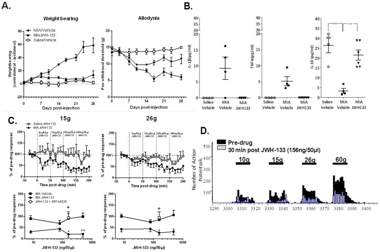
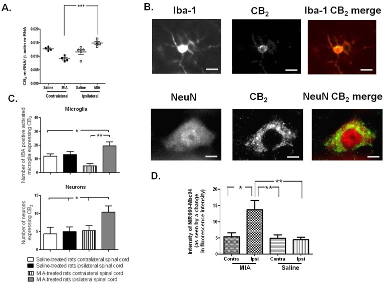
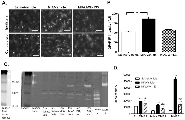
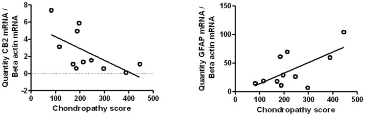
Osteoarthritis (OA) of the joint is a prevalent disease accompanied by chronic, debilitating pain. Recent clinical evidence has demonstrated that central sensitization contributes to OA pain. An improved understanding of how OA joint pathology impacts upon the central processing of pain is crucial for the identification of novel analgesic targets/new therapeutic strategies. Inhibitory cannabinoid 2 (CB2) receptors attenuate peripheral immune cell function and modulate central neuro-immune responses in models of neurodegeneration. Systemic administration of the CB2 receptor agonist JWH133 attenuated OA-induced pain behaviour, and the changes in circulating pro- and anti-inflammatory cytokines exhibited in this model. Electrophysiological studies revealed that spinal administration of JWH133 inhibited noxious-evoked responses of spinal neurones in the model of OA pain, but not in control rats, indicating a novel spinal role of this target. We further demonstrate dynamic changes in spinal CB2 receptor mRNA and protein expression in an OA pain model. The expression of CB2 receptor protein by both neurones and microglia in the spinal cord was significantly increased in the model of OA. Hallmarks of central sensitization, significant spinal astrogliosis and increases in activity of metalloproteases MMP-2 and MMP-9 in the spinal cord were evident in the model of OA pain. Systemic administration of JWH133 attenuated these markers of central sensitization, providing a neurobiological basis for analgesic effects of the CB2 receptor in this model of OA pain. Analysis of human spinal cord revealed a negative correlation between spinal cord CB2 receptor mRNA and macroscopic knee chondropathy. These data provide new clinically relevant evidence that joint damage and spinal CB2 receptor expression are correlated combined with converging pre-clinical evidence that activation of CB2 receptors inhibits central sensitization and its contribution to the manifestation of chronic OA pain. These findings suggest that targeting CB2 receptors may have therapeutic potential for treating OA pain.
Overview
The claims and statistics of this paper haven’t been checked yet.
Run the check — free, no account needed. It reads the open-access text and checks the sources it cites.
Integrity signals
Registry
Cites
Statistics
Figures
Verification
Claims the paper makes
The claims and statistics of this paper haven’t been checked yet.
Run the check — free, no account needed. It reads the open-access text and checks the sources it cites.
Claims other papers make about this one
Each is checked against this paper’s own text: does it say what the citing paper says it does?
In the citing paper
“Administration of MIA into the knee joint leads to upregulation of CB 2 and CB 1 receptor mRNA in the ipsilateral spinal cord of rats, suggesting that these receptors undergo compensatory changes in this osteoarthritis model, and may represent potential targets for novel osteoarthritis treatments (Burston et al, 2013;Malek et al, 2015).”
From The endogenous cannabinoid system: a budding source of targets for treating inflammatory and neuropathic pain (2018) · cites this paper as Burston et al, 2013
In the citing paper
“MIA induces chondrocyte death, cartilage degradation, and chronic nociception (Burston et al, 2013).”
From The endogenous cannabinoid system: a budding source of targets for treating inflammatory and neuropathic pain (2018) · cites this paper as Burston et al, 2013
In the citing paper
“suggesting that these receptors undergo compensatory changes in this osteoarthritis model, and may represent potential targets for novel osteoarthritis treatments (Burston et al, 2013;Malek et al, 2015).”
From The endogenous cannabinoid system: a budding source of targets for treating inflammatory and neuropathic pain (2018) · cites this paper as Burston et al, 2013
In the citing paper
“Furthermore, the CB 2 receptor agonist JWH133 attenuates MIA-induced allodynia and restores weight bearing on the arthritic joint (Burston et al, 2013).”
From The endogenous cannabinoid system: a budding source of targets for treating inflammatory and neuropathic pain (2018) · cites this paper as Burston et al, 2013
In the citing paper
“Intrathecal injection of JWH133 also attenuates the firing of wide dynamic range neurons after MIA injection, indicating CB 2 receptor activation in the spinal cord may attenuate MIA-induced allodynia (Burston et al, 2013).”
From The endogenous cannabinoid system: a budding source of targets for treating inflammatory and neuropathic pain (2018) · cites this paper as Burston et al, 2013
In the citing paper
“indicating CB 2 receptor activation in the spinal cord may attenuate MIA-induced allodynia (Burston et al, 2013).”
From The endogenous cannabinoid system: a budding source of targets for treating inflammatory and neuropathic pain (2018) · cites this paper as Burston et al, 2013
Self-check
Percentages, sums and reported differences re-computed from the paper’s own numbers.
The self-check needs the paper’s full text.
Citations
Cites
Cellular and Molecular Mechanisms of Pain
Basbaum, A. I., Bautista, D. M., Scherrer, G., & Julius, D. (2009) · Cell · doi:10.1016/j.cell.2009.09.028 · Cited by 4,344
Neuronal plasticity: Increasing the gain in pain
Woolf, C. J., & Salter, M. W. (2000) · Science · doi:10.1126/science.288.5472.1765 · Cited by 3,781 · cited 1×
“Central sensitization plays a pivotal role in the switch from acute to chronic pain mechanisms [5,6] and the”
BackgroundThe role of synovitis in pathophysiology and clinical symptoms of osteoarthritis
Sellam, J., & Berenbaum, F. (2010) · Nature Reviews Rheumatology · doi:10.1038/nrrheum.2010.159 · Cited by 1,500
Pathogenesis and management of pain in osteoarthritis
Dieppe, P. A., & Lohmander, S. L. (2005) · The Lancet · doi:10.1016/s0140-6736(05)71086-2 · Cited by 1,214
Kawasaki, Y., Zhang, L., Cheng, J.-K., & Ji, R.-R. (2008) · The Journal of Neuroscience · doi:10.1523/jneurosci.3338-07.2008 · Cited by 1,162
Models and Mechanisms of Hyperalgesia and Allodynia
Sandkühler, J. (2009) · Physiological Reviews · doi:10.1152/physrev.00025.2008 · Cited by 1,148
Sensitization in patients with painful knee osteoarthritis
Arendt‐Nielsen, L., Nie, H., Laursen, M. B., Laursen, B. S., Madeleine, P., Simonsen, O. H., & Graven‐Nielsen, T. (2010) · Pain · doi:10.1016/j.pain.2010.04.003 · Cited by 992
Zhuang, Z., Gerner, P., Woolf, C. J., & Ji, R. (2005) · Pain · doi:10.1016/j.pain.2004.12.022 · Cited by 774
Kawasaki, Y., Xu, Z., Wang, X., Park, J. Y., Zhuang, Z., Tan, P.-H., Gao, Y., Roy, K., Corfas, G., Lo, E. H., et al. (2008) · Nature Medicine · doi:10.1038/nm1723 · Cited by 759
Chemokines, neuronal–glial interactions, and central processing of neuropathic pain
Gao, Y., & Ji, R. (2010) · Pharmacology & Therapeutics · doi:10.1016/j.pharmthera.2010.01.002 · Cited by 636
Bove, S. E., Calcaterra, S., Brooker, R. M., Huber, C. M., Guzman, R. E., Juneau, P. L., Schrier, D. J., & Kilgore, K. S. (2003) · Osteoarthritis and Cartilage · doi:10.1016/s1063-4584(03)00163-8 · Cited by 508
Guzman, R. E., Evans, M. G., Bove, S. E., Morenko, B. J., & Kilgore, K. S. (2003) · Toxicologic Pathology · doi:10.1080/01926230390241800 · Cited by 464
Gwilym, S. E., Keltner, J. R., Warnaby, C. E., Carr, A. J., Chizh, B. A., Chessell, I. P., & Tracey, I. (2009) · Arthritis Care & Research · doi:10.1002/art.24837 · Cited by 431
Pain related behaviour in two models of osteoarthritis in the rat knee
Fernihough, J. K., Gentry, C., Malcangio, M., Fox, A., Rediske, J. J., Pellas, T. C., Kidd, B. L., Bevan, S., & Winter, J. (2004) · Pain · doi:10.1016/j.pain.2004.08.004 · Cited by 427
Sohn, D., Sokolove, J., Sharpe, O., Erhart, J., Chandra, P. E., Lahey, L., Lindstrom, T., Hwang, I., Boyer, K., Andriacchi, T., et al. (2012) · Arthritis Research & Therapy · doi:10.1186/ar3555 · Cited by 338
Colburn, R. W., DeLeo, J. A., Rickman, A. J., Yeager, M. P., Kwon, P., & Hickey, W. F. (1997) · Journal of Neuroimmunology · doi:10.1016/s0165-5728(97)00119-7 · Cited by 334
Osteoarthritis and its association with muscle hyperalgesia: an experimental controlled study
Bajaj, P., Bajaj, P., Graven‐Nielsen, T., & Arendt‐Nielsen, L. (2001) · Pain · doi:10.1016/s0304-3959(01)00300-1 · Cited by 317
Huffman, J. W., Liddle, J., Yu, S., Aung, M. M., Abood, M. E., Wiley, J. L., & Martin, B. R. (1999) · Bioorganic & Medicinal Chemistry · doi:10.1016/s0968-0896(99)00219-9 · Cited by 290
The monosodium iodoacetate model of osteoarthritis: a model of chronic nociceptive pain in rats?
Combe, R., Bramwell, S., & Field, M. J. (2004) · Neuroscience Letters · doi:10.1016/j.neulet.2004.08.023 · Cited by 228
CB cannabinoid receptor agonists: pain relief without psychoactive effects?
MALANJR, T., IBRAHIM, M., LAI, J., Vanderah, T. W., Makriyannis, A., & Porreca, F. (2003) · Current Opinion in Pharmacology · doi:10.1016/s1471-4892(02)00004-8 · Cited by 226
Orita, S., Ishikawa, T., Miyagi, M., Ochiai, N., Inoue, G., Eguchi, Y., Kamoda, H., Arai, G., Toyone, T., Aoki, Y., et al. (2011) · BMC Musculoskeletal Disorders · doi:10.1186/1471-2474-12-134 · Cited by 220
Racz, I., Nadal, X., Alferink, J., Baños, J. E., Rehnelt, J., Martín, M., Pintado, B., Gutierrez-Adan, A., Sanguino, E., Manzanares, J., et al. (2008) · The Journal of Neuroscience · doi:10.1523/jneurosci.3400-08.2008 · Cited by 195
Janusz, M. J., Hookfin, E. B., Heitmeyer, S. A., Woessner, J. F., Freemont, A. J., Hoyland, J. A., Brown, K. K., Hsieh, L. C., Almstead, N. G., De, B., et al. (2001) · Osteoarthritis and Cartilage · doi:10.1053/joca.2001.0472 · Cited by 194
Erhuma, A. M., Salter, A. M., Sculley, D., Langley‐Evans, S. C., & Bennett, A. J. (2007) · American Journal of Physiology-Endocrinology and Metabolism · doi:10.1152/ajpendo.00605.2006 · Cited by 171
Self-citationRomero-Sandoval, A., Nutile-McMenemy, N., & DeLeo, J. A. (2008) · Anesthesiology · doi:10.1097/aln.0b013e318167af74 · Cited by 168
The SFA system for assessing articular cartilage lesions at arthroscopy of the knee
Dougados, M., Ayral, X., Listrat, V., Guéguen, A., Bahuaud, J., Beaufils, P., Beguin, J., Bonvarlet, J. P., Boyer, T., Coudane, H., et al. (1994) · Arthroscopy The Journal of Arthroscopic and Related Surgery · doi:10.1016/s0749-8063(05)80295-6 · Cited by 155
Sagar, D. R., Kelly, S., Millns, P. J., O'Shaughnessey, C. T., Kendall, D. A., & Chapman, V. (2005) · European Journal of Neuroscience · doi:10.1111/j.1460-9568.2005.04206.x · Cited by 154
Self-citationSodium Iodoacetate-Induced Experimental Osteoarthritis and Associated Pain Model in Rats
Kobayashi, K., Imaizumi, R., Sumichika, H., Tanaka, H., Goda, M., Fukunari, A., & Komatsu, H. (2003) · Journal of Veterinary Medical Science · doi:10.1292/jvms.65.1195 · Cited by 149
Functional CB2 type cannabinoid receptors at CNS synapses
Morgan, N. H., Stanford, I. M., & Woodhall, G. L. (2009) · Neuropharmacology · doi:10.1016/j.neuropharm.2009.07.017 · Cited by 146
Neuroimmune interactions and pain: focus on glial-modulating targets.
Romero-Sandoval, E. A., Horvath, R. J., & DeLeo, J. A. (2008) · PubMed · Cited by 140
Arthritis and pain. Future targets to control osteoarthritis pain
Dray, A., & Read, S. (2007) · Arthritis Research & Therapy · doi:10.1186/ar2178 · Cited by 128
Development of cross-tolerance between delta 9-tetrahydrocannabinol, CP 55,940 and WIN 55,212.
Fan, F., Compton, D. M., Ward, S. J., Melvin, L. S., & Martin, B. R. (1994) · Journal of Pharmacology and Experimental Therapeutics · doi:10.1016/s0022-3565(25)24023-x · Cited by 126
Sagar, D. R., Burston, J. J., Hathway, G. J., Woodhams, S. G., Pearson, R. G., Bennett, A. J., Kendall, D. A., Scammell, B. E., & Chapman, V. (2011) · Molecular Pain · doi:10.1186/1744-8069-7-88 · Cited by 125
Self-citationSagar, D. R., Staniaszek, L. E., Okine, B. N., Woodhams, S. G., Norris, L. M., Pearson, R. G., Garle, M. J., Alexander, S. P. H., Bennett, A. J., Barrett, D. A., et al. (2010) · Arthritis & Rheumatism · doi:10.1002/art.27698 · Cited by 123
Self-citationLuongo, L., Palazzo, E., Tambaro, S., Giordano, C. S. D. S., Gatta, L., Scafuro, M. A., Rossi, F., Lazzari, P., Pani, L., de Novellis, V., et al. (2009) · Neurobiology of Disease · doi:10.1016/j.nbd.2009.09.021 · Cited by 116
Arthroscopic evaluation of chondropathy in osteoarthritis of the knee.
Ayral, X., Dougados, M., Listrat, V., Bonvarlet, J. P., Simonnet, J., & Amor, B. (1996) · PubMed · Cited by 95
Yamamoto, W., Mikami, T., & Iwamura, H. (2008) · European Journal of Pharmacology · doi:10.1016/j.ejphar.2008.01.010 · Cited by 89 · cited 1×
“It is well accepted that activation of spinal CB2 receptors attenuates pain behaviour in models of neuropathic pain [25,26], via modulation of microglia and astrocytic pro- and antiinflammatory responses [27,28].”
BackgroundChanges in cannabinoid receptor binding and mRNA levels in several brain regions of aged rats
Berrendero, F., Romero, J., Garcı́a-Gil, L., Suarez, I., la Cruz, P. D., Ramos, J. A., & Fernández‐Ruíz, J. (1998) · Biochimica et Biophysica Acta (BBA) - Molecular Basis of Disease · doi:10.1016/s0925-4439(98)00042-8 · Cited by 85
Dynamic changes to the endocannabinoid system in models of chronic pain
Sagar, D. R., Burston, J. J., Woodhams, S. G., & Chapman, V. (2012) · Philosophical Transactions of the Royal Society B Biological Sciences · doi:10.1098/rstb.2011.0390 · Cited by 77
Self-citationLa Porta, C., Bura, S. A., Aracil-Fernández, A., Manzanares, J., & Maldonado, R. (2013) · Pain · doi:10.1016/j.pain.2012.10.009 · Cited by 75
CB1 cannabinoid receptor expression in brain regions associated with zebra finch song control
Soderstrom, K., & Johnson, F. (2000) · Brain Research · doi:10.1016/s0006-8993(99)02393-8 · Cited by 63
Okine, B. N., Norris, L. M., Woodhams, S. G., Burston, J. J., Patel, A., Alexander, S. P. H., Barrett, D. A., Kendall, D. A., Bennett, A. J., & Chapman, V. (2012) · British Journal of Pharmacology · doi:10.1111/j.1476-5381.2012.02028.x · Cited by 56
Self-citationLee, Y., Pai, M., Brederson, J., Wilcox, D. M., Hsieh, G. C., Jarvis, M. F., & Bitner, R. S. (2011) · Molecular Pain · doi:10.1186/1744-8069-7-39 · Cited by 55
Osteoarthritis year 2010 in review: pharmacological therapies
Berenbaum, F. (2011) · Osteoarthritis and Cartilage · doi:10.1016/j.joca.2011.01.019 · Cited by 48
Walsh, D. A., Yousef, A., McWilliams, D. F., Hill, R., Hargin, E., & Wilson, D. (2008) · Osteoarthritis and Cartilage · doi:10.1016/j.joca.2008.07.016 · Cited by 35
Self-citationTakeshita, M., Nakamura, J., Ohtori, S., Inoue, G., Orita, S., Miyagi, M., Ishikawa, T., & Takahashi, K. (2012) · Rheumatology · doi:10.1093/rheumatology/kes173 · Cited by 33
Miller, T. R., Wetter, J. B., Jarvis, M. F., & Bitner, R. S. (2012) · European Journal of Pain · doi:10.1002/j.1532-2149.2012.00232.x · Cited by 22
Cited by
Sorted by citations
Donvito, G., Nass, S. R., Wilkerson, J. L., Curry, Z. A., Schurman, L. D., Kinsey, S. G., & Lichtman, A. H. (2018) · Neuropsychopharmacology · doi:10.1038/npp.2017.204 · Cited by 301
Self-citationBaron, E. P. (2018) · Headache The Journal of Head and Face Pain · doi:10.1111/head.13345 · Cited by 268
Mechanisms of Osteoarthritic Pain. Studies in Humans and Experimental Models
Eitner, A., Hofmann, G., & Schaible, H. (2017) · Frontiers in Molecular Neuroscience · doi:10.3389/fnmol.2017.00349 · Cited by 229
The Role of the Endocannabinoid System in Pain
Woodhams, S. G., Sagar, D. R., Burston, J. J., & Chapman, V. (2015) · Handbook of experimental pharmacology · doi:10.1007/978-3-662-46450-2_7 · Cited by 216
Self-citationDeng, L., Guindon, J., Cornett, B., Makriyannis, A., Mackie, K., & Hohmann, A. G. (2014) · Biological Psychiatry · doi:10.1016/j.biopsych.2014.04.009 · Cited by 200
Metadata
- Authors
- Burston, J. J., Sagar, D. R., Shao, P., Bai, M., King, E., Brailsford, L., Turner, J. M., Hathway, G. J., Bennett, A. J., Walsh, D. A., Kendall, D. A., Lichtman, A., & Chapman, V.
- Affiliations
- University of Nottingham (GB)
- University of Pittsburgh (US)
- Virginia Commonwealth University (US)
- Corresponding author
- Chapman, Victoria
- ORCID
- Venue
- PLoS ONE
- Publisher
- Public Library of Science (PLoS)
- Published
- 2013-11-25
- Volume
- 8
- Issue
- 11
- Pages
- e80440
- ISSN
- 1932-6203 (ISSN-L)
- PMC
- PMC3840025
- OpenAlex
- W2119352256
- Cited by
- 103
- References
- 47
- Citations by year
- 2026: 5 · 2025: 4 · 2024: 5 · 2023: 8 · 2022: 4 · 2021: 10 · 2020: 9 · 2019: 4 · 2018: 11 · 2017: 15 · 2016: 8 · 2015: 13 · 2014: 7
- FWCI
- 6.43
- Type
- journal article
- Language
- en
- Open access
- gold
- doi.org
- Licence
- Locations
- PLoS ONE (publishedVersion, cc-by)
- Europe PMC (PubMed Central) (publishedVersion, cc-by)
- PubMed Central (publishedVersion, cc-by)
- PubMed (publishedVersion)
- ORCA Online Research @Cardiff (Cardiff University) (acceptedVersion)
- D-Scholarship@Pitt (University of Pittsburgh) (acceptedVersion)
- VCU Scholars Compass (Virginia Commonwealth University) (submittedVersion, cc-by)
- DOAJ (DOAJ: Directory of Open Access Journals) (submittedVersion, cc-by-sa)
- Funders
- Virginia Commonwealth University
- Universidade Federal de Minas Gerais
- Versus Arthritis (18769)
- National Institutes of Health
- Medical Research Council (MR/K00414X/1)
- National Institute on Drug Abuse (P01DA009789, P50DA005274)
- Topics
- Health Sciences › Medicine › Physiology (medical) › Pain Mechanisms and Treatments
- Health Sciences › Medicine › Pharmacology (medical) › Cannabis and Cannabinoid Research
- Health Sciences › Medicine › Rheumatology › Osteoarthritis Treatment and Mechanisms
- Keywords
- CB2 receptor, osteoarthritis pain, central sensitization, JWH-133, spinal cord, microglia, astrocytes, MMP-2, MMP-9, neuroinflammation, analgesic targets, chronic pain
- MeSH terms
- Animals, Cannabinoids, Electrophysiology, Enzyme-Linked Immunosorbent Assay, Humans, Pain, Spinal Cord, Rats, Sprague-Dawley, Osteoarthritis, Knee, Matrix Metalloproteinase 2, Matrix Metalloproteinase 9, Receptor, Cannabinoid, CB2, Rats
About these checks
Registry, retraction, Cites and statistics results are computed, not judged, with one exception: a printed reference that no index lists is read once by a model before it counts as a citation error. Claims are extracted by a model and kept only when their quote appears verbatim in the text; each cited source is then checked, with a model’s help, for whether it says what the claim relies on. Those verdicts can be wrong — read the claim’s quote and the cited source before relying on one.
Statistics and arithmetic are recomputed, not judged.
Confidence bands are shown only for fields calibrated on our annotated test set. None is calibrated yet, so no band is shown.
What counts as a claim ›How we measure confidence ›How we check this ›
Are you an author, or do you see a problem with this page? Request a review
