

The DOI matches its Crossref record; 37 of the 37 references it cites were found in a registry or an index.
Circulating Exosomal microRNAs as Biomarkers of Colon Cancer
Open accessAbstract
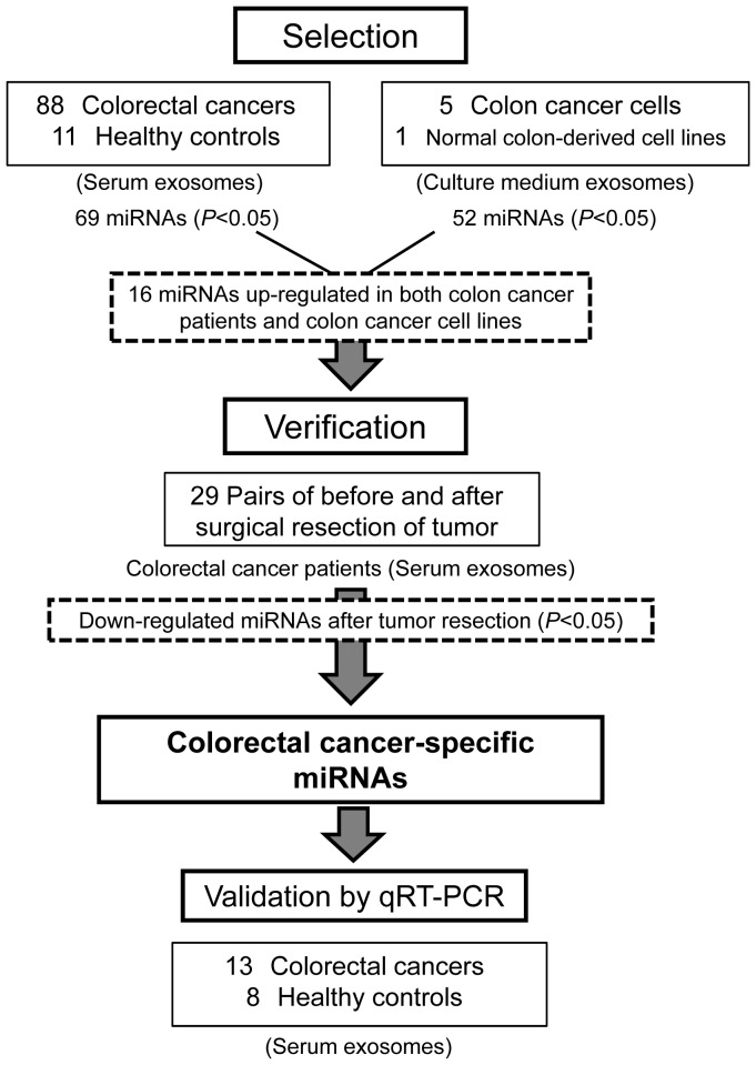
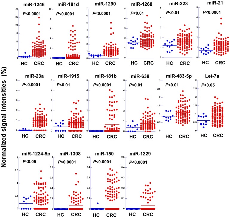
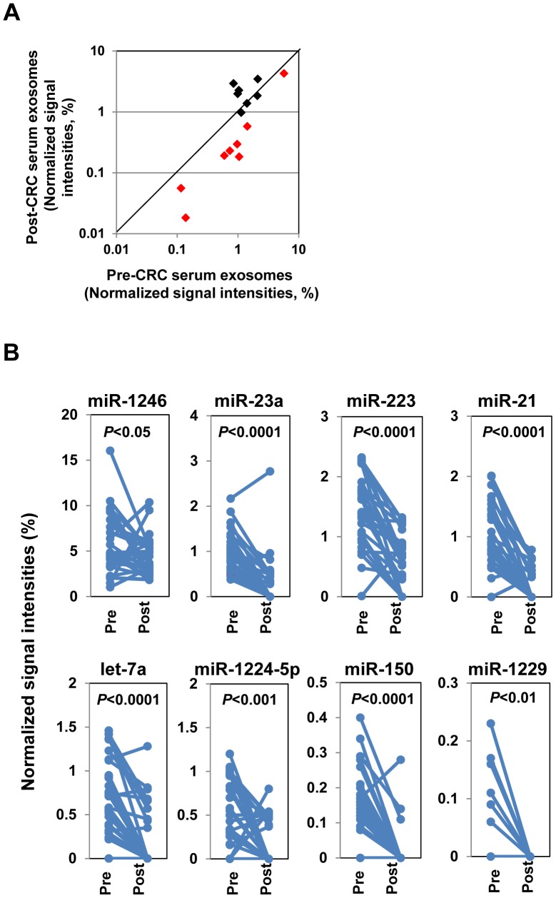
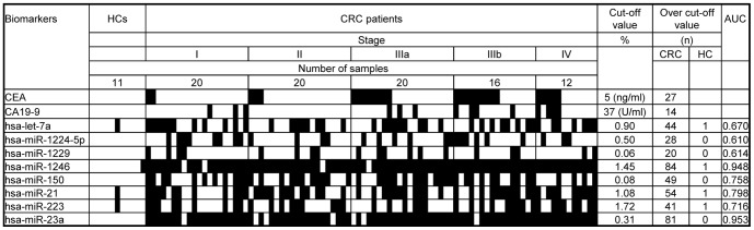
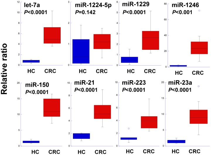
PURPOSE: Exosomal microRNAs (miRNAs) have been attracting major interest as potential diagnostic biomarkers of cancer. The aim of this study was to characterize the miRNA profiles of serum exosomes and to identify those that are altered in colorectal cancer (CRC). To evaluate their use as diagnostic biomarkers, the relationship between specific exosomal miRNA levels and pathological changes of patients, including disease stage and tumor resection, was examined. EXPERIMENTAL DESIGN: Microarray analyses of miRNAs in exosome-enriched fractions of serum samples from 88 primary CRC patients and 11 healthy controls were performed. The expression levels of miRNAs in the culture medium of five colon cancer cell lines were also compared with those in the culture medium of a normal colon-derived cell line. The expression profiles of miRNAs that were differentially expressed between CRC and control sample sets were verified using 29 paired samples from post-tumor resection patients. The sensitivities of selected miRNAs as biomarkers of CRC were evaluated and compared with those of known tumor markers (CA19-9 and CEA) using a receiver operating characteristic analysis. The expression levels of selected miRNAs were also validated by quantitative real-time RT-PCR analyses of an independent set of 13 CRC patients. RESULTS: The serum exosomal levels of seven miRNAs (let-7a, miR-1229, miR-1246, miR-150, miR-21, miR-223, and miR-23a) were significantly higher in primary CRC patients, even those with early stage disease, than in healthy controls, and were significantly down-regulated after surgical resection of tumors. These miRNAs were also secreted at significantly higher levels by colon cancer cell lines than by a normal colon-derived cell line. The high sensitivities of the seven selected exosomal miRNAs were confirmed by a receiver operating characteristic analysis. CONCLUSION: Exosomal miRNA signatures appear to mirror pathological changes of CRC patients and several miRNAs are promising biomarkers for non-invasive diagnosis of the disease.
Overview
The claims and statistics of this paper haven’t been checked yet.
Run the check — free, no account needed. It reads the open-access text and checks the sources it cites.
Integrity signals
Registry
Cites
Statistics
Figures
Verification
Claims the paper makes
The claims and statistics of this paper haven’t been checked yet.
Run the check — free, no account needed. It reads the open-access text and checks the sources it cites.
Claims other papers make about this one
Each is checked against this paper’s own text: does it say what the citing paper says it does?
The cited source says what the claim attributes to it.
In the citing paper
“microRNA's presence in exosomes have been already reported in different cells and cancer types (44–50).”
From Potential Therapeutic Effects of Exosomes Packed With a miR-21-Sponge Construct in a Rat Model of Glioblastoma (2019) · cites this paper as [48]
Self-check
Percentages, sums and reported differences re-computed from the paper’s own numbers.
The self-check needs the paper’s full text.
Citations
Cites
1 retracted
RETRACTED: MicroRNA signatures of tumor-derived exosomes as diagnostic biomarkers of ovarian cancer
Taylor, D. D., & Gerçel-Taylor, Ç. (2008) · Gynecologic Oncology · doi:10.1016/j.ygyno.2008.04.033 · Cited by 2,333
RetractedAnalysis of Relative Gene Expression Data Using Real-Time Quantitative PCR and the 2−ΔΔCT Method
Livak, K. J., & Schmittgen, T. D. (2001) · Methods · doi:10.1006/meth.2001.1262 · Cited by 180,860
Jemal, A., Bray, F., Ferlay, J., Ward, E., & Forman, D. (2011) · CA A Cancer Journal for Clinicians · doi:10.3322/caac.20107 · Cited by 55,077
Valadi, H., Ekström, K., Bossios, A., Sjöstrand, M., Lee, J. J., & Lötvall, J. O. (2007) · Nature Cell Biology · doi:10.1038/ncb1596 · Cited by 12,834
MicroRNA expression profiles classify human cancers
Lü, J., Getz, G. A., Miska, E. A., Alvarez-Saavedra, E., Lamb, J., Peck, D., Sweet-Cordero, A., Ebert, B. L., Mak, R. H., Ferrando, A. A., et al. (2005) · Nature · doi:10.1038/nature03702 · Cited by 9,637
Oncomirs—MicroRNAs with a role in cancer
Esquela-Kerscher, A., & Slack, F. J. (2006) · Nature Reviews Cancer · doi:10.1038/nrc1840 · Cited by 5,884
Skog, J., Würdinger, T., van Rijn, S., Meijer, D. H., Gainche, L., Curry, W. T., Carter, B. S., Krichevsky, A. M., & Breakefield, X. O. (2008) · Nature Cell Biology · doi:10.1038/ncb1800 · Cited by 5,001
Causes and consequences of microRNA dysregulation in cancer
Croce, C. M. (2009) · Nature Reviews Genetics · doi:10.1038/nrg2634 · Cited by 3,039
MicroRNA dysregulation in cancer: diagnostics, monitoring and therapeutics. A comprehensive review
Iorio, M. V., & Croce, C. M. (2012) · EMBO Molecular Medicine · doi:10.1002/emmm.201100209 · Cited by 1,778
Atkin, W. S., Edwards, R., Kralj‐Hans, I., Wooldrage, K., Hart, A. R., Northover, J. M. A., Parkin, D. M., Wardle, J., Duffy, S. W., & Cuzick, J. M. (2010) · The Lancet · doi:10.1016/s0140-6736(10)60551-x · Cited by 1,618
Cited by
Sorted by citations
Items 701–800 of 810
Trends in Precision Oncology and Precision Medicine 2.0
Hays, P. V. (2021) · Springer eBooks · doi:10.1007/978-3-030-80100-7_6 · Cited by 2
Progress of regulatory RNA in small extracellular vesicles in colorectal cancer
Hu, X., Lu, Y., Zhou, J., Wang, L., Zhang, M., Mao, Y., & Chen, Z. (2023) · Frontiers in Cell and Developmental Biology · doi:10.3389/fcell.2023.1225965 · Cited by 2
DIAGNOSTIC VALUE OF EXOSOMAL MIRNAS FOR COLORECTAL CANCER
Samsonov, R. B., Tarasov, M. A., Burdakov, V. S., Shtam, T. A., Guljaev, A. M., Tkachenko, O. B., Rybakov, E. G., Filatov, M. V., Aigner, A. M., & Malek, A. V. (2018) · Koloproktologia · doi:10.33878/2073-7556-2018-0-2-25-31 · Cited by 2
ILPMDA: Predicting miRNA–Disease Association Based on Improved Label Propagation
Wang, Y., Li, L., Ji, C., Zheng, C.-H., & Ni, J. (2021) · Frontiers in Genetics · doi:10.3389/fgene.2021.743665 · Cited by 2
MicroRNA Signatures as Biomarkers in Cancer
Adams, B. D., & Slack, F. J. (2015) · Wiley eBooks · doi:10.1002/9780470015902.a0025346 · Cited by 2
Extracellular Vesicles as Biomarkers for Cancer
Wang, S., Lin, Y., Hu, J., Fan, W., Yu, L., Hao, X., Huang, S., & Zhang, H. (2024) · Springer eBooks · doi:10.1007/978-981-99-8365-0_16 · Cited by 1
Role of Exosomal Non-Coding RNAs In Vivo and In Vitro Studies in Colorectal Cancer
Zaky, M. Y., & Hamdalla, H. M. (2025) · MicroRNA · doi:10.2174/0122115366331268250303031240 · Cited by 1
Nakayama, S., Umeda, M., Kobayashi, K., Nakano, Y., Hori, K., Umemura, T., & Kurokawa, H. (2025) · Frontiers in Oncology · doi:10.3389/fonc.2025.1619460 · Cited by 1
Circulating MicroRNA Expression in Glioblastoma: A Promising Diagnostic Panel
Macaione, V., Graziano, F., Polito, F., Scalia, G., Licitri, L., Khouyyi, M., Torre, M. M., Drago, S. F. A., Migliorato, A., Nicoletti, G. F., et al. (2025) · Oncology · doi:10.1159/000548121 · Cited by 1
Exosomal MicroRNAs in Cancer: Bridging Mechanistic Insights with Biomarker Potential
Kumar, S. K., Thalakanti, S., Patnam, S., Kanaparthi, N., & Venkata, S. M. (2026) · Advances in biochemistry in health and disease · doi:10.1007/978-3-032-08824-6_24 · Cited by 1
Gao, J., Cao, W., Zhang, Y., Zheng, D., & Zhu, H. (2026) · Annals of Medicine and Surgery · doi:10.1097/ms9.0000000000004777 · Cited by 1
Advances in microfluidic extracellular vesicle technology
Eigenfeld, M., & Schwaminger, S. P. (2026) · Advances in clinical chemistry · doi:10.1016/bs.acc.2026.03.004 · Cited by 1
Pernagallo, S., Tisato, V., & Gemmati, D. (2026) · Genes · doi:10.3390/genes17080847 · Cited by 1
Finger-tip size multiple reverse transcription device for early detection of diseases
Kimura, Y., Ikeuchi, M., & Ikuta, K. (2015) · International Conference on Solid-State Sensors, Actuators and Microsystems · doi:10.1109/transducers.2015.7181265 · Cited by 1
Prospective Advances in Medical Epigenetics
Xiao, J., & Li, G. (2016) · Elsevier eBooks · doi:10.1016/b978-0-12-803239-8.00045-4 · Cited by 1
Han, G., He, C., & Lian-qin, S. (2017) · Biomedical Research-tokyo · Cited by 1
Павлович, А. К., Ruksha, T. G., Бакшеева, С. Л., Владимировна, К. Т., Palkina, N. V., & Marugina, T. L. (2017) · В мире научных открытий · doi:10.12731/wsd-2017-4-77-95 · Cited by 1
Arora, M., & Kaul, D. (2018) · Springer eBooks · doi:10.1007/978-981-13-1568-8_4 · Cited by 1
Diagnosis of colorectal cancer based on imperialist competitive algorithm
Raju, B. R., Swamy, G. N., & Raju, K. P. (2020) · Journal of Intelligent & Fuzzy Systems · doi:10.3233/jifs-189021 · Cited by 1
Genetics of Colorectal Cancer Racial Disparities
Williams, J. L., Paredes, J. E., & Thaker, S. D. (2022) · IntechOpen eBooks · doi:10.5772/intechopen.103730 · Cited by 1
Prospective advances in microRNAs investigation
Shen, S., & Xiao, J. (2022) · Elsevier eBooks · doi:10.1016/b978-0-323-89774-7.00022-4 · Cited by 1
Translational Opportunities of Extracellular Vesicles in Biomedicine
Politis, M., Im, H., Carter, B. S., Skog, J., Balaj, L., Castro, C. M., & Lee, H. (2023) · Current cancer research · doi:10.1007/978-3-031-22903-9_4 · Cited by 1
Understanding the Potential of mRNA as Biomarker to Revolutionize Diagnosis of Colorectal Cancer
Das, R., Mehta, D. K., & Gupta, N. (2024) · Drug Research · doi:10.1055/a-2244-6572 · Cited by 1
Tumor microenvironment of colorectal cancer
Gupta, M. K., & Vadde, R. (2025) · Elsevier eBooks · doi:10.1016/b978-0-443-24742-2.00024-1 · Cited by 1
Ranjbarnejad, T., Ranjbarnejad, F., & Saidijam, M. (2019) · DOAJ (DOAJ: Directory of Open Access Journals) · Cited by 1
Liquid Biopsy: Translating Minimally Invasive Disease Profiling from the Lab to the Clinic
Zainfeld, D. E., Ghani, U., Kang, I. M., & Goldkorn, A. (2019) · Springer eBooks · doi:10.1007/978-3-030-23637-3_10 · Cited by 1
Identification of Key mRNAs and Pathways in Colorectal Cancer
Hou, X., Hou, N., Fu, J., He, X., Xiong, H., Xie, W., Jia, G., Zuo, X., Qin, X., & Pang, M. (2020) · Nutrition and Cancer · doi:10.1080/01635581.2020.1783328 · Cited by 1
Wang, H., Chen, X., Bao, L., & Zhang, X. (2020) · Medicine · doi:10.1097/md.0000000000022199 · Cited by 1
MicroRNA as Biomarkers in Cancer Diagnostics and Therapy
Sempere, L. F. (2019) · MDPI eBooks · doi:10.3390/books978-3-03921-250-7 · Cited by 1
Epigenetic Alterations in Colorectal Cancer
Ko, B., Hanna, M., Yu, M., & Grady, W. M. (2023) · Epigenetics and human health · doi:10.1007/978-3-031-42365-9_10 · Cited by 1
The role of extracellular vesicles as biomarkers in colorectal cancer
Lucchetti, D., Litta, F., Ratto, C., & Sgambato, A. (2018) · Techniques in Coloproctology · doi:10.1007/s10151-018-1880-4 · Cited by 1
Cancer exosomes harbor diverse hypoxia-targeted mRNAs and contribute toward tumor angiogenesisPreprint
Zhang, P., Lim, S. B., Jiang, K., Chew, T. W., Low, B. C., & Lim, C. T. (2020) · bioRxiv (Cold Spring Harbor Laboratory) · doi:10.1101/2020.06.12.147850 · Cited by 1
Espejo, C., Wilson, R., Woods, G. M., Willms, E., Hill, A. F., & Lyons, A. B. (2020) · bioRxiv (Cold Spring Harbor Laboratory) · doi:10.1101/2020.10.18.344721 · Cited by 1
Editorial: Advances in the Molecular Mechanisms in Gastrointestinal Tumorigenesis and Treatment
Zhang, R., Zhang, Y., & Qu, X. (2022) · Frontiers in Oncology · doi:10.3389/fonc.2021.827294 · Cited by 1
Pergoli, L. (2015) · Archivio Istituzionale della Ricerca (Universita Degli Studi Di Milano) · doi:10.13130/pergoli-laura-wanda_phd2015-02-24
Liquid Biopsy in Colorectal Cancer
Galvano, A., Peeters, M., Stefano, A. B. D., Castiglia, M., & Russo, A. D. (2017) · Current clinical pathology · doi:10.1007/978-3-319-55661-1_13
DNA and Histone Modifications in Cancer Diagnosis
Kinehara, M., Yamamoto, Y., Shiroma, Y., Ikuo, M., Shimamoto, A., & Tahara, H. (2017) · Cancer drug discovery and develogment/Cancer drug discovery and development · doi:10.1007/978-3-319-59786-7_19
The role of exosomes in diagnosis of non-haematological malignancies
Jain, D., Dhariwal, S., & Sachdeva, H. (2018) · Chronicles of Oncology · doi:10.33582/2638-4248/1002
Research Progress of Exosome Non-coding RNA in Cancer
Jin, J. (2019) · Science Discovery · doi:10.11648/j.sd.20190701.13
The Level of Thiol-Disulfide on Colorectal Cancer
Özdemir, A., Dilli, U. D., Özdemir, D., Neşelioğlu, S., & Erel, Ö. (2019) · Anadolu Kliniği Tıp Bilimleri Dergisi · doi:10.21673/anadoluklin.469201
Guillen, J. K. (2020) · ScholarSpace (University of Hawaii at Manoa)
Sun, L. J., & Meckes, D. G. (2020) · Research Square · doi:10.21203/rs.3.rs-125398/v1
Evaluation of exosome-encapsulated miR-23a/b in the diagnosis of human coronary heart diseasesPreprint
Xu, C., Xiao, H. F., Yi, Y., Zhu, D., Yue, X., & Xi, Y. (2020) · bioRxiv (Cold Spring Harbor Laboratory) · doi:10.1101/2020.12.27.424501
Rola miRNA w rozwoju wybranych nowotworów – potencjalne zastosowanie w diagnostyce*
Paciorek, P., Żuberek, M., & Grzelak, A. (2021) · Postępy Higieny i Medycyny Doświadczalnej · doi:10.5604/01.3001.0014.6578
Naseer, M., Hadi, S., Syed, A., Safdari, A., & Tahan, V. (2021) · World Journal of Meta-Analysis · doi:10.13105/wjma.v9.i1.12
Develop Micro/Nano Technologies for Cancer Diagnosis
Zheng, S. (2021) · IEEE International Conference on Micro Electro Mechanical Systems (MEMS) · doi:10.1109/mems51782.2021.9375338
Lee, B., Sahoo, A., Sawada, J., Marchica, J., Sahoo, S., Layng, F. I., Finlay, D., Mazar, J., Joshi, P. K., Komatsu, M., et al. (2019) · bioRxiv (Cold Spring Harbor Laboratory) · doi:10.1101/548818
Alashti, F. A., Goliaei, B., Karami, L., Vassiliev, S., Jooyan, N., & Minuchehr, Z. (2021) · Research Square · doi:10.21203/rs.3.rs-381601/v1
Tsukamoto, R., Hosoya, M., FUKAYA, M., Yokoyama, N., Kawano, S., Sugimoto, K., Takahashi, M., Sakamoto, K., & Komiyama, H. (2021) · Juntendō Igaku/Juntendo igaku · doi:10.14789/jmj.jmj20-oa12
Juzėnas, S., Lukoševičius, R., Šaltenienė, V., Kulokiene, U., Hemmrich‐Stanisak, G., Franke, A., Link, A., Ruzgys, P., Šatkauskas, S., Paužas, H., et al. (2021) · Research Square · doi:10.21203/rs.3.rs-558860/v1
Extracellular Vesicles in Colorectal Cancer Progression, Metastasis, Diagnosis, and Therapy
Merlin, M., Prabhakar, P. K., Shukla, D., Tiwari, A. K., & Saxena, S. (2021) · Springer eBooks · doi:10.1007/978-3-030-64668-4_17
Toprak, A., & Eryilmaz, E. (2021) · European Journal of Science and Technology · doi:10.31590/ejosat.980257
Exosomes as Radiation Biomarkers
Jella, K. K. (2022) · NATO science for peace and security series. A, Chemistry and biology · doi:10.1007/978-94-024-2101-9_8
EV (Extracellular Vesicle)‐associated miRNAs as Biomarkers of Toxicity
Ono, R., Yoshioka, Y., Furukawa, Y., Naruse, M., Kuwagata, M., Ochiya, T., Kitajima, S., & Hirabayashi, Y. (2022) · Wiley eBooks · doi:10.1002/9781119807704.ch3
Hernández-Bedolla, M. A., Alonso, D. E. A., Salguero, I. G. D., Reyes-Uribe, E., Barajas‐Espinosa, A. R., & Ochoa‐Cortés, F. (2021) · Ciencias Huasteca Boletín Científico de la Escuela Superior de Huejutla · doi:10.29057/esh.v9i18.6932
(2021) · Letters in Applied NanoBioScience · doi:10.33263/lianbs111.32323248
SALIVA BIOMARKERS IN PATHOLOGICAL CONDITIONS OF PERIODONTIUM
Ostrovskа, S. S., Герасимчук, П. Г., Krizhanivsky, D. G., L.О., L. О. Z., Tereshchenko, N. M., & Velikorodny, V. I. (2022) · Bulletin of Problems Biology and Medicine · doi:10.29254/2077-4214-2022-2-1-164-46-54
Ling, Q., Feng, S., Chen, R., Huang, H., Lin, Y., Jiang, J., Cui, J., & Liang, T. (2022) · Research Square · doi:10.21203/rs.3.rs-1540878/v2
Rapamycin Inhibits Human Oral Cancer Cell Growth via the Hsa-miR-199a-3p/mTOR Pathway
Hou, Y., Lv, F., Wenjing, Z., Wang, W.-N., Duan, Y., Liu, S., & Qiu, Y. (2022) · Science of Advanced Materials · doi:10.1166/sam.2022.4369
Kwok, Z. H., Zhang, B., Chew, X. H., Chan, J. J., Teh, V., Yang, H. H., Kappei, D., & Tay, Y. (2023) · doi:10.1158/0008-5472.c.6512451
Kwok, Z. H., Zhang, B., Chew, X. H., Chan, J. J., Teh, V., Yang, H. H., Kappei, D., & Tay, Y. (2023) · doi:10.1158/0008-5472.c.6512451.v1
Neuron Enriched Exosomal MicroRNA Expression Profiles as a Marker of Early Life Alcohol ConsumptionPreprint
Yakovlev, V. A., Lapato, D. M., Rana, P., Ghosh, P., Frye, R., & Roberson‐Nay, R. (2023) · bioRxiv (Cold Spring Harbor Laboratory) · doi:10.1101/2023.06.09.544235
Establishment of Premetatatic Niche Formation and Clincal Application
Cedric, K. K. (2020) · Archives of Orthopedics and Rheumatology · doi:10.22259/2639-3654.0302001
Exosome Detection for Early Cancer Diagnosis
Lee, R., & Park, Y. I. (2023) · Springer eBooks · doi:10.1007/978-3-030-80962-1_326-1
A multi-model study of extracellular vesicles in bowel cancer invasion and metastasis
Guarnerio, S. (2023) · Sheffield Hallam University · doi:10.7190/shu-thesis-00581
Potential role of exosomal miRNA in disease progression and therapeutics
Bhattacherjee, A. (2024) · Elsevier eBooks · doi:10.1016/b978-0-443-14008-2.00012-7
Ma, J., Wang, P., Huang, L., Qiao, J., & Li, J. (2021) · Figshare · doi:10.6084/m9.figshare.14131028
Ma, J., Wang, P., Huang, L., Qiao, J., & Li, J. (2021) · Figshare · doi:10.6084/m9.figshare.14131031
Saha, B., Adhikary, S., Banerjee, A., & Pathak, S. (2024) · Springer eBooks · doi:10.1007/978-981-97-2494-9_12
Extracellular Vesicles in Cancer Diagnosis
Unnikrishnan, K., Rames, A. D. U., & Kumar, R. (2024) · Springer eBooks · doi:10.1007/978-981-97-2494-9_10
DIAGNOSTIC POTENTIAL OF DETERMINING INDIVIDUAL EXTRACELLULAR VESICLES SUBSETS IN CLINICAL PRACTICE
Kudryavtsev, I. V., Головкин, А. С., & Totolyan, A. A. (2024) · Complex Issues of Cardiovascular Diseases · doi:10.17802/2306-1278-2024-13-3-200-214
Naeimi, S., Aliasgari, E., & Jamshidian, F. (2025) · Acta Scientiarum Health Sciences · doi:10.4025/actascihealthsci.v47i1.70248
Safiejko, K., Pruc, M., & Szarpak, Ł. (2025) · Indian Journal of Gastroenterology · doi:10.1007/s12664-025-01805-4
Exosomal microRNAs in Diagnosis of Colorectal Cancer
Serbaeva, M. S., Vorobyev, S. A., Ruksha, T. G., & Zukov, R. A. (2025) · Russian Journal of Oncology · doi:10.17816/onco643119
Efficient Isolation and Characterization Methods for Chicken Serum Extracellular Vesicles
Mukohda, H., Binici, M. M., Miyake, K., Sano, Y., Yamada, T., Terada, T., & Sugiyama, T. (2025) · Animal Science Journal · doi:10.1111/asj.70092
Ali, T., Degorre, C., & Tofilon, P. J. (2025) · International Journal of Molecular Sciences · doi:10.3390/ijms26178483
Isolation of Extracellular Vesicles and the Analysis of Their RNA Cargo
Nwachukwu, F., Gordon, M. E., Witek, M. A., & Soper, S. A. (2025) · Springer eBooks · doi:10.1007/978-3-031-89270-7_7
Exosome Detection for Early Cancer Diagnosis
Lee, R., & Park, Y. I. (2025) · Springer eBooks · doi:10.1007/978-3-032-00763-6_326
Diagnostics and monitoring of colorectal cancer with AuNP-APTASENSOR
Sharonova, T. V., Kadantseva, E. Y., Кацуба, К. Е., Kovalenko, I. M., Служев, М. И., Semiglazova, Т. Y., Burtcev, D. V., Dimitriadi, T. A., Евгеньевна, М. К., Mkrtchian, L. S., et al. (2025) · Koloproktologia · doi:10.33878/10.33878/2073-7556-2025-24-4-92-105
A Review of the Impact of Exosomal Non-Coding RNAs on the Development of Malignant Colorectal Tumors
Mansouri, P., & Shanehbandi, D. (2025) · مجله دانشگاه علوم پزشکی اردبیل · doi:10.61882/jarums.25.2.118
Xu, J., Pan, L., Wu, D., Yao, L., Jiang, W., Min, J., Xu, S., & Deng, Z. (2024) · Figshare · doi:10.6084/m9.figshare.26739657
Chen, K., Li, Y., Xu, L., Qian, Y., Liu, N., Zhou, C., Liu, J., Zhou, L., Xu, Z., Jia, R., et al. (2022) · Figshare · doi:10.6084/m9.figshare.20023111
Chen, K., Li, Y., Xu, L., Qian, Y., Liu, N., Zhou, C., Liu, J., Zhou, L., Xu, Z., Jia, R., et al. (2022) · Figshare · doi:10.6084/m9.figshare.20023108
Additional file 2 of Exosomal miRNA signatures of pancreatic lesions
Vicentini, C., Calore, F., Nigita, G., Fadda, P., Simbolo, M., Sperandio, N., Luchini, C., Lawlor, R. T., Croce, C. M., Vincenzo, C., et al. (2020) · Figshare · doi:10.6084/m9.figshare.12262508
Xie, B., Zhao, L., Zhang, Z., Zhou, C., Tian, Y., Kang, Y. Q., Chen, J., 魏虎来, & Li, L. (2023) · Figshare · doi:10.6084/m9.figshare.24277155
Xu, J., Pan, L., Wu, D., Yao, L., Jiang, W., Min, J., Xu, S., & Deng, Z. (2024) · Figshare · doi:10.6084/m9.figshare.26113036
Xu, J., Pan, L., Wu, D., Yao, L., Jiang, W., Min, J., Xu, S., & Deng, Z. (2024) · Figshare · doi:10.6084/m9.figshare.26113162
Xie, B., Zhao, L., Zhang, Z., Zhou, C., Tian, Y., Kang, Y., Chen, J., 魏虎来, & Li, L. (2024) · Figshare · doi:10.6084/m9.figshare.24277253
Impending Role of Exosomal miRNA as Substantial Players in Early Cancer Diagnostics
Prakash, R., Natesh, A., Kubendiran, K., Palakshappa, S. S., & Kannan, A. (2026) · Advances in biochemistry in health and disease · doi:10.1007/978-3-032-08824-6_6
MicroRNAs: Innovative Clinical Tools for Diagnosis, Prognosis, and Therapy in Colorectal Cancer
Long, F., Wang, W., Liu, Y., Yang, C., Ren, H., Leng, C., Xiong, P., Tang, X., Hu, L., Liu, L., et al. (2026) · Advances in biochemistry in health and disease · doi:10.1007/978-3-032-08824-6_22
Role of MicroRNAs in Intestinal Epithelial Transport and Its Mechanism Leading to Colorectal Cancer
Selvaraj, R., Kanakarajan, S., & Singh, S. (2026) · Advances in biochemistry in health and disease · doi:10.1007/978-3-032-08824-6_19
Microparticles and malignant pleurisy
Roca, E. (2017) · theses.fr (ABES) · doi:10.70675/d299922bz618dz4d4bz8e17z1d8f63493d9a
Effect of nasopharyngeal carcinoma derived exosomes on the induction of tolerogenic dendritic cells
Renaud, S. (2018) · theses.fr (ABES) · doi:10.70675/41db8e5ez4572z4453zb3efz2631b3820222
Sulikowski, M., Sulikowski, T., Kurzawski, M., Malinowski, D., Urasińska, E., & Rać, M. E. (2026) · International Journal of Molecular Sciences · doi:10.3390/ijms27083702
Jayaraman, V., Apthi, S., Datta, L., Palaniyandi, K., & Gnanasampanthapandian, D. (2026) · MicroRNA · doi:10.2174/0122115366340684251213003152
Moccia, V., Rovere, G. D., Minh, T. T. N., Zendrini, A., Kleinjan, M., Roelofs, M. C., Berto, P., Zeev‐Ben‐Mordehai, T., Zaal, E. A., Bergese, P., et al. (2026) · bioRxiv (Cold Spring Harbor Laboratory) · doi:10.64898/2026.05.29.727857
A bibliometric analysis of exosome research in colorectal cancer from 2010 to 2024
Chen, Z., Xu, X., & Ye, L. (2026) · Discover Oncology · doi:10.1007/s12672-026-05582-0
Extracellular Vesicles in Personalized Medicine: Precision Diagnostics, Tailored Therapies
Mohammadzadeh, S., Heidari, A., Dehghani, P., Mirzamohammadi, M., Hatkehlouei, S. A. T., Emami, M. H., & Maghool, F. (2026) · Molecular and translational medicine · doi:10.1007/978-3-032-23810-8_12
Hosseinzadeh, F., Kalhor, H., Fath‐Bayati, L., Kheirkhah, A. H., Jalise, S. Z., Yang, P., & Sheykhhasan, M. (2026) · Molecular and translational medicine · doi:10.1007/978-3-032-23810-8_9
Metadata
- Authors
- Ogata-Kawata, H., Izumiya, M., Kurioka, D., Honma, Y., Yamada, Y., Furuta, K., Gunji, T., Ohta, H., Okamoto, H., Sonoda, H., Watanabe, M., Nakagama, H., Yokota, J., Kohno, T., & Tsuchiya, N.
- Affiliations
- University of Tokyo Hospital (JP)
- Yokohama National University (JP)
- National Cancer Center Hospital (JP)
- NTT Medical Center (JP)
- Shionogi (Japan) (JP)
- National Cancer Center Research Institute (JP)
- Institute of Predictive and Personalized Medicine of Cancer (ES)
- Corresponding author
- Tsuchiya, Naoto
- ORCID
- Venue
- PLoS ONE
- Publisher
- Public Library of Science (PLoS)
- Published
- 2014-04-04
- Volume
- 9
- Issue
- 4
- Pages
- e92921
- ISSN
- 1932-6203 (ISSN-L)
- PMC
- PMC3976275
- OpenAlex
- W1992970284
- Cited by
- 809
- References
- 37
- Citations by year
- 2026: 28 · 2025: 37 · 2024: 51 · 2023: 45 · 2022: 76 · 2021: 96 · 2020: 89 · 2019: 81 · 2018: 76 · 2017: 76 · 2016: 78 · 2015: 60 · 2014: 15
- FWCI
- 31.32
- Type
- journal article
- Language
- en
- Open access
- gold
- doi.org
- Locations
- PLoS ONE (publishedVersion, cc-by)
- Europe PMC (PubMed Central) (publishedVersion, cc-by)
- PubMed Central (publishedVersion, cc-by)
- PubMed (publishedVersion)
- DOAJ (DOAJ: Directory of Open Access Journals) (submittedVersion)
- Figshare (submittedVersion, cc-by)
- Funders
- National Institute of Biomedical Innovation
- Japan Society for the Promotion of Science (23300359, 24790683)
- Topics
- Life Sciences › Biochemistry, Genetics and Molecular Biology › Molecular Biology › Extracellular vesicles in disease
- Life Sciences › Biochemistry, Genetics and Molecular Biology › Cancer Research › MicroRNA in disease regulation
- Life Sciences › Biochemistry, Genetics and Molecular Biology › Cancer Research › Cancer-related molecular mechanisms research
- Keywords
- exosomal microRNAs, colorectal cancer, serum exosomes, diagnostic biomarkers, non-invasive diagnosis, miR-21, miR-223, miR-23a, miR-150, miR-1246, let-7a, tumor resection, CA19-9, carcinoembryonic antigen
- MeSH terms
- Adult, Aged, Colonic Neoplasms, Female, Humans, Male, Middle Aged, Tumor Cells, Cultured, Biomarkers, Tumor, Gene Expression Regulation, Neoplastic, Case-Control Studies, HT29 Cells, Gene Expression Profiling, MicroRNAs, HCT116 Cells, Cell Line, Tumor, Microarray Analysis, Exosomes
About these checks
Registry, retraction, Cites and statistics results are computed, not judged, with one exception: a printed reference that no index lists is read once by a model before it counts as a citation error. Claims are extracted by a model and kept only when their quote appears verbatim in the text; each cited source is then checked, with a model’s help, for whether it says what the claim relies on. Those verdicts can be wrong — read the claim’s quote and the cited source before relying on one.
Statistics and arithmetic are recomputed, not judged.
Confidence bands are shown only for fields calibrated on our annotated test set. None is calibrated yet, so no band is shown.
What counts as a claim ›How we measure confidence ›How we check this ›
Are you an author, or do you see a problem with this page? Request a review
