

Conditioned Medium from Hypoxic Bone Marrow-Derived Mesenchymal Stem Cells Enhances Wound Healing in Mice
Open accessThe DOI matches its Crossref record; 32 of the 32 references it cites were found in a registry or an index; 1 of the 3 sources its claims cite were checked for support.
Abstract
Growing evidence indicates that bone marrow-derived mesenchymal stem cells (BM-MSCs) enhance wound repair via paracrine. Because the extent of environmental oxygenation affects the innate characteristics of BM-MSCs, including their stemness and migration capacity, the current study set out to elucidate and compare the impact of normoxic and hypoxic cell-culture conditions on the expression and secretion of BM-MSC-derived paracrine molecules (e.g., cytokines, growth factors and chemokines) that hypothetically contribute to cutaneous wound healing in vivo. Semi-quantitative reverse transcriptase polymerase chain reaction (RT-PCR) and enzyme-linked immunosorbent assay (ELISA) analyses of normoxic and hypoxic BM-MSCs and their conditioned medium fractions showed that the stem cells expressed and secreted significantly higher amounts of basic fibroblast growth factor (bFGF),vascular endothelial growth factor A (VEGF-A) interleukin 6 (IL-6) and interleukin 8 (IL-8) under hypoxic conditions. Moreover, hypoxic BM-MSC-derived conditioned medium (hypoCM) vs. normoxic BM-MSC-derived conditioned medium (norCM) or vehicle control medium significantly enhanced the proliferation of keratinocytes, fibroblasts and endothelial cells, the migration of keratinocytes, fibroblasts, endothelial cells and monocytes, and the formation of tubular structures by endothelial cells cultured on Matrigel matrix. Consistent with these in vitro results, skin wound contraction was significantly accelerated in Balb/c nude mice treated with topical hypoCM relative to norCM or the vehicle control. Notably increased in vivo cell proliferation, neovascularization as well as recruitment of inflammatory macrophages and evidently decreased collagen I, and collagen III were also found in the hypoCM-treated group. These findings suggest that BM-MSCs promote murine skin wound healing via hypoxia-enhanced paracrine.
Overview
CoChat Summary
## TL;DR
Hypoxic preconditioning increased BM-MSC proliferation and boosted secretion of several wound-healing factors, especially bFGF, VEGF-A, IL-6, and IL-8. In mice, hypoxic BM-MSC-conditioned medium (hypoCM) accelerated wound closure and increased cell proliferation, angiogenesis, and macrophage recruitment.
## Key findings
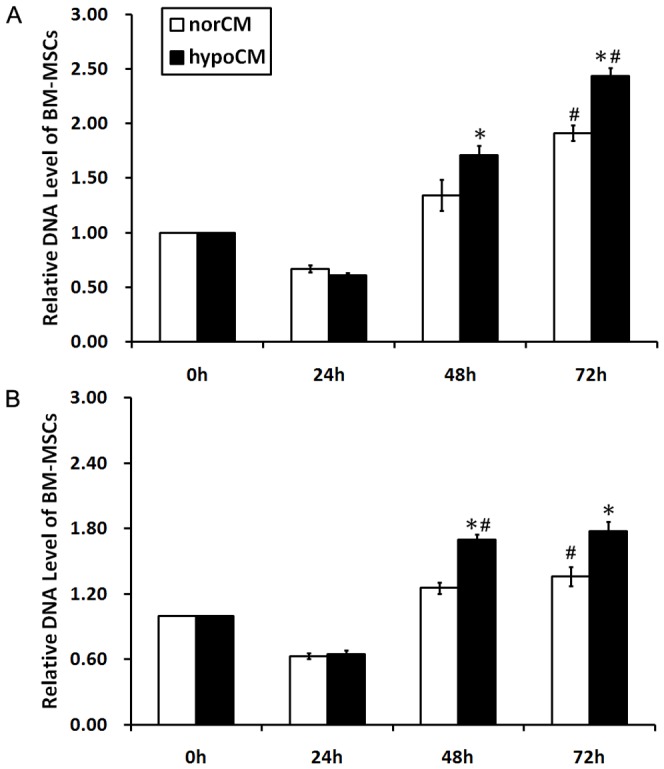
- Hypoxia increased BM-MSC proliferation at 48 h and 72 h in both complete medium and serum-free α-MEM; for example, in complete medium proliferation was 1.71±0.09 vs. 1.34±0.15 at 48 h and 2.43±0.08 vs. 1.91±0.07 at 72 h (p<0.05).
- Hypoxia up-regulated BM-MSC mRNA expression of several paracrine factors, including bFGF (3.33-fold), IGF-1 (2.91-fold), VEGF-A (2.56-fold), TGF-β3 (3.61-fold), IL-6 (2.18-fold), and IL-8 (2.10-fold) (p<0.05).
- ELISA of conditioned medium showed significantly higher levels of bFGF (1.95-fold), VEGF-A (1.27-fold), IL-6 (2.76-fold), and IL-8 (1.53-fold) in hypoCM vs. norCM (p<0.05).
- In vitro, hypoCM increased proliferation of keratinocytes, fibroblasts, and HUVECs, and increased migration of keratinocytes (28.84-fold vs vehicle; 2.01-fold vs norCM), fibroblasts (37.92-fold; 1.30-fold), HUVECs (14.87-fold; 1.44-fold), and CD14+ monocytes (6.54-fold; 1.60-fold) (p<0.05).
- In vivo, hypoCM-treated wounds closed faster; on day 7 wound area was 26.42±2.48% vs 45.00±1.97% (vehicle) and 37.92±2.44% (norCM), and on day 11 it was 8.75±1.25% vs 28.75±1.83% in vehicle controls (p<0.05).
- HypoCM also increased Ki67+ cells in dermis on day 11 (139.25±4.31 vs 56.50±3.12 vehicle and 61.75±4.13 norCM), F4/80+ macrophages (118.25±4.03 vs 64.75±5.12 and 75.50±6.70), and CD31+ vessels (55.50±2.78 vs 34.25±2.25 and 43.25±2.17) (p<0.05).
## Methods
- Human BM-MSCs were cultured under normoxic (20% O2) or hypoxic (2% O2) conditions; conditioned media from each were collected as norCM and hypoCM.
- In vitro analyses included BM-MSC proliferation measurement by DNA assay, semi-quantitative RT-PCR, ELISA, keratinocyte/fibroblast/HUVEC proliferation assays, transwell migration assays, and HUVEC tube formation assays.
- In vivo, Balb/c nude mice received 18 mm full-thickness dorsal wounds and were randomized into three groups (16 per group): vehicle control, norCM, or hypoCM; wound size and histology/IHC/IF outcomes were assessed over time.
- Statistics used Student’s t-test or one-way ANOVA with Bonferroni post-hoc test; significance was set at p<0.05.
## Limitations
- The paper states that the role of IL-6 is not fully resolved here and that they did not evaluate whether these factors act synergistically in wound healing.
- The authors note that the pathways involved in enhanced secretion of growth factors and cytokines by hypoxic BM-MSCs “still requires more information and further investigation.”
- Evident from the text: the study uses a mouse wound model and in vitro assays, so direct applicability to human wound healing is not tested here.
Model-written summary. Check it against the paper before relying on it.
Integrity signals
Registry
Cites
Statistics
Figures


Verification
Claims the paper makes
1 Valid · 0 Not valid · 2 Not checked · 25 Own results and interpretations
Every source the paper cites for this claim was checked and supports it.
In the paper
“the BM-MSCs appear to exert their principal therapeutic actions through the secretion of paracrine trophic signals [7].”
§ Conditioned Medium from Hypoxic Bone Marrow-Derived Mesenchymal Stem Cells Enhances Wound Healing in Mice > Introduction
What the sources say
- Supportedcites [7] AM Hocking, 2010, Mesenchymal stem cells: paracrine signaling and differentiation during cutaneous wound repair, Exp Cell Res source
The Introduction section 'The therapeutic benefits of MSCs for treating tissue injury: mechanisms of action' states that, given poor MSC engraftment/differentiation, 'MSC paracrine signaling is the primary mechanism accounting for the beneficial effects of MSCs', and the 'MSC paracrine signaling enhances wound repair' section says 'Growing evidence indicates that MSC paracrine signaling is the predominant mechanism responsible for enhanced wound repair'. The same sections attribute the effect to secreted mediators — proteomic analyses showing MSCs secrete growth factors, cytokines and chemokines, and MSC-conditioned medium recapitulating the benefit — and the paper frames its MSCs as bone marrow-derived multipotent mesenchymal stromal cells. The citing sentence's hedge ('appear to') matches the source's own hedged phrasing ('it has been proposed', 'growing evidence indicates'). (LLM, conf=0.88)
hedgedstates a cause- Asserted: passes — The authors put it forward as true.
- Checkable: passes — It can be checked against evidence.
- Load-bearing: passes — The paper’s conclusions depend on it.
- Based: passes — The paper gives a reason why it holds.
- Supported
A result the paper reports from its own data — it cites no other source for it, so there is none to check. The quote shows it is in the paper.
In the paper
“collagen I mRNA expression was reduced overtime (day 20 vs. day 40 for vehicle control group: 1.14-fold, P<0.05; for norCM group: 1.15-fold, P<0.05; for hypoCM group: 1.22-fold, P<0.05)”
§ Conditioned Medium from Hypoxic Bone Marrow-Derived Mesenchymal Stem Cells Enhances Wound Healing in Mice > Results > Hypoxia heightens the contribution of BM-MSC-mediated paracrine to cutaneous wound healing in vivo
plainstates a cause- Asserted: passes — The authors put it forward as true.
- Checkable: passes — It can be checked against evidence.
- Load-bearing: passes — The paper’s conclusions depend on it.
- Based: passes — The paper gives a reason why it holds.
A result the paper reports from its own data — it cites no other source for it, so there is none to check. The quote shows it is in the paper.
In the paper
“hypoxia differentially up-regulated the mRNA expression levels of basic fibroblast growth factor (bFGF, 3.33-fold), insulin-like growth factor 1 (IGF-1, 2.91-fold), vascular endothelial growth factor A (VEGF-A, 2.56-fold), transforming growth factor beta 2 (TGF β-2, 2.30-fold), transforming growth factor beta 3 (TGF β-3, 3.61-fold), interleukin 1 beta (IL-1β, 1.86-fold), interleukin 6 (IL-6, 2.18-”
§ Conditioned Medium from Hypoxic Bone Marrow-Derived Mesenchymal Stem Cells Enhances Wound Healing in Mice > Results > Hypoxia up-regulates the mRNA expression and protein secretion of BM-MSC-derived paracrine factors
plainstates a cause- Asserted: passes — The authors put it forward as true.
- Checkable: passes — It can be checked against evidence.
- Load-bearing: passes — The paper’s conclusions depend on it.
- Based: passes — The paper gives a reason why it holds.
A result the paper reports from its own data — it cites no other source for it, so there is none to check. The quote shows it is in the paper.
In the paper
“BM-MSC hypoCM significantly enhanced HUVEC tube formation ability on a Matrigel matrix compared with the vehicle control or norCM (Figs. 4, p<0.05).”
§ Conditioned Medium from Hypoxic Bone Marrow-Derived Mesenchymal Stem Cells Enhances Wound Healing in Mice > Results > Hypoxia enhances mitogenic, chemoattractive and angiogenic paracrine effects of BM-MSCs in vitro
plainstates a cause- Asserted: passes — The authors put it forward as true.
- Checkable: passes — It can be checked against evidence.
- Load-bearing: passes — The paper’s conclusions depend on it.
- Based: passes — The paper gives a reason why it holds.
A result the paper reports from its own data — it cites no other source for it, so there is none to check. The quote shows it is in the paper.
In the paper
“similar amounts of keratinocyte growth factor (KGF, 0.95-fold), monocyte chemotactic protein-1 (MCP-1, 1.03-fold), transforming growth factor beta 1 (TGF β-1, 0.93-fold), interleukin 10 (IL-10, 1.05-fold), tumor necrosis factor (TNF-α, 0.97-fold) and matrix metalloproteinases (MMP-1, 1.08-fold) were found in normoxic and hypoxic BM-MSCs (p>0.05).”
§ Conditioned Medium from Hypoxic Bone Marrow-Derived Mesenchymal Stem Cells Enhances Wound Healing in Mice > Results > Hypoxia up-regulates the mRNA expression and protein secretion of BM-MSC-derived paracrine factors
plain- Asserted: passes — The authors put it forward as true.
- Checkable: passes — It can be checked against evidence.
- Load-bearing: unclear — It is unclear whether the paper’s conclusions depend on it.
- Based: passes — The paper gives a reason why it holds.
13 more claims
Create a free account to see all 28 claims and to verify the 2 references still open.
- It may state two separate claims as one. 6
- Probably not a substantial claim. 5
Claims other papers make about this one
Each is checked against this paper’s own text: does it say what the citing paper says it does?
The source backs part of the claim, not all of it — or backs it for a different population, condition or comparison.
In the citing paper
“the secretome obtained from hypoxia preconditioned-BM-MSCs upregulated the levels of FGF-2, IGF-1, TGF-β, IL-1β, IL-6 [123] and IL-8 [122] and enhanced multiple key processes to promote wound healing, including keratinocyte, fibroblast and endothelial cells proliferation and migration, as well as angiogenesis (Fig. 2) [38], [120], [123].”
From Mesenchymal stem cell secretome for regenerative medicine: Where do we stand? (2025) · cites this paper as [123]
Experimental — accuracy not yet measured
Citations
Cites
Gurtner, G. C., Werner, S., Barrandon, Y., & Longaker, M. T. (2008) · Nature · doi:10.1038/nature07039 · Cited by 6,542
Singer, A. J., & Clark, R. A. F. (1999) · New England Journal of Medicine · doi:10.1056/nejm199909023411006 · Cited by 6,106
Wound Healing--Aiming for Perfect Skin Regeneration
Martin, P. (1997) · Science · doi:10.1126/science.276.5309.75 · Cited by 4,804 · cited 1×
“Epidermal growth factor (EGF), KGF, IGF-1 and VEGF-A are among the growth factors and cytokines known to enhance normal wound healing [17-19].”
BackgroundChen, L., Tredget, E. E., Wu, P. Y. G., & Wu, Y. (2008) · PLoS ONE · doi:10.1371/journal.pone.0001886 · Cited by 1,610 · cited 1×
“BM-MSC-mediated paracrine enhances wound healing through three predominant pathways, as evidenced by the demonstrated actions of BM-MSC-derived conditioned medium and secreted molecules: (1) BM-MSC-derived conditioned medium may act as a chemoattractant to recruit specific cell types to the wound, including epidermal…”
BackgroundSelf-citationKinnaird, T., Stabile, E., Burnett, M. S., Shou, M., Lee, C. W., Barr, S., Fuchs, S., & Epstein, S. E. (2004) · Circulation · doi:10.1161/01.cir.0000124062.31102.57 · Cited by 1,305 · cited 1×
“Consistent with previous reports [20,21], the current study demonstrated that BM-MSCs expressed and secreted VEGF-A, bFGF and other factors.”
ResultA Computational Tool for Quantitative Analysis of Vascular Networks
Zudaire, E., Gambardella, L., Kurcz, C., & Vermeren, S. (2011) · PLoS ONE · doi:10.1371/journal.pone.0027385 · Cited by 1,085 · cited 1×
“The number of tubular structures, tubule length, branches and nodes in 4 randomly chosen fields was quantified as previously described with the aid of Olympus Cell R imaging software [13].”
MethodKim, W.-S., Park, B.-S., Sung, J.-H., Yang, J.-M., Park, S.-B., Kwak, S.-J., & Park, J.-S. (2007) · Journal of Dermatological Science · doi:10.1016/j.jdermsci.2007.05.018 · Cited by 859
Djouad, F., Charbonnier, L.-M., Bouffi, C., Louis-Plence, P., Bony, C., Apparailly, F., Cantos, C., Jorgensen, C., & Noël, D. (2007) · Stem Cells · doi:10.1634/stemcells.2006-0548 · Cited by 667 · cited 1×
“However, it has been proposed as a proinflammatory cytokine involved in a lot of inflammatory diseases [28] or as an anti-inflammatory cytokine involved in the inhibition of T-cell proliferation by MSCs in diseases such as graft-versus-host disease [29].”
BackgroundHypoxia enhances proliferation and tissue formation of human mesenchymal stem cells
Grayson, W. L., Zhao, F., Bunnell, B., & Ma, T. (2007) · Biochemical and Biophysical Research Communications · doi:10.1016/j.bbrc.2007.05.054 · Cited by 521 · cited 1×
“Our current data are consistent with previous reports demonstrating the significantly increased proliferation of BM-MSCs under hypoxic conditions [26].”
ResultLee, E. Y., Xia, Y., Kim, W., Kim, M. H., Kim, T. H., Kim, K. J., Park, B., & Sung, J. (2009) · Wound Repair and Regeneration · doi:10.1111/j.1524-475x.2009.00499.x · Cited by 423
Cited by
Items 101–200 of 252
Magne, B., Lataillade, J., & Trouillas, M. (2018) · Stem Cells and Development · doi:10.1089/scd.2018.0094 · Cited by 31
“Accordingly, several studies have demonstrated that MSCs are able to stimulate acute wound closure [70] and neovascularization [71] through the paracrine release of growth factors in vivo.”
InfluentialAndalib, E., Kashfi, M., Mahmoudvand, G., Rezaei, E., Mahjoor, M., Torki, A., & Afkhami, H. (2023) · Frontiers in Microbiology · doi:10.3389/fmicb.2023.1251956 · Cited by 26
Hendudari, F., Piryaei, A., Hassani, S., Darbandi, H., & Bayat, M. (2016) · Lasers in Medical Science · doi:10.1007/s10103-016-1867-1 · Cited by 28
“In the last decade, CM has been applied successfully to treat wounds in non-diabetic animals, both in vivo and in vitro [25-30].”
BackgroundLi, M., Zhao, Y., Hao, H., Dong, L., Liu, J., Han, W., & Fu, X. (2017) · Cytotherapy · doi:10.1016/j.jcyt.2016.12.001 · Cited by 31
Ochiai, H., Kishi, K., Kubota, Y., Oka, A., Hirata, E., Yabuki, H., Iso, Y., Suzuki, H., & Umezawa, A. (2017) · Regenerative Therapy · doi:10.1016/j.reth.2017.06.003 · Cited by 26
“MSCs are able to promote the proliferation and activity of fibroblasts [27].”
BackgroundmiR‑10a‑5p inhibits osteogenic differentiation of bone marrow‑derived mesenchymal stem cells
Zhang, Y., Zhou, L., Zhang, Z., Ren, F., Chen, L., & Lan, Z. (2020) · Molecular Medicine Reports · doi:10.3892/mmr.2020.11110 · Cited by 25
Self-citationA New Chapter for Mesenchymal Stem Cells: Decellularized Extracellular Matrices
Anasiz, Y., Özgül, R. K., & Uçkan, D. (2017) · Stem Cell Reviews and Reports · doi:10.1007/s12015-017-9757-x · Cited by 28
Im, G., Kim, Y. H., Kim, Y., Kim, S. W., Jung, E., Jeong, G., Wang, K., Kim, J., Kim, D., Kim, T., et al. (2019) · International Journal of Molecular Sciences · doi:10.3390/ijms20194835 · Cited by 27
Takahashi, H., Ohnishi, S., Yamamoto, Y., Hayashi, T., Murao, N., Osawa, M., Maeda, T., Ishikawa, K., Sakamoto, N., & Funayama, E. (2021) · Plastic & Reconstructive Surgery · doi:10.1097/prs.0000000000007993 · Cited by 27
Bussche, L., Rauner, G., Antonyak, M. A., Syracuse, B., McDowell, M., Brown, A. M. C., Cerione, R. A., & de Walle, G. R. V. (2016) · Journal of Biological Chemistry · doi:10.1074/jbc.m116.726117 · Cited by 27
“109416, 1:1000) (52), and anti- -actin (Abcam, catalog no.”
Souza, C. M. C. D. O., de Souza, C. F., Mogharbel, B. F., Irioda, A. C., Franco, C. R. C., Sierakowski, M. R., & de Carvalho, K. A. T. (2021) · International Journal of Nanomedicine · doi:10.2147/ijn.s289868 · Cited by 24
Du, H., Jiang, L. H., Geng, W., Li, J., Zhang, R., Dang, J.-G., Shu, M., & Li, L. (2017) · BioMed Research International · doi:10.1155/2017/2578017 · Cited by 24
Khalid, R. S., Khan, I., Zaidi, M. B., Naeem, N., Haneef, K., Qazi, R., Habib, R., Malick, T. S., Ali, A., & Salim, A. (2019) · Wound Repair and Regeneration · doi:10.1111/wrr.12706 · Cited by 26
“Hypoxia-induced conditioned medium from MSCs has been shown to accelerate wound healing by increasing angiogenesis, cell proliferation, collagen regulation, and recruitment of resident macrophages to the injury site.(16) Genetic modification is another useful strategy that can be utilized to further increase the poten…”
BackgroundHan, K. H., Kim, A., Jeong, G., Jeon, H. R., Bhang, S. H., & Kim, D.-I. (2019) · International Journal of Stem Cells · doi:10.15283/ijsc19002 · Cited by 23
Pharmaceutical perspectives of impaired wound healing in diabetic foot ulcer
Lau, H.-C., & Kim, A. (2016) · Journal of Pharmaceutical Investigation · doi:10.1007/s40005-016-0268-6 · Cited by 25
“In addition to the reconstruction of skin tissues, the CMderived fromMSC (CM-MSCs) has also been found to carry the ability to promote the regeneration of skin tissues (Chen et al. 2014; Shen et al. 2015).”
BackgroundInfluentialCharacterization and Bioassays of Extracellular Vesicles Extracted by Tangential Flow Filtration
Zeng, X., Yi, X., Chen, L., Zhang, H., Zhou, R., Wu, J., Chen, Y., Huang, W., Zhang, L., Zheng, J., et al. (2022) · Regenerative Medicine · doi:10.2217/rme-2021-0038 · Cited by 26
Self-citationFaulknor, R., Olekson, M. A., Nativ, N. I., Ghodbane, M., Gray, A., & Berthiaume, F. (2015) · Biochemical and Biophysical Research Communications · doi:10.1016/j.bbrc.2015.01.013 · Cited by 23
Oh, S., Choi, K.-U., Choi, S., Kim, S., Kong, S., Lee, S., & Cho, K. (2020) · Stem Cells International · doi:10.1155/2020/8595940 · Cited by 24
Kim, J., Green, D. S., Ju, Y. M., Harrison, M. J., Vaughan, J. W., Atala, A., Lee, S. J., Jackson, J. D., Nykiforuk, C. L., & Yoo, J. J. (2022) · Frontiers in Bioengineering and Biotechnology · doi:10.3389/fbioe.2022.954682 · Cited by 22
“Several studies reported that stem cells secrete antiapoptotic, pro-proliferative, and pro-angiogenic factors under hypoxic conditions (Das et al., 2010; Chen et al., 2014; Jun et al., 2014).”
BackgroundKot, M., Musiał-Wysocka, A., Lasota, M., Ulman, A., & Majka, M. (2019) · Acta Biochimica Polonica · doi:10.18388/abp.2019_2895 · Cited by 25
“There is also evidence that the proliferation capacity of MSCs is significantly improved in an environment with low oxygen concentration (Lech et al., 2016; Choi et al., 2017).”
BackgroundHan, K.-H., Kim, A.-K., Kim, M.-H., Kim, D.-H., Go, H.-N., Kang, D., Chang, J. W., Choi, S. W., Kang, K.-S., & Kim, D.-I. (2017) · Tissue and Cell · doi:10.1016/j.tice.2017.09.006 · Cited by 20
Ghiasi, M., Hashemi, M., Salimi, A., Jadidi, K., Tavallaie, M., & Aghamollaei, H. (2023) · Tissue and Cell · doi:10.1016/j.tice.2023.102117 · Cited by 20
Mesenchymal stem cells accelerate liver regeneration in acute liver failure animal model
Putra, A., Antari, A. D., Kustiyah, A. R., Intan, Y. S. N., Sadyah, N. A. C., Wirawan, N., Astarina, S., Zubir, N., & Munir, D. (2018) · Biomedical Research and Therapy · doi:10.15419/bmrat.v5i11.498 · Cited by 25
Al-Rifai, R., Nguyên, P., Bouland, N., Terryn, C., Kanagaratnam, L., Poitevin, G., François-Fiquet, C., Boisson‐Vidal, C., Sevestre, M., & Tournois, C. (2019) · Journal of Translational Medicine · doi:10.1186/s12967-019-2003-3 · Cited by 20
Han, T. T. Y., & Flynn, L. E. (2020) · Journal of Tissue Engineering and Regenerative Medicine · doi:10.1002/term.3133 · Cited by 23
Lee, C. Y., Kang, J. Y., Lim, S., Ham, O., Chang, W., & Jang, D. (2016) · Stem Cell Research & Therapy · doi:10.1186/s13287-016-0296-1 · Cited by 22
“MSCs under hypoxia also showed increased proliferation and migration compared with those under normal growth conditions, and treatment with conditioned media from hypoxic MSCs exerted therapeutic effects on wound healing by enhancing the production of angiogenic paracrine factors including basic FGF, IGF, and VEGF [23…”
BackgroundAdvances in microneedles-based drug delivery system on promoting wound healing
Jose, J., Khot, K. B., Shastry, P., Thomas, S. P., Chopra, H., Gopan, G., Bandiwadekar, A., Babu, A., Ugare, S. R., Ashraf, G. M., et al. (2023) · Journal of Drug Delivery Science and Technology · doi:10.1016/j.jddst.2023.105163 · Cited by 20
Miceli, V. (2024) · World Journal of Stem Cells · doi:10.4252/wjsc.v16.i1.7 · Cited by 22
Serra, J., Alves, C. P. A., Brito, L., Monteiro, G. A., Cabral, J. M. S., Prazeres, D. M. F., & da Silva, C. L. (2018) · Human Gene Therapy · doi:10.1089/hum.2018.154 · Cited by 20
“…by using 8 l m pore transwell inserts (Millipore) previously coated with 10 l g/mL of fibronectin (Sigma–Aldrich) for 1 h at 37 (cid:4) C. 40,41 After a wash with PBS, 50,000 HUVEC were added to the transwells using 100 l l of EBM-2, which were then inserted and maintained on a 24-well…”
Mesenchymal Stem Cells Coated by the Extracellular Matrix Promote Wound Healing in Diabetic Rats
Wang, L., Wang, F., Zhao, L., Yang, W., Wan, X.-X., Yue, C., & Mo, Z. (2019) · Stem Cells International · doi:10.1155/2019/9564869 · Cited by 18
“It was reported that mesenchymal stem cells could enhance the wound healing, including diabetes-induced impaired wound healing [24].”
Zhao, Y., Zhang, M., Lu, G., Huang, B., Wang, D., Shao, Y., & Lu, M. (2020) · Frontiers in Cell and Developmental Biology · doi:10.3389/fcell.2020.580131 · Cited by 19
Miura, T., Kouno, T., Takano, M., Kuroda, T., Yamamoto, Y., Kusakawa, S., Morioka, M. S., Sugawara, T., Hirai, T., Yasuda, S., et al. (2023) · Stem Cells Translational Medicine · doi:10.1093/stcltm/szad029 · Cited by 19
“Consistent with previous reports, 23-25 the BM-MSC lines used in the current study also secreted VEGF in a low-oxygen environment.”
ResultCharacterization of Naturally Occurring Bioactive Factor Mixtures for Bone Regeneration
Bretschneider, H., Quade, M., Lode, A., Gelinsky, M., Rammelt, S., Zwingenberger, S., Schaser, K., & Vater, C. (2020) · International Journal of Molecular Sciences · doi:10.3390/ijms21041412 · Cited by 18
“, who reported a significantly higher number of tubes, branches and a larger tubule length when seeding HUVEC onto a Matrigel matrix functionalized with conditioned medium derived from hypoxia-cultured BM-MSC compared to normoxia-cultured cells [25].”
BackgroundGabrielyan, A., Quade, M., Gelinsky, M., & Rösen‐Wolff, A. (2020) · Stem Cell Research · doi:10.1016/j.scr.2020.101814 · Cited by 19
Mathen, C., Sawant, M. G., Gupta, R., Dsouza, W., & Krishna, S. G. (2021) · Cells Tissues Organs · doi:10.1159/000513895 · Cited by 22
“…the conditioned media or stem cell secretome is rapidly emerging as a an attractive therapeutic option as it circumvents the numerous challenges related to storage, logistics, and engraftment of transplanted MSCs [Chen et al., 2014; Kusindarta et al., 2016; Im et al., 2019; Joseph et al., 2020].”
BackgroundMeppelink, A. M., Wang, X., Bradica, G., Barron, K., Hiltz, K., Liu, X., Goldman, S., Vacanti, J. P., Keating, A., & Hoganson, D. M. (2016) · Cytotherapy · doi:10.1016/j.jcyt.2016.03.291 · Cited by 18
“These cells generate extracellular matrix early and secrete chemokines and cytokines [2–4], including vascular endothelial growth factor and basic fibroblast growth factor, which promote the growth, migration and recruitment of cells.”
BackgroundSelf-citationFunctional characterisation of bone marrow-derived mesenchymal stromal cells from COPD patients
Broekman, W., Roelofs, H., Zarcone, M. C., Taube, C., Stolk, J. N., & Hiemstra, P. S. (2016) · ERJ Open Research · doi:10.1183/23120541.00045-2015 · Cited by 18
Hwang, S. J., Cho, T. H., Lee, B., & Kim, I. S. (2017) · Journal of Biomedical Materials Research Part A · doi:10.1002/jbm.a.36224 · Cited by 19
Unravelling Heterogeneity of Amplified Human Amniotic Fluid Stem Cells Sub-Populations
Casciaro, F., Zia, S., Forcato, M., Zavatti, M., Beretti, F., Bertucci, E., Zattoni, A., Reschiglian, P., Alviano, F., Bonsi, L., et al. (2021) · Cells · doi:10.3390/cells10010158 · Cited by 20
“Mesenchymal stem cells (MSCs) have the ability to support other cell types during tissue regeneration [1], to secrete various growth factors and cytokines with pro-regenerative properties [2] and to modulate immune responses [3].”
BackgroundSun, B., Guo, S., Xu, F., Wang, B., Liu, X., Zhang, Y., & Xu, Y. (2014) · International Scholarly Research Notices · doi:10.1155/2014/652713 · Cited by 17
“Recent research showed that the low oxygen concentration could improve the effects of paracrine of the bone mesenchymal stem cells on murine skin wound healing [5].”
BackgroundSelf-citationVawda, R., Badner, A., Hong, J., Mikhail, M., Dragas, R., Xhima, K., Jose, A., & Fehlings, M. G. (2020) · Stem Cells and Development · doi:10.1089/scd.2020.0079 · Cited by 17
Mesenchymal Stem Cells Regenerate Diabetic Foot Ulcers: A Review Article
Verdi, J., Shirian, S., Saleh, M., Haghighian, H. K., & Kavianpour, M. (2022) · WORLD JOURNAL OF PLASTIC SURGERY · doi:10.52547/wjps.11.1.12 · Cited by 18
Zeng, X., Lin, J., Wu, H., Yu, J., Tu, M., Cheang, L. H., & Zhang, J. (2020) · International Journal of Stem Cells · doi:10.15283/ijsc20027 · Cited by 16
“Recent studies have suggested that MSCs can promote the viability of NP cells in animal models and in preclinical studies (20, 21).”
Kira, T., Omokawa, S., Akahane, M., Shimizu, T., Nakano, K., Nakanishi, Y., Onishi, T., Kido, A., Inagaki, Y., & Tanaka, Y. (2015) · Plastic & Reconstructive Surgery · doi:10.1097/prs.0000000000001679 · Cited by 15
Hidaka, K., Mikuni‐Takagaki, Y., Wada-Takahashi, S., Saita, M., Kawamata, R., Sato, T., Kawata, A., Miyamoto, C., Maehata, Y., Watabe, H., et al. (2019) · Ultrasound in Medicine & Biology · doi:10.1016/j.ultrasmedbio.2019.02.015 · Cited by 15
Drawina, P., Putra, A. B., Nasihun, T., Prajoko, Y. W., Dirja, B. T., & Amalina, N. D. (2022) · Indonesian Journal of Biotechnology · doi:10.22146/ijbiotech.64021 · Cited by 21
“Another study also reports that the HMSC-CM can enhance wound healing through an increase in vascular endothelial growth factor (VEGF) and macrophage recruitment to wound sites triggering wound closure acceleration (Chen et al. 2014).”
BackgroundPutra, A. B., Widyatmoko, A., Ibrahim, S., Amansyah, F., Amansyah, F., Berlian, M. A., Retnaningsih, R., Pasongka, Z., Sari, F. E., & Rachmad, B. (2021) · F1000Research · doi:10.12688/f1000research.51191.3 · Cited by 23
“S-MSCs contain several anti-inflammatory cytokines, including IL-10 and TGF-β , to control the overactivated immunity and hamper the storm’s excessive cytokines.”
InfluentialAslam, N., Abusharieh, E., Abuarqoub, D. A., Ali, D., Alhattab, D. M., Wehaibi, S., Al-Kurdi, B., Jamali, F., Alshaer, W., Jafar, H. D., et al. (2020) · Molecular Therapy — Oncolytics · doi:10.1016/j.omto.2020.11.005 · Cited by 15
Putra, A. B., Widyatmoko, A., Ibrahim, S., Amansyah, F., Amansyah, F., Berlian, M. A., Retnaningsih, R., Pasongka, Z., Sari, F. E., & Rachmad, B. (2021) · F1000Research · doi:10.12688/f1000research.51191.1 · Cited by 14
Furuhata, Y., Kikuchi, Y., Tomita, S., & Yoshimoto, K. (2016) · Genes to Cells · doi:10.1111/gtc.12448 · Cited by 16
“…their wound-healing functions, in vitro hypoxic culture of MSCs in monolayer using a hypoxia incubator has recently been proposed (Lee et al. 2009; Chen et al. 2014), indicating that a hypoxic environment is an important factor in improving the healing functions of MSCs. Three-dimensional (3D)…”
BackgroundMesenchymal stem cells from a hypoxic culture improve nerve regeneration
Wang, J., Liao, Y., Wu, S., Chiang, E., Hsu, S., Tseng, T.-C., & Hung, S. (2020) · Journal of Tissue Engineering and Regenerative Medicine · doi:10.1002/term.3136 · Cited by 14
“The conditioned media derived from hypoxic BMSCs had also been applied to stimulate wound healing (L. Chen et al., 2014), tendon repair (Huang et al., 2013), fracture healing (Wang et al., 2012), and traumatic brain injury repair (Chang et al., 2013).”
MethodZhang, J., Guo, F., Mi, J., & Zhang, Z. (2014) · BioMed Research International · doi:10.1155/2014/410167 · Cited by 14
“It has been reported that MSCs expressed and secreted significant amounts of basic fibroblast growth factor (bFGF), vascular endothelial growth factor A (VEGF-A), and interleukin 6 (IL-6) and interleukin 8 (IL-8) [27].”
BackgroundSelf-citationRibeiro, T. O., Silveira, B. M. D., Meira, M. C., Carreira, A. C. O., Sogayar, M. C., Meyer, R., & Fortuna, V. (2019) · PLoS ONE · doi:10.1371/journal.pone.0222093 · Cited by 17
Ge, S., He, W., Zhang, L., Lin, S., Luo, Y., Chen, Q., & Zeng, M. (2022) · Molecular and Cellular Endocrinology · doi:10.1016/j.mce.2022.111612 · Cited by 12
Barbosa, R. D. L., Rocha, N. R. S., Lourenço, E. S., Lima, V. H. D. S., Mavropoulos, E., Mello-Machado, R. C., Spiegel, C. N., Mourão, C. F., & Alves, G. G. (2024) · Journal of Functional Biomaterials · doi:10.3390/jfb15010018 · Cited by 12
“While such interactions may be interesting for the controlled delivery of biological mediators in some settings [51,52], the insufficient control over the release profile of GFs may be an issue in the applicability of composite materials such as Alb-ncHA-PRF, as they could cause the loss of…”
BackgroundWiśniewska, J., Słyszewska, M., Stałanowska, K., Walendzik, K., Kopcewicz, M. M., Machcińska-Zielińska, S., & Gawrońska‐Kozak, B. (2021) · International Journal of Molecular Sciences · doi:10.3390/ijms22115469 · Cited by 15
“Similar results for bFGF and MCP-1 were obtained in studies on hASCs and mouse bone-marrow mesenchymal stem cells (BM-MSCs), respectively [35,38].”
ResultZhou, W., Zhao, X., Shi, X., Chen, C., Cao, Y., & Liu, J. (2022) · Stem Cells International · doi:10.1155/2022/2976185 · Cited by 16
“In addition, the conditioned media from hypoxia cultured MSCs is more effective for enhancing wound healing in comparison with normoxia-cultured MSCs because it contains more bioactive substance beneficial for cell survival and tube formation [24].”
BackgroundCsöbönyeiová, M., Smolinská, V., Harsányi, Š., Ivantysyn, M., & Klein, M. (2023) · Biomedicines · doi:10.3390/biomedicines11061736 · Cited by 11
Chenari, A., Hazrati, A., Hosseini, A. Z., Motiee, M., & Soudi, S. (2023) · Life Sciences · doi:10.1016/j.lfs.2023.121465 · Cited by 13
Wang, S., Mundada, L. V., Colomb, E. L., Ohye, R. G., & Si, M. (2015) · Stem Cells International · doi:10.1155/2016/5098747 · Cited by 10
“Hypoxia has also been shown to enhance the proangiogenic properties of the MSC secretome [16–19].”
Menéndez-Menéndez, Y., Otero, J., Vega, J. A., Pérez‐Basterrechea, M., Pérez-López, S., Álvarez‐Viejo, M., & Ferrero-Gutiérrez, A. (2017) · Cellular & Molecular Biology Letters · doi:10.1186/s11658-017-0055-z · Cited by 12
“This is in line with previous experiments demonstrating enhanced proliferation of cutaneous fibroblasts by BM-MSCs and adipose MSCs in hypoxic conditions [33, 34].”
BackgroundResultInfluentialBehind the Scenes of Extracellular Vesicle Therapy for Skin Injuries and Disorders
Subhan, B. S., Ki, M., Verzella, A. N., Shankar, S., & Rabbani, P. S. (2021) · Advances in Wound Care · doi:10.1089/wound.2021.0066 · Cited by 10
Ramos-González, G., Salazar, L., Wittig, O., Diaz‐Solano, D., & Cardier, J. E. (2022) · Archives of Dermatological Research · doi:10.1007/s00403-022-02451-y · Cited by 10
“Due to the importance of the growth factors in the wound repair process, we evaluated one of the most relevant of them, bFGF [30, 41].”
BackgroundA Comprehensive Review: Advances in Mesenchymal Stem Cell Applications for Burn Wound Repair
Zhang, H.-J., Ming, J.-J., Zhang, H.-X., Fang, S.-Y.-H., Liu, Q., & Zhang, H.-Y. (2025) · Stem Cells International · doi:10.1155/sci/6683745 · Cited by 8
“Moreover, in vivo transplantation of BM-MSCs leads to accelerated wound contraction, cell proliferation, and neovascularization in mouse skin, all of which are attributed to the bene fi cial effects of hypoxia [91].”
Reina, C., Cardella, C., Pinto, M. L., Pucci, G., Acuto, S., Maggio, A., & Cavalieri, V. (2023) · International Journal of Molecular Sciences · doi:10.3390/ijms241713191 · Cited by 10
Stem Cell Therapies for Wound Healing
Grada, A. A., & Falanga, V. (2018) · Recent clinical techniques, results, and research in wounds · doi:10.1007/15695_2017_100 · Cited by 9
Alijani-Ghazyani, Z., Roushandeh, A. M., Sabzevari, R., Salari, A., Toosi, M. T. R., Jahanian‐Najafabadi, A., & Roudkenar, M. H. (2020) · The International Journal of Biochemistry & Cell Biology · doi:10.1016/j.biocel.2020.105897 · Cited by 9
“Concentration of human vascular endothelial growth factor A (VEGF-A) in the prepared CMs was determined by Human VEGF-A ELISA assay Kit (Abnova, Taiwan) according to the manufacture’s protocol.”
BackgroundInfluentialYamaguchi, Y., Hosokawa, S., Haraguchi, G., Kajikawa, Y., Sakurai, M., Ishii, T., Ando, N., Morio, T., Doi, S., & Furukawa, T. (2023) · Journal of Pharmacology and Experimental Therapeutics · doi:10.1124/jpet.122.001399 · Cited by 7
Hypoxic Postconditioning Promotes Angiogenesis After Ischemic Stroke
Shi, W., Ren, C., Zhang, W., Chen, G., Yu, W., Ji, X., & Chang, L. (2023) · Neuroscience · doi:10.1016/j.neuroscience.2023.06.009 · Cited by 7
“However, it is whether hypoxic conditioning has a (promoting or inhibiting) effect on angiogenesis, as there are few studies on the effect and mechanism of hypoxic postconditioning on this process, except for a few studies suggesting that hypoxic preconditioning can promote angiogenesis (Chen et al., 2015; Han et al.,…”
BackgroundMesenchymal Stem Cell-derived Extracellular Vesicles for Skin Wound Healing
Kim, S., Park, J., & Kim, T. M. (2021) · Advances in experimental medicine and biology · doi:10.1007/978-981-33-6064-8_18 · Cited by 7
Alif, I., Amalina, N. D., Nazar, M. A., Fredianto, M., Herman, H., Ismiarto, Y. D., & Putra, A. B. (2022) · Medicinski Glasnik · doi:10.17392/1605-23 · Cited by 7
Vu, B. T., Le, H. T., Nguyen, K. N., & Pham, P. V. (2021) · Advances in experimental medicine and biology · doi:10.1007/5584_2021_672 · Cited by 8
Abdelrahman, S. A., Abdelrahman, A. A., Samy, W. M., Dessouky, A. A., & Ahmed, S. M. (2022) · Cell and Tissue Research · doi:10.1007/s00441-022-03627-8 · Cited by 6
Li, D., Lu, G., Liang, Y.-D., Liang, Z.-J., Huang, M., Peng, Q., Zou, D., Gu, R., Xu, F.-T., Gao, H., et al. (2017) · Oncotarget · doi:10.18632/oncotarget.16777 · Cited by 6
“Angiogenesis is not the only process affected by paracrine signals; previous studies demonstrated that paracrine signals from MSCs promote cell proliferation, migration, and secretion independently of promoting angiogenesis [44].”
BackgroundSelf-citationMesenchymal Stem Cells for Cutaneous Wound Healing
Camões, S. P., Santos, J. M., Carvalho, F., & Miranda, J. P. (2020) · Learning Materials in Biosciences · doi:10.1007/978-3-030-43939-2_13 · Cited by 6
Ramos-González, G., Wittig, O., Diaz‐Solano, D., Salazar, L., Ayala‐Grosso, C. A., & Cardier, J. E. (2020) · Bioscience Reports · doi:10.1042/bsr20200461 · Cited by 6
Kishta, M. S., Youssef, A. M., El‐Khonezy, M. I., Kotob, S. E., Hassan, N. E., & Abd-Rabou, A. A. (2025) · Inflammopharmacology · doi:10.1007/s10787-025-02020-5 · Cited by 5
“These findings comply with the International Society for Cellular Therapy (ISCT) criteria for MSC definition (Dominici et al. 2006) and align with similar morpho - logical and immunophenotypic profiles reported for rat BM-MSCs (Chen et al. 2014).”
Wang, W., Guo, Q., & Li, C. (2025) · Cell Regeneration · doi:10.1186/s13619-025-00233-1 · Cited by 4
“Chen et al. (2014) have shown that higher levels of Col 3 yield smaller, reticular structures with more cross-linking than Col 1, and contributes towards scarless wound healing.”
Functional impacts of lactylation in Hypoxia‒primed mesenchymal stromal cells
Zhao, F., Tang, Q., & Liu, J. (2025) · Frontiers in Cell and Developmental Biology · doi:10.3389/fcell.2025.1678282 · Cited by 5
Zhu, Z., Zhu, X., Miao, S., Wang, B., Li, Z., Zhang, D., Zou, S., Zhang, Y., Zhang, Q., & Hu, K. (2025) · Scientific Reports · doi:10.1038/s41598-025-86716-2 · Cited by 5
“…factors, including epidermal growth factor (EGF), basic fibroblast growth factor (bFGF), vascular endothelial growth factor (VEGF), and PDGF, have been employed to stimulate endothelial cell activation and to facilitate neovascularization, with the objective of increasing GF concentrations 16 .”
Lei, R., da Silva, T. B., Cui, Z., & Ye, H. (2025) · Scientific Reports · doi:10.1038/s41598-025-04057-6 · Cited by 4
“…conditioned medium derived from BM-MSCs significantly enhanced wound closure by accelerating the migration of keratinocytes, fibroblasts and endothelial cells, with identified wound healing mediators from MSC-CM, including TGF-β1, the chemokines IL-6, IL-8, collagen type I and fibronectin 18–20 .”
Sadeghi-Avalshahr, A., Nazarnezhad, S., Noughabi, M. K., Hassanzadeh, H., Namaei‐Ghasemnia, N., & Jalali, M. (2025) · International Journal of Biological Macromolecules · doi:10.1016/j.ijbiomac.2025.145725 · Cited by 5
Likhitha, B. N., Deshpande, A. D., Shukla, S., Emmanuel, R. S., Singh, A., Thirupathi, Y., Singh, V., Saikumar, G., Chandra, V., & Sharma, G. T. (2024) · American Journal of Reproductive Immunology · doi:10.1111/aji.13853 · Cited by 4
Engineered Biomaterials for Chronic Wound Healing
Davenport, M., & Dickinson, L. E. (2018) · Recent clinical techniques, results, and research in wounds · doi:10.1007/15695_2017_92 · Cited by 5
Serra, J., Alves, C. P. A., Cabral, J. M. S., Monteiro, G. A., da Silva, C. L., & Prazeres, D. M. F. (2021) · The Journal of Gene Medicine · doi:10.1002/jgm.3342 · Cited by 4
“Briefly, CM collected from MSC transfected cultures was used as a stimulus and the capacity of HUVECs to migrate was quantified.(40,41) The cells were stained with crystal violet 0.”
MethodDam, P. T. M., Nguyen, D. D., Ngo, A. T. T., Tran, A. M., Nguyen, T. T., Nguyen, P. V., Nguyen, C. Q., Nguyen, A. T. T., Pham, Q.-T. T., & Than, U. T. T. (2026) · World Journal of Stem Cells · doi:10.4252/wjsc.v18.i1.110470 · Cited by 4
“This FGF-2 has also been detected in MSC secretome associated with bone marrow, adipose tissue, and umbilical cord[33-35].”
Evaluation of MSC‐Secretome Effects in an Ex Vivo Compartmentalized Osteochondral Interface Model
Cadelano, F., Giannasi, C., Rossi, N., Morte, E. D., Niada, S., Talò, G., Mistretta, D. A., Moretti, M., Peretti, G. M., Mangiavini, L., et al. (2026) · Stem Cells International · doi:10.1155/sci/3275855 · Cited by 3
Doğan, A., Okumuş, E. B., & Şenkal, S. (2024) · Molecular Biology Reports · doi:10.1007/s11033-024-09375-y · Cited by 3
Widhiastuti, S. S. (2020) · Journal of Biota · doi:10.24002/biota.v5i1.2963 · Cited by 3
Huang, Q., You, R., Tan, M., Cai, D., Zou, H., Zhang, S., & Huang, H. (2023) · PROTOPLASMA · doi:10.1007/s00709-023-01920-z · Cited by 3
“It has been initially found increased secretion of IL-8 in BMSCs under hypoxic environment (Chen et al. 2014).”
Harnessing MSC Immunomodulation in Orthopedics: Clinical Insights for Comorbidities
Rasouli, M., Reynolds, S. T., & Wenke, J. C. (2025) · Stem Cell Reviews and Reports · doi:10.1007/s12015-025-11040-1 · Cited by 3
“Mouse studies have shown that MSCs and their conditioned media under hypoxia produce higher levels of bFGF, VEGF-A, IL-6, and IL-8, promoting endothelial and mac - rophage proliferation and migration [167 ].”
Rinendyaputri, R., Dany, F., Mariya, S. S., Panjaitan, N. S. D., Nikmah, U. A., Nareswara, H. A., Dumingan, A., Sonny, S., Sanjaya, R. H., Malik, A., et al. (2026) · Journal of Medical Primatology · doi:10.1111/jmp.70056 · Cited by 2
“Under hypoxic circumstances, UC-MSCs rely on coordinated AKT and NF-κB signaling pathways to preserve viability, boost proliferation, improve paracrine activity, control immunological responses, and enable migration [17, 18].”
Vu, N. B., Vu, B. T., Nguyen, K. N., & Pham, P. V. (2021) · Biomedical Research and Therapy · doi:10.15419/bmrat.v8i6.680 · Cited by 3
“Chen et al. (2014) demonstrated that CM from the hypoxia culture of MSCs enhanced wound healing20.”
BackgroundThe Potential of Mesenchymal Stem Cells for the Treatment of Cytokine Storm due to COVID‐19
Li, X., Yan, M., Chen, J., & Luo, Y. (2021) · BioMed Research International · doi:10.1155/2021/3178796 · Cited by 3
“Moreover, MSCconditioned media also act as a chemoattractant for recruiting macrophages and endothelial cells into wound tissue to enhance the healing process or decrease the cardiac infarct size [66, 72].”
BackgroundThe application of human Wharton's jelly mesenchymal stem cells in wound healing: A narrative review
Rezaee, R., Verdi, J., Sadeghi, M., Soleymanha, M., Mirzaei, M., Mobayen, M., & Kianoush, A. (2022) · Journal of Current Biomedical Reports · doi:10.52547/jcbior.3.1.1 · Cited by 2
“These studies showed significant contributions to tissue regeneration and wound healing [5, 119, 132-135].”
BackgroundConditioned Media Therapy in Alzheimer's Disease: Current Findings and Future Challenges
Firoozi, A., Shadi, M., Aghaei, Z., & Namavar, M. R. (2023) · Current Stem Cell Research & Therapy · doi:10.2174/1574888x18666230523155659 · Cited by 2
Moratin, H., Mache, I., Goncalves, M., Kasemo, T. E., Stöth, M., Meyer, T. J., Hackenberg, S., Scherzad, A., & Herrmann, M. (2024) · International Journal of Molecular Sciences · doi:10.3390/ijms26010293 · Cited by 2
“The secretome of MSCs has pro-regenerative effects caused by inhibition of cell death and fibrosis [23], stimulation of vascularization [24], promotion of tissue remodeling, immunomodulation, and recruitment of other cells [25].”
Sutrisman, I. P., Antari, A. D., Putra, A. B., Irawan, R. C. S., & Handoyo, F. E. (2022) · International Journal of Cell and Biomedical Science · doi:10.59278/cbs.v1i2.18 · Cited by 2
“While, VEGF is an angiogenic potent that is responsible for vascular permeability, angiogenesis, endothelial cell growth, and apoptosis inhibition 35 T1DM is a local autoimmune disorder that involves various factors and immune cells 4,9 .”
Stem Cell and Stem Cell-Derived Molecular Therapies to Enhance Dermal Wound Healing
Antonyshyn, J. A., & Fitzpatrick, L. E. (2015) · Springer eBooks · doi:10.1007/978-3-319-20726-1_6 · Cited by 2
Mesenchymal Stem/Stromal Cells as Biological Factories
Angulski, A. B. B., Correa, A., & Stimamiglio, M. A. (2016) · Elsevier eBooks · doi:10.1016/b978-0-12-803102-5.00005-7 · Cited by 2
Metadata
- Authors
- Chen, L., Xu, Y., Zhao, J., Zhang, Z., Yang, R., Xie, J., Liu, X., & Qi, S.
- Affiliations
- The First Affiliated Hospital, Sun Yat-sen University (CN)
- Sixth Affiliated Hospital of Sun Yat-sen University (CN)
- First People's Hospital of Foshan (CN)
- Corresponding author
- Qi, Shaohai
- ORCID
- Venue
- PLoS ONE
- Publisher
- Public Library of Science (PLoS)
- Published
- 2014-04-29
- Volume
- 9
- Issue
- 4
- Pages
- e96161
- ISSN
- 1932-6203 (ISSN-L)
- PMC
- PMC4004560
- OpenAlex
- W4239894970
- Semantic Scholar
- b818b82b42ef7ae7423a30f97f31ae72620ee92b
- Cited by
- 251
- References
- 32
- Citations by year
- 2026: 9 · 2025: 10 · 2024: 11 · 2023: 25 · 2022: 28 · 2021: 28 · 2020: 35 · 2019: 17 · 2018: 22 · 2017: 19 · 2016: 24 · 2015: 17 · 2014: 5
- FWCI
- 21.04
- Type
- journal article
- Language
- en
- Update notices
- correction (2015-12-18)
- Open access
- gold
- doi.org
- Locations
- PLoS ONE (publishedVersion, cc-by)
- Europe PMC (PubMed Central) (publishedVersion, other-oa)
- PubMed Central (publishedVersion, cc-by)
- PubMed (publishedVersion)
- DOAJ (DOAJ: Directory of Open Access Journals) (submittedVersion, cc-by-sa)
- Funders
- National Natural Science Foundation of China
- Natural Science Foundation of Guangdong Province
- Topics
- Health Sciences › Medicine › Surgery › Wound Healing and Treatments
- Life Sciences › Biochemistry, Genetics and Molecular Biology › Cell Biology › Mesenchymal stem cell research
- Health Sciences › Dentistry › Periodontics › Periodontal Regeneration and Treatments
- Keywords
- bone marrow-derived mesenchymal stem cells, hypoxia, conditioned medium, paracrine signaling, skin wound healing, basic fibroblast growth factor, VEGF-A, interleukin-6, interleukin-8, keratinocyte proliferation, cell migration, neovascularization, macrophage recruitment, collagen deposition, topical treatment
- MeSH terms
- Animals, Base Sequence, Bone Marrow Cells, Cells, Cultured, Humans, Wound Healing, Cell Hypoxia, Culture Media, Conditioned, DNA Primers, Reverse Transcriptase Polymerase Chain Reaction, Cell Proliferation, Mice, Mesenchymal Stem Cells
About these checks
Registry, retraction, Cites and statistics results are computed, not judged, with one exception: a printed reference that no index lists is read once by a model before it counts as a citation error. Claims are extracted by a model and kept only when their quote appears verbatim in the text; each cited source is then checked, with a model’s help, for whether it says what the claim relies on. Those verdicts can be wrong — read the claim’s quote and the cited source before relying on one.
A language model drew the claims from the paper on 2026-10-11, under version 3 of the claim definition (model deepseek-v4.1-flash, prompt extract-v3).
A second model labelled each claim on 2026-10-11 (model jev-1.13-20260917, definition revision gold-guideline-v0.2).
Cited sources are checked with a language model’s help (qwen3.8-omni-flash).
Statistics and arithmetic are recomputed, not judged.
Confidence bands are shown only for fields calibrated on our annotated test set. None is calibrated yet, so no band is shown.
What counts as a claim ›How we measure confidence ›How we check this ›
Are you an author, or do you see a problem with this page? Request a review
Data sources
Bibliographic data: Semantic Scholar, OpenAlex, Crossref.
Contains information from the Semantic Scholar Academic Graph Datasets, made available under the ODC Attribution License (ODC-BY 1.0).
Semantic Scholar’s data is described in: Kinney, R., et al. (2023). The Semantic Scholar Open Data Platform. arXiv:2301.10140.
