

Conditioned Medium from Hypoxic Bone Marrow-Derived Mesenchymal Stem Cells Enhances Wound Healing in Mice
Open accessThe DOI matches its Crossref record; 32 of the 32 references it cites were found in a registry or an index; 1 of the 3 sources its claims cite were checked for support.
Abstract
Growing evidence indicates that bone marrow-derived mesenchymal stem cells (BM-MSCs) enhance wound repair via paracrine. Because the extent of environmental oxygenation affects the innate characteristics of BM-MSCs, including their stemness and migration capacity, the current study set out to elucidate and compare the impact of normoxic and hypoxic cell-culture conditions on the expression and secretion of BM-MSC-derived paracrine molecules (e.g., cytokines, growth factors and chemokines) that hypothetically contribute to cutaneous wound healing in vivo. Semi-quantitative reverse transcriptase polymerase chain reaction (RT-PCR) and enzyme-linked immunosorbent assay (ELISA) analyses of normoxic and hypoxic BM-MSCs and their conditioned medium fractions showed that the stem cells expressed and secreted significantly higher amounts of basic fibroblast growth factor (bFGF),vascular endothelial growth factor A (VEGF-A) interleukin 6 (IL-6) and interleukin 8 (IL-8) under hypoxic conditions. Moreover, hypoxic BM-MSC-derived conditioned medium (hypoCM) vs. normoxic BM-MSC-derived conditioned medium (norCM) or vehicle control medium significantly enhanced the proliferation of keratinocytes, fibroblasts and endothelial cells, the migration of keratinocytes, fibroblasts, endothelial cells and monocytes, and the formation of tubular structures by endothelial cells cultured on Matrigel matrix. Consistent with these in vitro results, skin wound contraction was significantly accelerated in Balb/c nude mice treated with topical hypoCM relative to norCM or the vehicle control. Notably increased in vivo cell proliferation, neovascularization as well as recruitment of inflammatory macrophages and evidently decreased collagen I, and collagen III were also found in the hypoCM-treated group. These findings suggest that BM-MSCs promote murine skin wound healing via hypoxia-enhanced paracrine.
Overview
CoChat Summary
## TL;DR
Hypoxic preconditioning increased BM-MSC proliferation and boosted secretion of several wound-healing factors, especially bFGF, VEGF-A, IL-6, and IL-8. In mice, hypoxic BM-MSC-conditioned medium (hypoCM) accelerated wound closure and increased cell proliferation, angiogenesis, and macrophage recruitment.
## Key findings
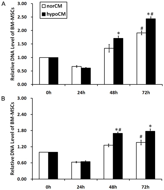
- Hypoxia increased BM-MSC proliferation at 48 h and 72 h in both complete medium and serum-free α-MEM; for example, in complete medium proliferation was 1.71±0.09 vs. 1.34±0.15 at 48 h and 2.43±0.08 vs. 1.91±0.07 at 72 h (p<0.05).
- Hypoxia up-regulated BM-MSC mRNA expression of several paracrine factors, including bFGF (3.33-fold), IGF-1 (2.91-fold), VEGF-A (2.56-fold), TGF-β3 (3.61-fold), IL-6 (2.18-fold), and IL-8 (2.10-fold) (p<0.05).
- ELISA of conditioned medium showed significantly higher levels of bFGF (1.95-fold), VEGF-A (1.27-fold), IL-6 (2.76-fold), and IL-8 (1.53-fold) in hypoCM vs. norCM (p<0.05).
- In vitro, hypoCM increased proliferation of keratinocytes, fibroblasts, and HUVECs, and increased migration of keratinocytes (28.84-fold vs vehicle; 2.01-fold vs norCM), fibroblasts (37.92-fold; 1.30-fold), HUVECs (14.87-fold; 1.44-fold), and CD14+ monocytes (6.54-fold; 1.60-fold) (p<0.05).
- In vivo, hypoCM-treated wounds closed faster; on day 7 wound area was 26.42±2.48% vs 45.00±1.97% (vehicle) and 37.92±2.44% (norCM), and on day 11 it was 8.75±1.25% vs 28.75±1.83% in vehicle controls (p<0.05).
- HypoCM also increased Ki67+ cells in dermis on day 11 (139.25±4.31 vs 56.50±3.12 vehicle and 61.75±4.13 norCM), F4/80+ macrophages (118.25±4.03 vs 64.75±5.12 and 75.50±6.70), and CD31+ vessels (55.50±2.78 vs 34.25±2.25 and 43.25±2.17) (p<0.05).
## Methods
- Human BM-MSCs were cultured under normoxic (20% O2) or hypoxic (2% O2) conditions; conditioned media from each were collected as norCM and hypoCM.
- In vitro analyses included BM-MSC proliferation measurement by DNA assay, semi-quantitative RT-PCR, ELISA, keratinocyte/fibroblast/HUVEC proliferation assays, transwell migration assays, and HUVEC tube formation assays.
- In vivo, Balb/c nude mice received 18 mm full-thickness dorsal wounds and were randomized into three groups (16 per group): vehicle control, norCM, or hypoCM; wound size and histology/IHC/IF outcomes were assessed over time.
- Statistics used Student’s t-test or one-way ANOVA with Bonferroni post-hoc test; significance was set at p<0.05.
## Limitations
- The paper states that the role of IL-6 is not fully resolved here and that they did not evaluate whether these factors act synergistically in wound healing.
- The authors note that the pathways involved in enhanced secretion of growth factors and cytokines by hypoxic BM-MSCs “still requires more information and further investigation.”
- Evident from the text: the study uses a mouse wound model and in vitro assays, so direct applicability to human wound healing is not tested here.
Model-written summary. Check it against the paper before relying on it.
Integrity signals
Registry
Cites
Statistics
Figures


Verification
Claims the paper makes
1 Valid · 0 Not valid · 2 Not checked · 25 Own results and interpretations
Every source the paper cites for this claim was checked and supports it.
In the paper
“the BM-MSCs appear to exert their principal therapeutic actions through the secretion of paracrine trophic signals [7].”
§ Conditioned Medium from Hypoxic Bone Marrow-Derived Mesenchymal Stem Cells Enhances Wound Healing in Mice > Introduction
What the sources say
- Supportedcites [7] AM Hocking, 2010, Mesenchymal stem cells: paracrine signaling and differentiation during cutaneous wound repair, Exp Cell Res source
The Introduction section 'The therapeutic benefits of MSCs for treating tissue injury: mechanisms of action' states that, given poor MSC engraftment/differentiation, 'MSC paracrine signaling is the primary mechanism accounting for the beneficial effects of MSCs', and the 'MSC paracrine signaling enhances wound repair' section says 'Growing evidence indicates that MSC paracrine signaling is the predominant mechanism responsible for enhanced wound repair'. The same sections attribute the effect to secreted mediators — proteomic analyses showing MSCs secrete growth factors, cytokines and chemokines, and MSC-conditioned medium recapitulating the benefit — and the paper frames its MSCs as bone marrow-derived multipotent mesenchymal stromal cells. The citing sentence's hedge ('appear to') matches the source's own hedged phrasing ('it has been proposed', 'growing evidence indicates'). (LLM, conf=0.88)
hedgedstates a cause- Asserted: passes — The authors put it forward as true.
- Checkable: passes — It can be checked against evidence.
- Load-bearing: passes — The paper’s conclusions depend on it.
- Based: passes — The paper gives a reason why it holds.
- Supported
A result the paper reports from its own data — it cites no other source for it, so there is none to check. The quote shows it is in the paper.
In the paper
“collagen I mRNA expression was reduced overtime (day 20 vs. day 40 for vehicle control group: 1.14-fold, P<0.05; for norCM group: 1.15-fold, P<0.05; for hypoCM group: 1.22-fold, P<0.05)”
§ Conditioned Medium from Hypoxic Bone Marrow-Derived Mesenchymal Stem Cells Enhances Wound Healing in Mice > Results > Hypoxia heightens the contribution of BM-MSC-mediated paracrine to cutaneous wound healing in vivo
plainstates a cause- Asserted: passes — The authors put it forward as true.
- Checkable: passes — It can be checked against evidence.
- Load-bearing: passes — The paper’s conclusions depend on it.
- Based: passes — The paper gives a reason why it holds.
A result the paper reports from its own data — it cites no other source for it, so there is none to check. The quote shows it is in the paper.
In the paper
“hypoxia differentially up-regulated the mRNA expression levels of basic fibroblast growth factor (bFGF, 3.33-fold), insulin-like growth factor 1 (IGF-1, 2.91-fold), vascular endothelial growth factor A (VEGF-A, 2.56-fold), transforming growth factor beta 2 (TGF β-2, 2.30-fold), transforming growth factor beta 3 (TGF β-3, 3.61-fold), interleukin 1 beta (IL-1β, 1.86-fold), interleukin 6 (IL-6, 2.18-”
§ Conditioned Medium from Hypoxic Bone Marrow-Derived Mesenchymal Stem Cells Enhances Wound Healing in Mice > Results > Hypoxia up-regulates the mRNA expression and protein secretion of BM-MSC-derived paracrine factors
plainstates a cause- Asserted: passes — The authors put it forward as true.
- Checkable: passes — It can be checked against evidence.
- Load-bearing: passes — The paper’s conclusions depend on it.
- Based: passes — The paper gives a reason why it holds.
A result the paper reports from its own data — it cites no other source for it, so there is none to check. The quote shows it is in the paper.
In the paper
“BM-MSC hypoCM significantly enhanced HUVEC tube formation ability on a Matrigel matrix compared with the vehicle control or norCM (Figs. 4, p<0.05).”
§ Conditioned Medium from Hypoxic Bone Marrow-Derived Mesenchymal Stem Cells Enhances Wound Healing in Mice > Results > Hypoxia enhances mitogenic, chemoattractive and angiogenic paracrine effects of BM-MSCs in vitro
plainstates a cause- Asserted: passes — The authors put it forward as true.
- Checkable: passes — It can be checked against evidence.
- Load-bearing: passes — The paper’s conclusions depend on it.
- Based: passes — The paper gives a reason why it holds.
A result the paper reports from its own data — it cites no other source for it, so there is none to check. The quote shows it is in the paper.
In the paper
“similar amounts of keratinocyte growth factor (KGF, 0.95-fold), monocyte chemotactic protein-1 (MCP-1, 1.03-fold), transforming growth factor beta 1 (TGF β-1, 0.93-fold), interleukin 10 (IL-10, 1.05-fold), tumor necrosis factor (TNF-α, 0.97-fold) and matrix metalloproteinases (MMP-1, 1.08-fold) were found in normoxic and hypoxic BM-MSCs (p>0.05).”
§ Conditioned Medium from Hypoxic Bone Marrow-Derived Mesenchymal Stem Cells Enhances Wound Healing in Mice > Results > Hypoxia up-regulates the mRNA expression and protein secretion of BM-MSC-derived paracrine factors
plain- Asserted: passes — The authors put it forward as true.
- Checkable: passes — It can be checked against evidence.
- Load-bearing: unclear — It is unclear whether the paper’s conclusions depend on it.
- Based: passes — The paper gives a reason why it holds.
13 more claims
Create a free account to see all 28 claims and to verify the 2 references still open.
- It may state two separate claims as one. 6
- Probably not a substantial claim. 5
Claims other papers make about this one
Each is checked against this paper’s own text: does it say what the citing paper says it does?
The source backs part of the claim, not all of it — or backs it for a different population, condition or comparison.
In the citing paper
“the secretome obtained from hypoxia preconditioned-BM-MSCs upregulated the levels of FGF-2, IGF-1, TGF-β, IL-1β, IL-6 [123] and IL-8 [122] and enhanced multiple key processes to promote wound healing, including keratinocyte, fibroblast and endothelial cells proliferation and migration, as well as angiogenesis (Fig. 2) [38], [120], [123].”
From Mesenchymal stem cell secretome for regenerative medicine: Where do we stand? (2025) · cites this paper as [123]
Experimental — accuracy not yet measured
Citations
Cites
Gurtner, G. C., Werner, S., Barrandon, Y., & Longaker, M. T. (2008) · Nature · doi:10.1038/nature07039 · Cited by 6,542
Singer, A. J., & Clark, R. A. F. (1999) · New England Journal of Medicine · doi:10.1056/nejm199909023411006 · Cited by 6,106
Wound Healing--Aiming for Perfect Skin Regeneration
Martin, P. (1997) · Science · doi:10.1126/science.276.5309.75 · Cited by 4,804 · cited 1×
“Epidermal growth factor (EGF), KGF, IGF-1 and VEGF-A are among the growth factors and cytokines known to enhance normal wound healing [17-19].”
BackgroundChen, L., Tredget, E. E., Wu, P. Y. G., & Wu, Y. (2008) · PLoS ONE · doi:10.1371/journal.pone.0001886 · Cited by 1,610 · cited 1×
“BM-MSC-mediated paracrine enhances wound healing through three predominant pathways, as evidenced by the demonstrated actions of BM-MSC-derived conditioned medium and secreted molecules: (1) BM-MSC-derived conditioned medium may act as a chemoattractant to recruit specific cell types to the wound, including epidermal…”
BackgroundSelf-citationKinnaird, T., Stabile, E., Burnett, M. S., Shou, M., Lee, C. W., Barr, S., Fuchs, S., & Epstein, S. E. (2004) · Circulation · doi:10.1161/01.cir.0000124062.31102.57 · Cited by 1,305 · cited 1×
“Consistent with previous reports [20,21], the current study demonstrated that BM-MSCs expressed and secreted VEGF-A, bFGF and other factors.”
ResultA Computational Tool for Quantitative Analysis of Vascular Networks
Zudaire, E., Gambardella, L., Kurcz, C., & Vermeren, S. (2011) · PLoS ONE · doi:10.1371/journal.pone.0027385 · Cited by 1,085 · cited 1×
“The number of tubular structures, tubule length, branches and nodes in 4 randomly chosen fields was quantified as previously described with the aid of Olympus Cell R imaging software [13].”
MethodKim, W.-S., Park, B.-S., Sung, J.-H., Yang, J.-M., Park, S.-B., Kwak, S.-J., & Park, J.-S. (2007) · Journal of Dermatological Science · doi:10.1016/j.jdermsci.2007.05.018 · Cited by 859
Djouad, F., Charbonnier, L.-M., Bouffi, C., Louis-Plence, P., Bony, C., Apparailly, F., Cantos, C., Jorgensen, C., & Noël, D. (2007) · Stem Cells · doi:10.1634/stemcells.2006-0548 · Cited by 667 · cited 1×
“However, it has been proposed as a proinflammatory cytokine involved in a lot of inflammatory diseases [28] or as an anti-inflammatory cytokine involved in the inhibition of T-cell proliferation by MSCs in diseases such as graft-versus-host disease [29].”
BackgroundHypoxia enhances proliferation and tissue formation of human mesenchymal stem cells
Grayson, W. L., Zhao, F., Bunnell, B., & Ma, T. (2007) · Biochemical and Biophysical Research Communications · doi:10.1016/j.bbrc.2007.05.054 · Cited by 521 · cited 1×
“Our current data are consistent with previous reports demonstrating the significantly increased proliferation of BM-MSCs under hypoxic conditions [26].”
ResultLee, E. Y., Xia, Y., Kim, W., Kim, M. H., Kim, T. H., Kim, K. J., Park, B., & Sung, J. (2009) · Wound Repair and Regeneration · doi:10.1111/j.1524-475x.2009.00499.x · Cited by 423
Cited by
Items 201–252 of 252
Mesenchymal stromal cells as a resource for regeneration of damaged skin
Domaratskaya, E. I., & Payushina, O. V. (2017) · Biology Bulletin Reviews · doi:10.1134/s207908641704003x · Cited by 2
“Under these conditions, MSCs produce greater amounts of bFGF, VEGF, IL-6, and IL-8 (Chen et al., 2014); they become more resistant to ischemia and enhance the proliferation and migration of endothelial cells in vitro (Bader et al.”
BackgroundInfluentialSolarte-David, V. A., Becerra‐Bayona, S. M., Sanchez-Aranguren, L. C., Sossa, C., Meana, Á., Lloves, J. M., & Arango-Rodríguez, M. L. (2018) · Journal of Stem Cell Research & Therapy · doi:10.4172/2157-7633.1000423 · Cited by 2
“In contrast, other studies indicate that transplanted MSCs do not necessarily have to be in close proximity to the damaged tissue in order to promote wound repair and functional recovery, since the secretion of paracrine factors appears to be the main MSC therapeutic action involved in skin disorder repair [115,116].”
ResultPrenatal Mesenchymal Stem Cell Secretome and Its Clinical Implication
Liang, L. (2019) · Springer eBooks · doi:10.1007/978-981-13-2703-2_13 · Cited by 2
Ajit, A., Ramakrishnan, R., Santhoshkumar, T. R., Senan, M., & Krishnan, L. K. (2020) · Journal of Biomedical Materials Research Part B Applied Biomaterials · doi:10.1002/jbm.b.34611 · Cited by 2
Widowati, W., Jasaputra, D. K., Kusuma, H. S. W., Rizal, R., Laksmıtawatı, D. R., Artie, D. S., Sholihah, I. A., Yusepany, D. T., LESTARI, R., Handayani, R. A. S., et al. (2021) · Walailak Journal of Science and Technology (WJST) · doi:10.48048/wjst.2021.9270 · Cited by 2
“ATG-5 : Autophagy Related 5 gene BMMSCs : Bone marrow-derived Mesenchymal Stem Cells BAX : Bcl-2-associated X protein BCL2 : B-cell lymphoma 2 CD : Cluster of differentiation EC : Endothelial cell EPCs : Endothelial Progenitor Cells FACS : Fluorescence Activated Cell Sorting FGF-2 : Fibroblast Growth Factor-2 HeLa : h…”
BackgroundHypoxia-driven Dental Pulp Stem Cells as a Promising Strategy for Tissue Regeneration
Liao, C., Chen, L., Xiang, M., Long, S., Xiang, M., Xiao, L., Long, Q., Liu, J., & Guan, X. (2024) · Current Stem Cell Research & Therapy · doi:10.2174/011574888x312892240625091241 · Cited by 1
Self-citationJafari, Z., Hamzehpour, Z., Faghihi, M., Derazi, R. A., Makoui, M. H., Makoui, R. H., Fekri, S. M., Ansari, N. E., & Athari, S. S. (2025) · Modern Rheumatology · doi:10.1093/mr/roaf112 · Cited by 1
Effects of hypoxia on the biological characteristics of urine-derived stem cells
Xiao, Y., Guo, Z., Shao, W., Hq, X., & Huang, Y. (2026) · Frontiers in Cell and Developmental Biology · doi:10.3389/fcell.2026.1851623 · Cited by 1
“…have reported heterogeneous effects of hypoxia on the paracrine secretion of stem/stromal cells, which may vary depending on oxygen tension, cell source, and culture conditions (Zhao et al., 2022; Zhang et al., 2020; Chang et al., 2013; Quade et al., 2020; Xiang and Xie, 2018; Chen et al., 2014).”
The role of mesenchymal stromal cells in skin wound healing
Teng, M., & Hengshu, Z. (2016) · Wiley eBooks · doi:10.1002/9781118907474.ch59 · Cited by 1
Натрус, Л. В., Verko, N. P., Ryzhko, I. N., Panova, T. I., & Osadchuk, Y. (2018) · Medical Science of Ukraine (MSU) · doi:10.32345/2664-4738.1-2.2018.01 · Cited by 1
Peltzer, J., & Lataillade, J. (2018) · Elsevier eBooks · doi:10.1016/b978-0-12-812015-6.00008-x · Cited by 1
Rinendyaputri, R., Noviantari, A., Zainuri, M., Nikmah, U. A., Dany, F., & Intan, P. R. (2023) · AIP conference proceedings · doi:10.1063/5.0118570 · Cited by 1
Mesenchymal Stromal Cells for Wound Healing Therapy: From Expectations to Reality
Rosca, A., Ţuţuianu, R., Ghetu, D. M., & Titorencu, I. D. (2024) · Springer eBooks · doi:10.1007/978-981-99-7119-0_53 · Cited by 1
Kustiyah, A. R., Fatimah, N., Rizkiyani, E. M., Ramadhanti, O. W., Istiqomah, D. A. F., Hidayah, N., & Wilaksono, B. (2022) · International Journal of Cell and Biomedical Science · doi:10.59278/cbs.v1i1.9 · Cited by 1
“In addition, several studies have also confirmed that under hypoxic culture conditions, MSCs can increase the secretion of cytokines, including IL-10 and TGF-β 31–34 .”
Chemokine Pathway Can Be the Potential Therapeutic Target for Hypertrophic Scar
Umeyama, H., Ding, J., & Tredget, E. E. (2019) · European Medical Journal · doi:10.33590/emj/10312169 · Cited by 1
Regenerative therapy for the nonhealing cutaneous wounds
Orlovska, I. V., Yakovenko, I., Haidak, A., Zmejkoski, D. Z., & Kozyrovska, N. (2018) · Biopolymers and Cell · doi:10.7124/bc.000979
Transfection of mesenchymal stem cells at physiological oxygen concentrations
Shuvalova, N. S., Toporova, O. K., & Кордюм, В. А. (2020) · Biopolymers and Cell · doi:10.7124/bc.000a41
“Additionally, the enhancement of MSC para crine activity in conditions of mild hypoxia is detected [17].”
Background616 テストの出来ばえに関する評価の動機づけ機能(測定・評価6-3,600 測定・評価)
福島, 脩., & 宗内, 敦. (1975) · 日本教育心理学会総会発表論文集 · doi:10.20587/pamjaep.17.0_572
Jones, A. R. (2014) · Aquila Digital Community (University of Southern Mississippi)
The Role of Mesenchymal Stem Cell in Controlling Liver Function on Liver Fibrosis
Antari, A. D., Prasetio, A., & Nugroho, B. A. (2021) · Journal of Science and Technology Research for Pharmacy · doi:10.15294/jstrp.v1i2.45699
fallah, F., Pourhassan, P., Khani, M., Burke, B., Ebrahimi, M., & Sotoodehnejadnematalahi, F. (2022) · Research Square · doi:10.21203/rs.3.rs-1222431/v1 · Cited by 1
(2017) · ISRN Tissue Engineering · doi:10.1155/8592
Aslam, N., Abusharieh, E., Abuarqoub, D. A., Ali, D., Alhattab, D. M., Wehaibi, S., Al-Kurdi, B., Jamali, F., Alshaer, W., Jafar, H. D., et al. (2020) · Research Square · doi:10.21203/rs.3.rs-38696/v1
A preview of selected articles
Atkinson, S. P. (2020) · Stem Cells Translational Medicine · doi:10.1002/sctm.20-0395
Chouw, A., Sartika, C. R., Milanda, T., & Faried, A. (2022) · Regenerative Engineering and Translational Medicine · doi:10.1007/s40883-022-00259-w
Mesenchymal Stromal Cells for Wound Healing Therapy: From Expectations to Reality
Rosca, A., Ţuţuianu, R., Ghetu, D. M., & Titorencu, I. D. (2023) · Springer eBooks · doi:10.1007/978-981-99-0846-2_53-1
Efeitos da hipóxia em células-tronco mesenquimais sobre-expressando BMP-9
PAZ, J. E. R. (2023) · Digital Library of Theses and Dissertations (Universidade de São Paulo) · doi:10.11606/d.58.2023.tde-23102023-153320
Yang, S., Lian, Y., Song, G., Ni, H., Jia, B., Qian, X., He, G., Ullah, A., & Qi, X. (2024) · Science of Advanced Materials · doi:10.1166/sam.2024.4727
Accelerating Wound Repair: The Multifaceted Role of Mesenchymal Stem CellsPreprint
Abdul Ghani, N. I., Nurhidayatul, M. N., Khairul, A. K., Hasmad, H. N., Afifa, N. T., Helmi, B. S., Aida Juliana, A. J., Farisha, M. N., Balqish, M. R., Kh, C., et al. (2024) · Preprints.org · doi:10.20944/preprints202409.1422.v1 · Cited by 1
Amellia, N. L., Antari, A. D., Nazar, M. A., Husain, S. A., & Arifin, N. A. (2023) · International Journal of Cell and Biomedical Science · doi:10.59278/cbs.v1i3.23
Elnour, M. S., Abdelrazeg, S., Mohamed, R., Lakshmanan, M., Sudesh, K., & Shaharuddin, B. (2024) · Malaysian Journal of Medicine and Health Sciences · doi:10.47836/mjmhs.20.4.29
Advanced Drug Delivery TechnologiesPreprint
Çetin, E. Ö., Erisik, D., TAŞKIRAN, A., Çalışkan, E. E., Demirtürk, E., AYDIN, Y. Ç., & Uyanıkgil, Y. (2025) · SSRN Electronic Journal · doi:10.2139/ssrn.5080254 · Cited by 1
Liu, C., Xu, Y., Lu, Y., Du, P., Li, X., Wang, C., Guo, P., Diao, L., & Lu, G. (2022) · Figshare · doi:10.6084/m9.figshare.20486702
Self-citationLiu, C., Xu, Y., Lu, Y., Du, P., Li, X., Wang, C., Guo, P., Diao, L., & Lu, G. (2022) · Figshare · doi:10.6084/m9.figshare.20486711
Self-citationLiu, C., Xu, Y., Lu, Y., Du, P., Li, X., Wang, C., Guo, P., Diao, L., & Lu, G. (2022) · Figshare · doi:10.6084/m9.figshare.20486717
Self-citationLiu, C., Xu, Y., Lu, Y., Du, P., Li, X., Wang, C., Guo, P., Diao, L., & Lu, G. (2022) · Figshare · doi:10.6084/m9.figshare.20486699
Self-citationLiu, C., Xu, Y., Lu, Y., Du, P., Li, X., Wang, C., Guo, P., Diao, L., & Lu, G. (2022) · Figshare · doi:10.6084/m9.figshare.20486693
Self-citationZhang, X.-R., Huang, Y.-Z., Gao, H.-W., Jiang, Y.-L., Hu, J.-G., Pi, J.-K., Chen, A.-J., Zhang, Y., Zhou, L., & Xie, H.-Q. (2020) · Figshare · doi:10.6084/m9.figshare.12096414
Liu, C., Xu, Y., Lu, Y., Du, P., Li, X., Wang, C., Guo, P., Diao, L., & Lu, G. (2022) · Figshare · doi:10.6084/m9.figshare.20486696
Self-citationLiu, C., Xu, Y., Lu, Y., Du, P., Li, X., Wang, C., Guo, P., Diao, L., & Lu, G. (2022) · Figshare · doi:10.6084/m9.figshare.20486708
Self-citationLiu, C., Xu, Y., Lu, Y., Du, P., Li, X., Wang, C., Guo, P., Diao, L., & Lu, G. (2022) · Figshare · doi:10.6084/m9.figshare.20486714
Self-citationZhang, X.-R., Huang, Y.-Z., Gao, H.-W., Jiang, Y.-L., Hu, J.-G., Pi, J.-K., Chen, A.-J., Zhang, Y., Zhou, L., & Xie, H.-Q. (2020) · Figshare · doi:10.6084/m9.figshare.12096420
Liu, C., Xu, Y., Lu, Y., Du, P., Li, X., Wang, C., Guo, P., Diao, L., & Lu, G. (2022) · Figshare · doi:10.6084/m9.figshare.20486705
Self-citationSmart Drug Delivery Solutions for Wound Management: Innovations and Implementation Barriers
Yadav, P., Kumar, S., & Kumar, D. (2026) · Drug Metabolism and Bioanalysis · doi:10.2174/0118723128406161251208194426 · Cited by 1
Elaboration d’un épiderme de culture et évaluation non clinique sur un modèle de brûlure cutanée
Alexaline, M. M. (2015) · theses.fr (ABES) · doi:10.70675/30308059z3871z47aaza4a9z005bc8d8f8f5
Dias, T. D., & Anderson, D. E. (2026) · Cells · doi:10.3390/cells15090815 · Cited by 1
“Hypoxia also increases secretion of ECM remodeling factors, including matrix metallopeptidase (MMP)-2 and MMP-9 [165,166], and mediators of the inflammatory response such as IL-6 and IL-8 [167].”
Nasiri, H., Khosravi, F., Eskandari, F., Manoochehrabadi, T., Samadikuchaksaraei, A., Lotfi, E., Majidi, J., Rafati, F., & Ghorbani, Z. (2026) · Clinical and Translational Discovery · doi:10.1002/ctd2.70172
Purnamasari, Y., Mahyudin, F., & Edward, M. (2026) · Scripta Medica · doi:10.5937/scriptamed57-60517
“5, 6 Recent molecular studies have detailed how bioactive peptides and growth factors—particularly TGF-β and PDGF—modulate osteogenic differentiation and vascular remodeling.”
Gunawan, V. A., Sumarawati, T., Putra, A., Setiawan, E., Trisnadi, S., Chodijah, C., & Amalina, N. D. (2026) · Journal of Research in Pharmacy · doi:10.12991/jrespharm.1684891
Metadata
- Authors
- Chen, L., Xu, Y., Zhao, J., Zhang, Z., Yang, R., Xie, J., Liu, X., & Qi, S.
- Affiliations
- The First Affiliated Hospital, Sun Yat-sen University (CN)
- Sixth Affiliated Hospital of Sun Yat-sen University (CN)
- First People's Hospital of Foshan (CN)
- Corresponding author
- Qi, Shaohai
- ORCID
- Venue
- PLoS ONE
- Publisher
- Public Library of Science (PLoS)
- Published
- 2014-04-29
- Volume
- 9
- Issue
- 4
- Pages
- e96161
- ISSN
- 1932-6203 (ISSN-L)
- PMC
- PMC4004560
- OpenAlex
- W4239894970
- Semantic Scholar
- b818b82b42ef7ae7423a30f97f31ae72620ee92b
- Cited by
- 251
- References
- 32
- Citations by year
- 2026: 9 · 2025: 10 · 2024: 11 · 2023: 25 · 2022: 28 · 2021: 28 · 2020: 35 · 2019: 17 · 2018: 22 · 2017: 19 · 2016: 24 · 2015: 17 · 2014: 5
- FWCI
- 21.04
- Type
- journal article
- Language
- en
- Update notices
- correction (2015-12-18)
- Open access
- gold
- doi.org
- Locations
- PLoS ONE (publishedVersion, cc-by)
- Europe PMC (PubMed Central) (publishedVersion, other-oa)
- PubMed Central (publishedVersion, cc-by)
- PubMed (publishedVersion)
- DOAJ (DOAJ: Directory of Open Access Journals) (submittedVersion, cc-by-sa)
- Funders
- National Natural Science Foundation of China
- Natural Science Foundation of Guangdong Province
- Topics
- Health Sciences › Medicine › Surgery › Wound Healing and Treatments
- Life Sciences › Biochemistry, Genetics and Molecular Biology › Cell Biology › Mesenchymal stem cell research
- Health Sciences › Dentistry › Periodontics › Periodontal Regeneration and Treatments
- Keywords
- bone marrow-derived mesenchymal stem cells, hypoxia, conditioned medium, paracrine signaling, skin wound healing, basic fibroblast growth factor, VEGF-A, interleukin-6, interleukin-8, keratinocyte proliferation, cell migration, neovascularization, macrophage recruitment, collagen deposition, topical treatment
- MeSH terms
- Animals, Base Sequence, Bone Marrow Cells, Cells, Cultured, Humans, Wound Healing, Cell Hypoxia, Culture Media, Conditioned, DNA Primers, Reverse Transcriptase Polymerase Chain Reaction, Cell Proliferation, Mice, Mesenchymal Stem Cells
About these checks
Registry, retraction, Cites and statistics results are computed, not judged, with one exception: a printed reference that no index lists is read once by a model before it counts as a citation error. Claims are extracted by a model and kept only when their quote appears verbatim in the text; each cited source is then checked, with a model’s help, for whether it says what the claim relies on. Those verdicts can be wrong — read the claim’s quote and the cited source before relying on one.
A language model drew the claims from the paper on 2026-10-11, under version 3 of the claim definition (model deepseek-v4.1-flash, prompt extract-v3).
A second model labelled each claim on 2026-10-11 (model jev-1.13-20260917, definition revision gold-guideline-v0.2).
Cited sources are checked with a language model’s help (qwen3.8-omni-flash).
Statistics and arithmetic are recomputed, not judged.
Confidence bands are shown only for fields calibrated on our annotated test set. None is calibrated yet, so no band is shown.
What counts as a claim ›How we measure confidence ›How we check this ›
Are you an author, or do you see a problem with this page? Request a review
Data sources
Bibliographic data: Semantic Scholar, OpenAlex, Crossref.
Contains information from the Semantic Scholar Academic Graph Datasets, made available under the ODC Attribution License (ODC-BY 1.0).
Semantic Scholar’s data is described in: Kinney, R., et al. (2023). The Semantic Scholar Open Data Platform. arXiv:2301.10140.
