

The DOI matches its Crossref record; 61 of the 62 references it cites were found in a registry or an index.
Muscle Releases Alpha-Sarcoglycan Positive Extracellular Vesicles Carrying miRNAs in the Bloodstream
Open accessAbstract
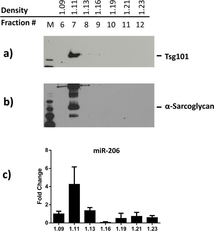
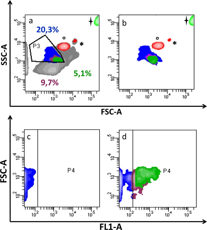
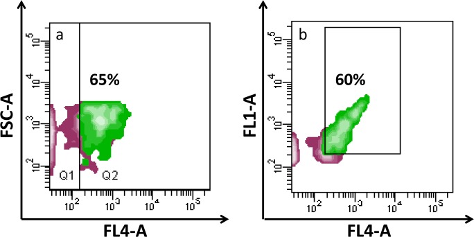
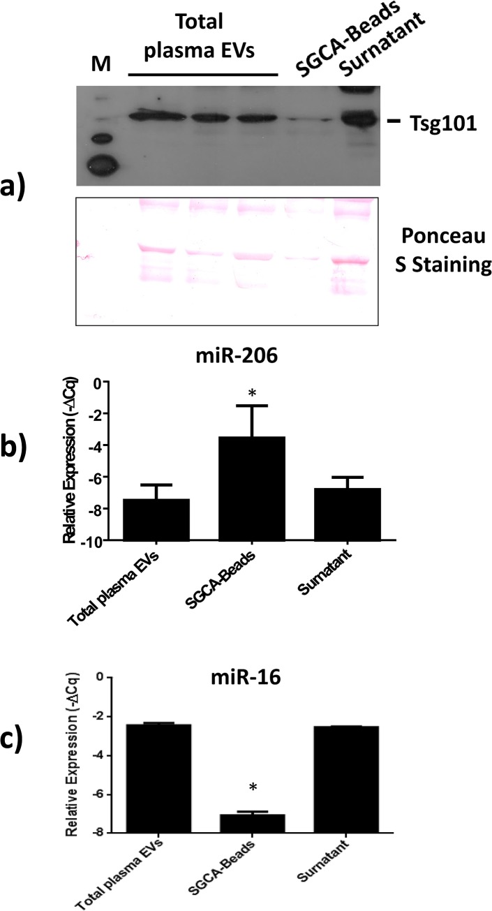
In the past few years, skeletal muscle has emerged as an important secretory organ producing soluble factors, called myokines, that exert either autocrine, paracrine or endocrine effects. Moreover, recent studies have shown that muscle releases microRNAs into the bloodstream in response to physical exercise. These microRNAs affect target cells, such as hormones and cytokines. The mechanisms underlying microRNA secretion are poorly characterized at present. Here, we investigated whether muscle tissue releases extracellular vesicles (EVs), which carry microRNAs in the bloodstream under physiological conditions such as physical exercise. Using density gradient separation of plasma from sedentary and physically fit young men we found EVs positive for TSG101 and alpha-sarcoglycan (SGCA), and enriched for miR-206. Cytometric analysis showed that the SGCA+ EVs account for 1-5% of the total and that 60-65% of these EVs were also positive for the exosomal marker CD81. Furthermore, the SGCA-immuno captured sub-population of EVs exhibited higher levels of the miR-206/miR16 ratio compared to total plasma EVs. Finally, a significant positive correlation was found between the aerobic fitness and muscle-specific miRNAs and EV miR-133b and -181a-5p were significantly up-regulated after acute exercise. Thus, our study proposes EVs as a novel means of muscle communication potentially involved in muscle remodeling and homeostasis.
Overview
The claims and statistics of this paper haven’t been checked yet.
Run the check — free, no account needed. It reads the open-access text and checks the sources it cites.
Integrity signals
Registry
Cites
Statistics
Figures
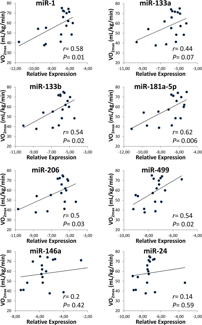
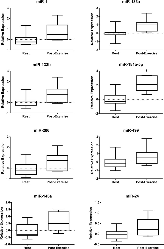
Verification
Claims the paper makes
The claims and statistics of this paper haven’t been checked yet.
Run the check — free, no account needed. It reads the open-access text and checks the sources it cites.
Claims other papers make about this one
Each is checked against this paper’s own text: does it say what the citing paper says it does?
In the citing paper
“These ELVs were also positive for CD81, TSG101 and miR-206”
From Skeletal Muscle-Released Extracellular Vesicles: State of the Art (2019) · cites this paper as Guescini et al., 2015
In the citing paper
“ELVs containing alpha-sarcoglycan (SGCA) a component of the dystrophin-glycoprotein complex involved in the stability of muscle fiber membranes, were found in human plasma”
From Skeletal Muscle-Released Extracellular Vesicles: State of the Art (2019) · cites this paper as Guescini et al., 2015
Self-check
Percentages, sums and reported differences re-computed from the paper’s own numbers.
The self-check needs the paper’s full text.
Citations
Cites
Bradford, M. M. (1976) · Analytical Biochemistry · doi:10.1016/0003-2697(76)90527-3 · Cited by 255,302 · cited 1×
“After centrifugation at 18,000 g, protein concentration was determined by the Bradford assay [35].”
MethodAnalysis of Relative Gene Expression Data Using Real-Time Quantitative PCR and the 2−ΔΔCT Method
Livak, K. J., & Schmittgen, T. D. (2001) · Methods · doi:10.1006/meth.2001.1262 · Cited by 180,860 · cited 1×
“Quantification was performed according to the ΔCq method [34], miR-16 or the exogenously added spike-in cel-miR-39 were used as a reference.”
MethodValadi, H., Ekström, K., Bossios, A., Sjöstrand, M., Lee, J. J., & Lötvall, J. O. (2007) · Nature Cell Biology · doi:10.1038/ncb1596 · Cited by 12,834 · cited 1×
“Numerous studies indicate that circulating miRNAs may be protected from degradation by several complementary mechanisms, including their incorporation into secreted membrane vesicles such as microvesicles, exosomes or apoptotic bodies [16–18], or via the formation of RNA-binding protein complexes [19].”
Background(2014) · Medical Entomology and Zoology · Cited by 9,400
Skog, J., Würdinger, T., van Rijn, S., Meijer, D. H., Gainche, L., Curry, W. T., Carter, B. S., Krichevsky, A. M., & Breakefield, X. O. (2008) · Nature Cell Biology · doi:10.1038/ncb1800 · Cited by 5,001
Mutation of the mouse klotho gene leads to a syndrome resembling ageing
Kuro-o, M., Matsumura, Y., Aizawa, H., Kawaguchi, H., Suga, T., Utsugi, T., Ohyama, Y., Kurabayashi, M., Kaname, T., Kume, E., et al. (1997) · Nature · doi:10.1038/36285 · Cited by 3,837
Arroyo, J. D., Chevillet, J. R., Kroh, E. M., Ruf, I. K., Pritchard, C. C., Gibson, D. F., Mitchell, P. S., Bennett, C. F., Pogosova-Agadjanyan, E. L., Stirewalt, D. L., et al. (2011) · Proceedings of the National Academy of Sciences · doi:10.1073/pnas.1019055108 · Cited by 3,294 · cited 1×
“Numerous studies indicate that circulating miRNAs may be protected from degradation by several complementary mechanisms, including their incorporation into secreted membrane vesicles such as microvesicles, exosomes or apoptotic bodies [16–18], or via the formation of RNA-binding protein complexes [19].”
BackgroundMuscles, exercise and obesity: skeletal muscle as a secretory organ
Pedersen, B. K., & Febbraio, M. A. (2012) · Nature Reviews Endocrinology · doi:10.1038/nrendo.2012.49 · Cited by 2,909 · cited 1×
“Therefore, the discovery that contracting muscle is a myokine-producing organ has opened up a whole new field of research on skeletal muscle [6].”
BackgroundMicroRNAs are transported in plasma and delivered to recipient cells by high-density lipoproteins
Vickers, K. C., Palmisano, B. T., Shoucri, B. M., Shamburek, R. D., & Remaley, A. T. (2011) · Nature Cell Biology · doi:10.1038/ncb2210 · Cited by 2,741 · cited 2×
“In the extracellular environment, miRNAs could be stabilized by EVs (such as exosomes, microvesicles or apoptotic bodies), HDL/LDLs, or protected by RNA-binding proteins (reviewed in [14,45]).”
BackgroundExosomes: Extracellular organelles important in intercellular communication
Mathivanan, S., Ji, H. L., & Simpson, R. J. (2010) · Journal of Proteomics · doi:10.1016/j.jprot.2010.06.006 · Cited by 2,581 · cited 1×
“11 g/mL, as previously described [37].”
Cited by
Sorted by citations
Extracellular Vesicles Provide a Means for Tissue Crosstalk during Exercise
Whitham, M., Parker, B. L., Friedrichsen, M. H., Hingst, J. R., Hjorth, M., Hughes, W. E., Egan, C. L., Cron, L., Watt, K. I., Kuchel, R. P., et al. (2018) · Cell Metabolism · doi:10.1016/j.cmet.2017.12.001 · Cited by 700
Brahmer, A., Neuberger, E. W. I., Esch-Heisser, L., Haller, N., Jørgensen, M. M., Bæk, R., Möbius, W., Simon, P. D., & Krämer‐Albers, E. (2019) · Journal of Extracellular Vesicles · doi:10.1080/20013078.2019.1615820 · Cited by 251
“exercise-related cell types such as muscle cells [alphasarcoglycan, [39], Figure 4(a)] or hepatocytes (asialoglycoprotein receptor, not shown) could not be detected.”
BackgroundFulzele, S., Mendhe, B., Khayrullin, A., Johnson, M. H., Kaiser, H., Liu, Y., Isales, C. M., & Hamrick, M. W. (2019) · Aging · doi:10.18632/aging.101874 · Cited by 220
Bei, Y., Xu, T., Lv, D., Yu, P., Xu, J., Che, L., Das, A., Tigges, J., Toxavidis, V., Ghiran, I. C., et al. (2017) · Basic Research in Cardiology · doi:10.1007/s00395-017-0628-z · Cited by 201
“In this regard, exercise-induced EVs derived from skeletal muscle deserve further investigation [1, 21, 40].”
Extracellular Vesicles and Exosomes: Insights From Exercise Science
Nederveen, J. P., Warnier, G., Carlo, A. D., Nilsson, M. I., & Tarnopolsky, M. A. (2021) · Frontiers in Physiology · doi:10.3389/fphys.2020.604274 · Cited by 200
“…of anaerobic threshold (Sossdorf et al., 2010, 2011), ∼70% of VO2max (Lansford et al., 2016) and a single bout of high intensity interval exercise (Guiraud et al., 2013) all appear to increase PMVs. Work by Wilhelm et al. (2016) examined PMVs during a 1 h bout of heavy intensity exercise (∼65%…”
BackgroundExtracellular Vesicles in Blood: Sources, Effects, and Applications
Alberro, A., Iparraguirre, L., Fernandes, A., & Otaegui, D. (2021) · International Journal of Molecular Sciences · doi:10.3390/ijms22158163 · Cited by 170
“In addition, the muscle tissue also secretes EVs and, indeed, striated muscle-specific miRNAs or myo-miRNAs have been found in human plasma EVs [75,76].”
BackgroundYates, A. G., Pink, R. C., Erdbrügger, U., Siljander, P. R. M., Dellar, E. R., Pantazi, P., Akbar, N., Cooke, W. R., Vatish, M., Dias‐Neto, E., et al. (2022) · Journal of Extracellular Vesicles · doi:10.1002/jev2.12151 · Cited by 187
“One of the most abundant miRNA in muscle-derived EVs is miR-206, which induces the development and differentiation of tissue, especially after muscle injury during exercise (Guescini et al., 2015).”
MicroRNAs as Important Regulators of Exercise Adaptation
da Silva, G. J. J., Bye, A., el Azzouzi, H., & Wisløff, U. (2017) · Progress in Cardiovascular Diseases · doi:10.1016/j.pcad.2017.06.003 · Cited by 158
Castaño, C., Mirasierra, M., Vallejo, M., Novials, A., & Párrizas, M. (2020) · Proceedings of the National Academy of Sciences · doi:10.1073/pnas.2016112117 · Cited by 156
“In particular, we observed up-regulation of miR-133a, miR-133b, and miR-206, three myomiRs whose circulating levels in vesicles have been shown by others to be increased in correlation with VO2 max in humans (41).”
BackgroundBurtscher, J., Millet, G. P., Place, N., Kayser, B., & Zanou, N. (2021) · International Journal of Molecular Sciences · doi:10.3390/ijms22126479 · Cited by 164
“Many of these molecules travel systemically (in blood or lymph) via extracellular vesicles that are today considered as key messengers of paracrine exercise signals [116–119], as they have been described to be released from skeletal muscle only in 2015 [120].”
BackgroundAgostini, D., Donati Zeppa, S., Lucertini, F., Annibalini, G., Gervasi, M., Ferri Marini, C., Piccoli, G., Stocchi, V., Barbieri, E., & Sestili, P. (2018) · Nutrients · doi:10.3390/nu10081103 · Cited by 151
“Furthermore, skeletal muscle expresses high levels of several microRNAs that can be delivered by exosomes [54].”
BackgroundThe role of extracellular vesicles in skeletal muscle and systematic adaptation to exercise
Vechetti, I. J., Valentino, T., Mobley, C. B., & McCarthy, J. J. (2021) · The Journal of Physiology · doi:10.1113/jp278929 · Cited by 141
“These authors found that after an acute bout of endurance exercise there was a significant increase in the abundance of EV miR-181a-5p but no change in the abundance of miR-1, miR-133b, miR-206, miR-499 or miR-146a within EVs (Guescini et al. 2015).”
BackgroundInfluentialSkeletal Muscle-Released Extracellular Vesicles: State of the Art
Rome, S., Forterre, A., Mizgier, M. L., & Bouzakri, K. (2019) · Frontiers in Physiology · doi:10.3389/fphys.2019.00929 · Cited by 139
Circulating myomiRs: a new class of biomarkers to monitor skeletal muscle in physiology and medicine
Siracusa, J., Koulmann, N., & Banzet, S. (2017) · Journal of Cachexia Sarcopenia and Muscle · doi:10.1002/jcsm.12227 · Cited by 135
“Whether exercise triggers the release of extracellular vesicles from muscle tissues is unclear in this study, because running exercise induced no changes in circulating myomiRs.(27)”
BackgroundMethodResultInfluentialEmerging role of extracellular vesicles in musculoskeletal diseases
Murphy, C., Withrow, J. S., Hunter, M., Liu, Y., Tang, Y., Fulzele, S., & Hamrick, M. W. (2017) · Molecular Aspects of Medicine · doi:10.1016/j.mam.2017.09.006 · Cited by 123
“Not surprisingly, EVs released from skeletal muscle are enriched in these miRNAs, and circulating muscle-derived EVs transport these miRNAs [26].”
BackgroundBone and Muscle Crosstalk in Aging
He, C., He, W., Hou, J., Chen, K., Huang, M., Yang, M., Luo, X., & Li, C. (2020) · Frontiers in Cell and Developmental Biology · doi:10.3389/fcell.2020.585644 · Cited by 129
Yu, A., Zhang, T., Duan, H., Pan, Y., Zhang, X., Yang, G., Wang, J., Deng, Y., & Yang, Z. (2017) · Immunology Letters · doi:10.1016/j.imlet.2016.12.003 · Cited by 107
Extracellular Vesicles: Delivery Vehicles of Myokines
Trovato, E., Felice, V. D., & Barone, R. (2019) · Frontiers in Physiology · doi:10.3389/fphys.2019.00522 · Cited by 115
Rigamonti, A. E., Bollati, V., Pergoli, L., Iodice, S., Col, A. D., Tamini, S., Cicolini, S., Tringali, G., Micheli, R. D., Cella, S. G., et al. (2019) · International Journal of Obesity · doi:10.1038/s41366-019-0460-7 · Cited by 110
de Oliveira, G. P., Porto, W. F., Palu, C. C., Pereira, L. M., Petriz, B. A., de Almeida, J. A., Viana, J., Filho, N. N. A., Franco, O. L., & Pereira, R. W. (2018) · Frontiers in Physiology · doi:10.3389/fphys.2018.00532 · Cited by 108
“…vesicles include vesicles found in all fluids investigated so far, such as urine (Gomes et al., 2014), saliva (Goodarzi et al., 2016), milk (Guescini et al., 2015), seminal (Gupta and Knowlton, 2007) and amniotic fluids (Gutiérrez-Vázquez et al., 2013), serum (Hawley and Holloszy, 2009)…”
Circulating microRNAs in acute and chronic exercise: more than mere biomarkers
Sapp, R. M., Shill, D. D., Roth, S. M., & Hagberg, J. M. (2016) · Journal of Applied Physiology · doi:10.1152/japplphysiol.00982.2016 · Cited by 109
“1 ↔ (9) Endurance running Healthy men ↔ ↔ ↔ ↔ ↔ (40) Half marathon Healthy, active men 1 1 1 (39) Marathon and 10 km run Healthy, trained men ↔ ↔ ↔ ↔ (25) Marathon Healthy, trained men 1 1 1 1 (8) Marathon Healthy, trained men 1 1 1 1 1 (75) Marathon Healthy, trained men 1 (20) Marathon Trained men and women with hype…”
BackgroundRong, S., Wang, L., Peng, Z., Liao, Y., Li, D., Yang, X., Nussler, A. K., Liu, L., Bao, W., & Yang, W. Q. (2020) · Journal of Cachexia Sarcopenia and Muscle · doi:10.1002/jcsm.12536 · Cited by 110
“For instance, EVs-miR-133b and EVs-miR181a-5p were observed to be significantly up-regulated after exercise, which possibly may be related to muscle remodelling and homeostasis.(140) Recently, it has been discovered that muscle-derived EVs carrying senescence-associated miRNAs, such as miR-34a-5p (miR-34a), were able…”
BackgroundWang, W., Li, M., Chen, Z., Xu, L., Chang, M., Wang, K., Deng, C., Gu, Y., Zhou, S., Shen, Y., et al. (2022) · Biochemical Pharmacology · doi:10.1016/j.bcp.2022.114954 · Cited by 110
Exosomes and Extracellular RNA in Muscle and Bone Aging and Crosstalk
Qin, W., & Dallas, S. L. (2019) · Current Osteoporosis Reports · doi:10.1007/s11914-019-00537-7 · Cited by 99
Prattichizzo, F., Matacchione, G., Giuliani, A., Sabbatinelli, J., Olivieri, F., de Candia, P., Nigris, V. D., & Ceriello, A. (2020) · Theranostics · doi:10.7150/thno.51605 · Cited by 99
“As expected, some of the EVs released during acute physical activity derive from the skeletal muscles, as evidenced by a large positivity for alpha-sarcoglycan [118], but also platelet, endothelial, and immune cells have been reported to contribute to the released EV pool [24].”
BackgroundEffects of exercise on exosome release and cargo in in vivo and ex vivo models: A systematic review
Estébanez, B., Jiménez‐Pavón, D., Huang, C., Cuevas, M. J., & González‐Gallego, J. (2020) · Journal of Cellular Physiology · doi:10.1002/jcp.30094 · Cited by 94
Nair, V. D., Ge, Y., Li, S., Pinças, H., Jain, N., Seenarine, N., Amper, M. A. S., Goodpaster, B. H., Walsh, M. J., Coen, P. M., et al. (2020) · Frontiers in Physiology · doi:10.3389/fphys.2020.00605 · Cited by 92
Lovett, J. A. C., Durcan, P., & Myburgh, K. H. (2018) · Frontiers in Physiology · doi:10.3389/fphys.2018.01149 · Cited by 85
“Both in vivo and in vitro studies have shown that miRs are selectively packaged into EVs (Forterre et al., 2014b; Guescini et al., 2015; GarciaContreras et al., 2017).”
BackgroundD’Souza, R. F., Markworth, J. F., Aasen, K. M. M., Zeng, N., Cameron‐Smith, D., & Mitchell, C. J. (2017) · PLoS ONE · doi:10.1371/journal.pone.0181594 · Cited by 86
Huang, Y., Zhang, X., Zhan, J., Yan, Z., Chen, D., Xue, X., & Pan, X. (2021) · Journal of Cellular and Molecular Medicine · doi:10.1111/jcmm.16654 · Cited by 82
Watanabe, S., Sudo, Y., Makino, T., Kimura, S., Tomita, K., Noguchi, M., Sakurai, H., Shimizu, M., Takahashi, Y., Sato, R., et al. (2022) · PNAS Nexus · doi:10.1093/pnasnexus/pgac173 · Cited by 81
“These results may be consistent with previous reports showing that SGCA-positive EVs constitute only 1% to 5% of total circulating EVs (46) and that most circulating exosomal miRNAs are derived from adipose tissue (47).”
BackgroundResultmicroRNAs: Modulators of the underlying pathophysiology of sarcopenia?
Brown, D. M., & Goljanek‐Whysall, K. (2015) · Ageing Research Reviews · doi:10.1016/j.arr.2015.08.007 · Cited by 77
Muscle-derived small extracellular vesicles induce liver fibrosis during overtraining
Liu, Y., Zhou, R., Guo, Y., Hu, B., Xie, L., An, Y., Wen, J., Liu, Z., Zhou, M., Kuang, W., et al. (2025) · Cell Metabolism · doi:10.1016/j.cmet.2024.12.005 · Cited by 78
De Gasperi, R., Hamidi, S., Harlow, L. M., Ksiezak-Reding, H., Bauman, W. A., & Cardozo, C. P. (2017) · Scientific Reports · doi:10.1038/s41598-017-13105-9 · Cited by 75
Bone and muscle crosstalk in ageing and disease
Kirk, B., Lombardi, G., & Duque, G. (2025) · Nature Reviews Endocrinology · doi:10.1038/s41574-025-01088-x · Cited by 89
“In mice, 4–5% of circulating extracellular vesicles are muscle-derived (for example, tetraspanin + (such as CD9 + , CD63 + and CD81 + )) 150 , as they are in humans 151 (Fig.”
The impact of obesity on adipocyte-derived extracellular vesicles
Kwan, H. Y., Chen, M., Xu, K., & Chen, B. (2021) · Cellular and Molecular Life Sciences · doi:10.1007/s00018-021-03973-w · Cited by 78
The secretome of skeletal muscle cells: A systematic review
Florin, A., Lambert, C., Sanchez, C., Zappia, J., Durieux, N., Tieppo, A. M., Mobasheri, A., & Henrotin, Y. E. (2020) · Osteoarthritis and Cartilage Open · doi:10.1016/j.ocarto.2019.100019 · Cited by 73
Effects of Physical Exercise on the Expression of MicroRNAs: A Systematic Review
da Silva, F. C., Iop, R. D. R., Andrade, A., Costa, V. P., Filho, P. J. B. G., & da Silva, R. (2019) · The Journal of Strength and Conditioning Research · doi:10.1519/jsc.0000000000003103 · Cited by 68
Just, J., Yan, Y., Farup, J., Sieljacks, P., Sloth, M., Venø, M. T., Gu, T., de Paoli, F. V., Nyengaard, J. R., Bæk, R., et al. (2020) · Scientific Reports · doi:10.1038/s41598-020-62456-3 · Cited by 65
Annibalini, G., Contarelli, S., Lucertini, F., Guescini, M., Maggio, S., Ceccaroli, P., Gervasi, M., Marini, C. F., Fardetti, F., Grassi, E., et al. (2019) · Frontiers in Physiology · doi:10.3389/fphys.2019.00554 · Cited by 63
“Moreover, these results are consistent with those of recent studies demonstrating the potential relevance of exercise-derived EVs as markers of muscle function and exercise adaptation (Guescini et al., 2015; Whitham et al., 2018).”
BackgroundResultInfluentialLisi, V., Senesi, G., Bertola, N., Pecoraro, M., Bolis, S., Gualerzi, A., Picciolini, S., Raimondi, A., Fantini, C., Moretti, E., et al. (2023) · Redox Biology · doi:10.1016/j.redox.2023.102737 · Cited by 64
“Some of the miRNAs carried by circulating EVs obtained post-PE belong to the group of myomiRs, indicating the involvement of EVs in muscle regeneration processes following exercise [34].”
BackgroundFuller, O. K., Whitham, M., Mathivanan, S., & Febbraio, M. A. (2020) · Cells · doi:10.3390/cells9102182 · Cited by 60
“A number of proteins have been observed in exercise-induced EVs, including CD36, flotillin-1, alpha-sarcoglycan, HSP72 and Aβ; however, the biological functions of these various proteins are currently unknown [192].”
BackgroundGuescini, M., Maggio, S., Ceccaroli, P., Battistelli, M., Annibalini, G., Piccoli, G., Sestili, P., & Stocchi, V. (2017) · International Journal of Molecular Sciences · doi:10.3390/ijms18112488 · Cited by 59
“Our and other reports opened up the possibility that EVs convey signalling molecules allowing for intercellular regulation of gene expression [5,25,36].”
ResultRoles of Skeletal Muscle-Derived Exosomes in Organ Metabolic and Immunological Communication
Aoi, W., & Tanimura, Y. (2021) · Frontiers in Endocrinology · doi:10.3389/fendo.2021.697204 · Cited by 57
“(31) reported that muscle-enriched miR-133b andmiR-181a-5p in EVs were elevated after acute exercise, and a positive correlation was found between aerobic fitness and circulating muscle-enriched miRNAs.”
BackgroundPlasma microRNA profiling distinguishes patients with frontotemporal dementia from healthy subjects
Grasso, M., Piscopo, P., Talarico, G., Ricci, L., Crestini, A., Tosto, G., Gasparini, M., Bruno, G., Denti, M. A., & Confaloni, A. (2019) · Neurobiology of Aging · doi:10.1016/j.neurobiolaging.2019.01.024 · Cited by 54
Extracellular Vesicles as Communicators of Senescence in Musculoskeletal Aging
Alfonzo, M. C., Saedi, A. A., Fulzele, S., & Hamrick, M. W. (2022) · JBMR Plus · doi:10.1002/jbm4.10686 · Cited by 60
“EV secretion is increased with exercise,((20,21)) possibly through membrane (sarcolemma) damage that leads to calcium influx and exosome release.”
BackgroundXhuti, D., Nilsson, M. I., Manta, K., Tarnopolsky, M. A., & Nederveen, J. P. (2023) · The Journal of Physiology · doi:10.1113/jp282663 · Cited by 63
“Herein, our findings confirm previous reports from Guescini et al. (2015) who observed the presence of TSG101 and muscle-specific miRNAs in α -SGCA + captured ELVs.”
BackgroundResultHuang, H., Ma, S., Xing, X., Su, X., Xu, X., Tang, Q., Gao, X. Q., Yang, J., Li, M., Cheng, L., et al. (2022) · Acta Biomaterialia · doi:10.1016/j.actbio.2022.12.019 · Cited by 54
Doncheva, A. I., Romero, S., Ramírez‐Garrastacho, M., Lee-Ødegård, S., Kolnes, K. J., Tangen, D. S., Olsén, T., Drevon, C. A., Llorente, A., Dalen, K. T., et al. (2022) · Acta Physiologica · doi:10.1111/apha.13862 · Cited by 49
Circulating microRNA as Emerging Biomarkers of Exercise
Fernández‐Sanjurjo, M., de Gonzalo‐Calvo, D., Fernández‐Garcia, B., Díez‐Robles, S., Martínez-Canal, Á., Olmedillas, H., Dávalos, A., & Iglesias‐Gutiérrez, E. (2018) · Exercise and Sport Sciences Reviews · doi:10.1249/jes.0000000000000148 · Cited by 49
Darragh, I. A. J., O’Driscoll, L., & Egan, B. (2021) · Frontiers in Physiology · doi:10.3389/fphys.2021.738333 · Cited by 48
Extracellular vesicle microRNAs mediate skeletal muscle myogenesis and disease
Wang, H., & Wang, B. (2016) · Biomedical Reports · doi:10.3892/br.2016.725 · Cited by 44
“Guescini et al (66) investigated muscle tissue release of EVs carrying miRNAs in the bloodstream during physical exercise.”
Qin, M., Xing, L., Wu, J., Wen, S., Luo, J., Jiajie, S., Chen, T., Fan, Y., Zhu, J., Yang, L., et al. (2023) · International Journal of Molecular Sciences · doi:10.3390/ijms24054561 · Cited by 44
“They can also enter the circulatory system, such as the blood, and may act on distant tissues [14,15].”
BackgroundHuang, K., Upadhyay, G. K., Ahn, Y., Sakakura, M., Pagan‐Diaz, G. J., Cho, Y., Weiss, A. C., Huang, C., Mitchell, J. W., Li, J., et al. (2024) · Proceedings of the National Academy of Sciences · doi:10.1073/pnas.2313590121 · Cited by 42
“Several in vivo experiments reported that up to 5% of the exosomes in plasma were derived from skeletal muscle (53).”
BackgroundMaggio, S., Canonico, B., Ceccaroli, P., Polidori, E., Cioccoloni, A., Giacomelli, L., Marini, C. F., Annibalini, G., Gervasi, M., Benelli, P., et al. (2023) · International Journal of Molecular Sciences · doi:10.3390/ijms24033039 · Cited by 41
“For example, EV-miR-133 is upregulated following a single bout of moderate aerobic exercise [17] or after high-intensity interval exercise [34].”
BackgroundInfluentialCirculating miRNAs as Biomarkers of Acute Muscle Damage in Rats
Siracusa, J., Koulmann, N., Bourdon, S., Goriot, M., & Banzet, S. (2016) · American Journal Of Pathology · doi:10.1016/j.ajpath.2016.01.007 · Cited by 37
Gao, L., Wang, H., Tian, C., & Zucker, I. H. (2021) · Exercise and Sport Sciences Reviews · doi:10.1249/jes.0000000000000257 · Cited by 39
Conkright, W. R., Beckner, M. E., Sterczala, A. J., Mi, Q., Lovalekar, M. T., Sahu, A., Krajewski, K. T., Martin, B. J., Flanagan, S. D., Greeves, J. P., et al. (2022) · Physiological Genomics · doi:10.1152/physiolgenomics.00171.2021 · Cited by 39
Denham, J., & Spencer, S. J. (2020) · American Journal of Physiology-Endocrinology and Metabolism · doi:10.1152/ajpendo.00215.2020 · Cited by 36
Maggio, S., Ceccaroli, P., Polidori, E., Cioccoloni, A., Stocchi, V., & Guescini, M. (2019) · International Journal of Molecular Sciences · doi:10.3390/ijms20112804 · Cited by 34
“The findings that muscle cells release in vitro [83] and in vivo [84] exosomes containing miRNAs [84,85] are in agreement with the idea that EV-miRNAs can function as signaling molecules at the NMJ.”
ResultSilver, J. L., Alexander, S. E., Dillon, H. T., Lamon, S., & Wadley, G. D. (2020) · Physiological Reports · doi:10.14814/phy2.14520 · Cited by 30
“Another normalization strategy used in previous human EV miRNA studies is to normalize to miR-16 (Guescini et al., 2015).”
BackgroundMethodResultInfluentialGevaert, A. B., Wood, N., Boen, J. R. A., Davos, C. H., Hansen, D., Hanssen, H., Krenning, G., Moholdt, T. T., Osto, E., Paneni, F., et al. (2022) · European Journal of Preventive Cardiology · doi:10.1093/eurjpc/zwac179 · Cited by 34
Non-coding RNAs in exercise immunology: A systematic review
Kotewitsch, M., Heimer, M., Schmitz, B., & Mooren, F. C. (2023) · Journal of sport and health science/Journal of Sport and Health Science · doi:10.1016/j.jshs.2023.11.001 · Cited by 37
Muscle–bone interactions: movement in the field of mechano–humoral coupling of muscle and bone
Cardozo, C. P., & Graham, Z. (2017) · Annals of the New York Academy of Sciences · doi:10.1111/nyas.13411 · Cited by 32
Warnier, G., Groote, E. D., Britto, F. A., Delcorte, O., Nederveen, J. P., Nilsson, M. I., Pierreux, C. E., Tarnopolsky, M. A., & Deldicque, L. (2021) · American Journal of Physiology-Regulatory, Integrative and Comparative Physiology · doi:10.1152/ajpregu.00220.2021 · Cited by 32
Chalchat, É., Charlot, K., García-Vicencio, S., Hertert, P., Baugé, S., Bourdon, S., Bompard, J., Farges, C., Martin, V., Bourrilhon, C., et al. (2021) · Scandinavian Journal of Medicine and Science in Sports · doi:10.1111/sms.14000 · Cited by 32
Silva, F. D., Rode, M. P., Vietta, G. G., Iop, R. D. R., Creczynski‐Pasa, T. B., Martin, A., & da Silva, R. (2021) · Molecular Medicine Reports · doi:10.3892/mmr.2021.12257 · Cited by 32
Muscle-Derived Extracellular Vesicles Influence Motor Neuron Regeneration Accuracy
Madison, R. D., & Robinson, G. A. (2019) · Neuroscience · doi:10.1016/j.neuroscience.2019.08.028 · Cited by 32
Gomes, C. P. C., de Gonzalo‐Calvo, D., Toro, R., Fernandes, T. M., Theisen, D., Wang, D., Devaux, Y., & on behalf of the Cardiolinc™ network (2018) · Clinical Science · doi:10.1042/cs20171463 · Cited by 31
Nielsen, M. H., Sabaratnam, R., Pedersen, A. J. T., Højlund, K., & Handberg, A. (2019) · The Journal of Clinical Endocrinology & Metabolism · doi:10.1210/jc.2018-02547 · Cited by 30
Extracellular Vesicles in Neurodegenerative Diseases: An Update
Sigdel, S., Swenson, S., & Wang, J. (2023) · International Journal of Molecular Sciences · doi:10.3390/ijms241713161 · Cited by 31
Burke, B. I., Ismaeel, A., Long, D. E., Depa, L. A., Coburn, P. T., Goh, J. Z., Saliu, T. P., Walton, B. J., Vechetti, I. J., Peck, B. D., et al. (2024) · JCI Insight · doi:10.1172/jci.insight.182589 · Cited by 33
Detection and Isolation of Tissue‐Specific Extracellular Vesicles From the Blood
Newman, L. A., & Rowland, A. (2025) · Journal of Extracellular Biology · doi:10.1002/jex2.70059 · Cited by 28
“In healthy humans, for instance, one study suggested up to 20% may come from hepatocytes (Povero et al. 2020), 1%–5% from skeletal muscle (Guescini et al. 2015) and 1%–4% from the colon (Nazarova et al. 2021) and in pregnant women, up to 15% may arise from the placenta (Li et al. 2023).”
Yoga for heart failure: A review and future research
Pullen, P. R., Seffens, W. S., & Thompson, W. (2018) · International Journal of Yoga · doi:10.4103/ijoy.ijoy_24_17 · Cited by 28
“[34] Recently, circulating muscle‐specific miRNAs increased in blood samples obtained following prolonged aerobic exercise in humans,[35] and after stretching in animal models.”
BackgroundMiR-199-3p enhances muscle regeneration and ameliorates aged muscle and muscular dystrophy
Fukuoka, M., Fujita, H., Numao, K., Nakamura, Y., Shimizu, H., Sekiguchi, M., & Hohjoh, H. (2021) · Communications Biology · doi:10.1038/s42003-021-01952-2 · Cited by 30
Considerations for the Analysis of Small Extracellular Vesicles in Physical Exercise
Brahmer, A., Neuberger, E. W. I., Simon, P. D., & Krämer‐Albers, E. (2020) · Frontiers in Physiology · doi:10.3389/fphys.2020.576150 · Cited by 28
“, 2018), dUC (Chaturvedi et al., 2015; Frühbeis et al., 2015; Guescini et al., 2015; Ma et al., 2018; Hou et al., 2019; Rigamonti et al., 2019), chemical precipitation (Bei et al.”
BackgroundExercise, healthy ageing, and the potential role of small extracellular vesicles
McIlvenna, L. C., & Whitham, M. (2022) · The Journal of Physiology · doi:10.1113/jp282468 · Cited by 27
“It has been estimated that a total of ∼ 5% of the circulating sEV pool is derived from skeletal muscle (Estrada et al., 2022; Guescini et al., 2015).”
BackgroundGuescini, M., Tiano, L., Genova, M. L., Polidori, E., Silvestri, S., Orlando, P., Fimognari, C., Calcabrini, C., Stocchi, V., & Sestili, P. (2017) · Oxidative Medicine and Cellular Longevity · doi:10.1155/2017/7083049 · Cited by 32
“Muscle niche is regulated by growth and trophic factors, cytokines, and extracellular miRNAs carried by exosomes [62].”
BackgroundKargl, C. K., Sterczala, A. J., Santucci, D., Conkright, W. R., Krajewski, K. T., Martin, B. J., Greeves, J. P., O’Leary, T. J., Wardle, S. L., Sahu, A., et al. (2024) · Physiological Reports · doi:10.14814/phy2.16016 · Cited by 26
“…muscle-derived (SGCA + ), which is consistent with a growing number of studies (Annibalini et al., 2019; Brahmer et al., 2019; Estrada et al., 2021; Guescini et al., 2015; Ismaeel et al., 2023) demonstrating that only a small fraction of circulating EVs at rest or following exercise originate from…”
BackgroundInfluentialNon-coding RNAs as Putative Biomarkers of Cancer-Associated Cachexia
Donzelli, S., Farneti, A., Marucci, L., Ganci, F., Sacconi, A., Strano, S., Sanguineti, G., & Blandino, G. (2020) · Frontiers in Cell and Developmental Biology · doi:10.3389/fcell.2020.00257 · Cited by 17
McIlvenna, L. C., Parker, H., Seabright, A. P., Sale, B., Anghileri, G., Weaver, S. R., Lucas, S. J. E., & Whitham, M. (2023) · The Journal of Physiology · doi:10.1113/jp284047 · Cited by 29
“…by experimental approaches that aimed to quantify changes, during exercise, in protein markers (Brahmer et al., 2019; Fruhbeis et al., 2015; Guescini et al., 2015; Helmig et al., 2015; Just et al., 2020) or microRNAs (D’souza et al., 2018; Guescini et al., 2015; Just et al., 2020; Lovett…”
BackgroundLlorente, A., Brokāne, A., Mlynska, A., Puurand, M., Sagini, K., Folkmane, S., Hjorth, M., Martín-Gracia, B., Romero, S., Skorinkina, D., et al. (2024) · Journal of Extracellular Vesicles · doi:10.1002/jev2.12500 · Cited by 25
“Skeletal muscle indeed produces EVs that are enriched in muscle-specific proteins such as alpha-sarcoglycan (SGCA) (Guescini et al., 2015), ATP2A1, β -enolase, and desmin (Watanabe et al., 2022).”
BackgroundInfluentialCatitti, G., Bellis, D. D., Vespa, S., Simeone, P., Canonico, B., & Lanuti, P. (2022) · International Journal of Molecular Sciences · doi:10.3390/ijms232214098 · Cited by 26
Fischetti, F., Poli, L., de Tommaso, M., Paolicelli, D., Greco, G., & Cataldi, S. (2023) · Frontiers in Physiology · doi:10.3389/fphys.2023.1241010 · Cited by 23
“, 2019); interestingly miRNA expression changes after exercise (Guescini et al., 2015; D’Souza et al., 2018).”
BackgroundFu, G., Wang, Z., & Hu, S. (2024) · Frontiers in Cardiovascular Medicine · doi:10.3389/fcvm.2024.1323329 · Cited by 24
Wang, Y., & Ding, S. (2024) · Journal of Translational Medicine · doi:10.1186/s12967-024-05266-9 · Cited by 25
“They are particularly interesting for their possible correlation with muscle remodeling and homeostasis [218].”
BackgroundMetabolic regulation of aging and age-related disease
Hamrick, M. W., & Stranahan, A. M. (2020) · Ageing Research Reviews · doi:10.1016/j.arr.2020.101175 · Cited by 27
“In humans, exercise stimulates the release of EVs from skeletal muscle (Guescini et al., 2015; Whitham et al., 2018; Frühbeis et al., 2015).”
BackgroundNoncoding RNAs regulating cardiac muscle mass
Wadley, G. D., Lamon, S., Alexander, S. E., McMullen, J. R., & Bernardo, B. C. (2018) · Journal of Applied Physiology · doi:10.1152/japplphysiol.00904.2018 · Cited by 23
“Exosomes released from skeletal muscle also contain many miRNA species that are implicated in cardiac hypertrophy, including miR-1, -133a/b, -222, and -208a (30, 52).”
BackgroundLombardo, M., Aiello, G., Fratantonio, D., Karav, S., & Baldelli, S. (2024) · Nutrients · doi:10.3390/nu16183097 · Cited by 24
“A significant correlation was highlighted between aerobic capacity and plasma-isolated EVs (miRNA-133b and miRNA-181a-5p), which were found upregulated after acute PE [92]. miRNA-486-5p, miRNA-215-5p, and miRNA-941 levels were also found to be higher following regular PE.”
BackgroundGu, T., Just, J., Stenz, K. T., Yan, Y., Sieljacks, P., Wang, J., Groennebaek, T., Jakobsgaard, J. E., Rindom, E., Herskind, J., et al. (2022) · International Journal of Molecular Sciences · doi:10.3390/ijms23063334 · Cited by 22
“However, muscle-specific EVs have been described to swiftly increase during aerobic exercise and account for 1–5% of the total EV pool [29].”
BackgroundAdvance in Drug Delivery for Ageing Skeletal Muscle
Li, Y., Chen, M., Zhao, Y., Li, M., Qin, Y., Cheng, S., Yang, Y., Yin, P., Zhang, L., & Tang, P. (2020) · Frontiers in Pharmacology · doi:10.3389/fphar.2020.01016 · Cited by 22
“Guescini, M found similar results and further showed that muscle-specific miRNAs in EVs were significantly upregulated (Guescini et al., 2015).”
ResultSoares, E. F. P., Reis, J., Rodrigues, M. F., Ribeiro, C., & Pereira, F. C. (2021) · International Journal of Molecular Sciences · doi:10.3390/ijms22020542 · Cited by 22
“However, a FC analysis revealed that only 1–5% of total circulating EVs were SGCA+ [91].”
BackgroundMethodInfluentialThe effect of the menstrual cycle on the circulating microRNA pool in human plasma: a pilot study
Lamon, S., Carré, J. L., Petito, G., Duong, H. P., Lüthi, F., Hiam, D., & Léger, B. (2022) · Human Reproduction · doi:10.1093/humrep/deac243 · Cited by 21
“A number of studies suggest highly selective and tightly regulated processes (Alexander et al. , 2015; Thomou et al. , 2017; Whitham et al. , 2018), which may underpin a whole level of cross-tissue communication (Guescini et al. , 2015; Whitham et al. , 2018).”
InfluentialMontanari, M., Guescini, M., Gundogdu, O., Luchetti, F., Lanuti, P., Ciacci, C., Burattini, S., Campana, R., Ortolani, C., Papa, S., et al. (2022) · International Journal of Molecular Sciences · doi:10.3390/ijms24010487 · Cited by 20
Ismaeel, A., Pelt, D. W. V., Hettinger, Z. R., Fu, X., Richards, C. I., Butterfield, T. A., Petrocelli, J. J., Vechetti, I. J., Confides, A. L., Drummond, M. J., et al. (2023) · Skeletal Muscle · doi:10.1186/s13395-023-00315-1 · Cited by 23
“first reported that EVs positive for α-sarcoglycan, an integral membrane protein localized to the sarcolemma of skeletal muscle, could be detected in 1–5% of EVs found in the plasma [16].”
BackgroundFernández‐Rhodes, M., Adlou, B., Williams, S., Lees, R., Peacock, B., Aubert, D., Jalal, A. R., Lewis, M. P., & Davies, O. G. (2023) · Journal of Extracellular Biology · doi:10.1002/jex2.85 · Cited by 22
“…have applied differential centrifugation (dUC) and density gradient centrifugation (Baci et al., 2020; Davies et al., 2021; Forterre et al., 2014; Guescini et al., 2010, 2015; Kim et al., 2018; Le Bihan et al., 2012; Romancino et al., 2013; Sork et al., 2018; Takafuji et al., 2020; Vumbaca et…”
BackgroundMethodEmerging role of extracellular vesicles in the regulation of skeletal muscle adaptation
Vechetti, I. J. (2019) · Journal of Applied Physiology · doi:10.1152/japplphysiol.00914.2018 · Cited by 21
Energetic Stress‐Induced Metabolic Regulation by Extracellular Vesicles
Crewe, C. (2023) · Comprehensive physiology · doi:10.1002/cphy.c230001 · Cited by 48
Conkright, W. R., Beckner, M. E., Sahu, A., Mi, Q., Clemens, Z. J., Lovalekar, M. T., Flanagan, S. D., Martin, B. J., Ferrarelli, F., Ambrosio, F., et al. (2022) · Journal of Applied Physiology · doi:10.1152/japplphysiol.00664.2021 · Cited by 20
Obi, P. O., Souza, T. F. G., Özerkliğ, B., Seif, S., Bydak, B., Klassen, N., Duhamel, T. A., West, A. R., Gordon, J. W., & Saleem, A. (2025) · Journal of Extracellular Vesicles · doi:10.1002/jev2.70045 · Cited by 22
“EVs in circulation largely originate from endothelial cells, platelets, erythrocytes, leukocytes, (Frühbeis et al. 2015, Brahmer et al. 2019), as well as skeletal muscle, albeit at very low levels ( ∼ 5% of total EVs) (Guescini et al. 2015).”
Influential
Metadata
- Authors
- Guescini, M., Canonico, B., Lucertini, F., Maggio, S., Annibalini, G., Barbieri, E., Luchetti, F., Papa, S., & Stocchi, V.
- Affiliations
- University of Urbino (IT)
- Corresponding author
- Guescini, Michele
- ORCID
- Venue
- PLoS ONE
- Publisher
- Public Library of Science (PLoS)
- Published
- 2015-05-08
- Volume
- 10
- Issue
- 5
- Pages
- e0125094
- ISSN
- 1932-6203 (ISSN-L)
- PMC
- PMC4425492
- OpenAlex
- W219060072
- Cited by
- 228
- References
- 62
- Citations by year
- 2026: 19 · 2025: 31 · 2024: 26 · 2023: 27 · 2022: 33 · 2021: 24 · 2020: 22 · 2019: 18 · 2018: 11 · 2017: 11 · 2016: 5 · 2015: 1
- FWCI
- 4.05
- Type
- journal article
- Language
- en
- Open access
- gold
- doi.org
- Licence
- Locations
- PLoS ONE (publishedVersion, cc-by)
- Europe PMC (PubMed Central) (publishedVersion, cc-by)
- PubMed Central (publishedVersion, cc-by)
- PubMed (publishedVersion)
- DOAJ (DOAJ: Directory of Open Access Journals) (submittedVersion, cc-by-sa)
- Figshare (submittedVersion, cc-by)
- CINECA IRIS Institutional Research information system (University of Urbino) (submittedVersion, cc-by)
- Funders
- Regione Marche
- Università degli Studi di Urbino Carlo Bo
- Topics
- Life Sciences › Biochemistry, Genetics and Molecular Biology › Molecular Biology › Extracellular vesicles in disease
- Health Sciences › Medicine › Physiology (medical) › Exercise and Physiological Responses
- Life Sciences › Biochemistry, Genetics and Molecular Biology › Molecular Biology › Muscle Physiology and Disorders
- Keywords
- extracellular vesicles, alpha-sarcoglycan, miR-206, microRNA secretion, myokines, skeletal muscle, physical activity, cardiorespiratory fitness, miR-133b, miR-181a-5p, TSG101, CD81, plasma vesicles, muscle remodeling, muscle homeostasis
- MeSH terms
- Extracellular Vesicles, Tsg101 Protein, Adult, Cell Communication, Centrifugation, Density Gradient, DNA-Binding Proteins, Gene Expression Regulation, Humans, Male, Transcription Factors, Signal Transduction, Exercise, Muscle, Skeletal, MicroRNAs, Sarcoglycans, Exosomes, Endosomal Sorting Complexes Required for Transport, Sedentary Behavior, Tetraspanin 28
About these checks
Registry, retraction, Cites and statistics results are computed, not judged, with one exception: a printed reference that no index lists is read once by a model before it counts as a citation error. Claims are extracted by a model and kept only when their quote appears verbatim in the text; each cited source is then checked, with a model’s help, for whether it says what the claim relies on. Those verdicts can be wrong — read the claim’s quote and the cited source before relying on one.
Statistics and arithmetic are recomputed, not judged.
Confidence bands are shown only for fields calibrated on our annotated test set. None is calibrated yet, so no band is shown.
What counts as a claim ›How we measure confidence ›How we check this ›
Are you an author, or do you see a problem with this page? Request a review
