

The DOI matches its Crossref record; 53 of the 53 references it cites were found in a registry or an index.
Altered resting-state intra- and inter- network functional connectivity in patients with persistent somatoform pain disorder
Open accessAbstract
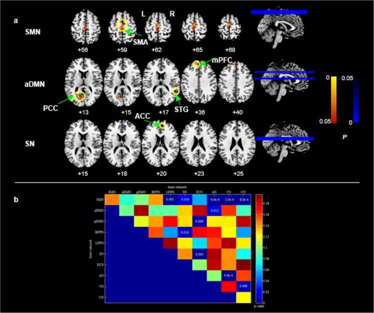
Patients with persistent somatoform pain disorder (PSPD) usually experience various functional impairments in pain, emotion, and cognition, which cannot be fully explained by a physiological process or a physical disorder. However, it is still not clear for the mechanism underlying the pathogenesis of PSPD. The present study aimed to explore the intra- and inter-network functional connectivity (FC) differences between PSPD patients and healthy controls (HCs). Functional magnetic resonance imaging (fMRI) was performed in 13 PSPD patients and 23 age- and gender-matched HCs. We used independent component analysis on resting-state fMRI data to calculate intra- and inter-network FCs, and we used the two-sample t-test to detect the FC differences between groups. Spearman correlation analysis was employed to evaluate the correlations between FCs and clinical assessments. As compared to HCs, PSPD patients showed decreased coactivations in the right superior temporal gyrus within the anterior default-mode network and the anterior cingulate cortex within the salience network, and increased coactivations in the bilateral supplementary motor areas within the sensorimotor network and both the left posterior cingulate cortex and the medial prefrontal cortex within the anterior default-mode network. In addition, we found that the PSPD patients showed decreased FNCs between sensorimotor network and audio network as well as visual network, between default-mode network and executive control network as well as audio network and between salience network and executive control network as well as right frontoparietal network, and increased FNCs between sensorimotor network and left frontoparietal network, salience network as well as cerebellum network, which were negatively correlated with the clinical assessments in PSPD patients. Our findings suggest that PSPD patients experience large-scale reorganization at the level of the functional networks, which suggests a possible mechanism underlying the pathogenesis of PSPD.
Overview
The claims and statistics of this paper haven’t been checked yet.
Free: it reads the open-access text and checks the first 5 claims that cite a source we can check. Create a free account to check every claim.
Integrity signals
Registry
Cites
Statistics
Figures


Verification
Claims the paper makes
The claims and statistics of this paper haven’t been checked yet.
Free: it reads the open-access text and checks the first 5 claims that cite a source we can check. Create a free account to check every claim.
Claims other papers make about this one
Each is checked against this paper’s own text: does it say what the citing paper says it does?
The cited source doesn’t support the claim as written.
In the citing paper
“Not only does each network independently demonstrate changes in functional connectivity in response to different types of chronic pain stimuli, it has been demonstrated that there is elevated functional connectivity among the DMN, FPN, and SN in individuals suffering from CMP [12,13,14,15,16,17,18].”
From Graph theory analysis of induced neural plasticity post-Acceptance and Commitment Therapy for chronic pain (2020) · cites this paper as [18]
Checked by a reasoning model — may be wrong.
Experimental — accuracy not yet measured
Self-check
Percentages, sums and reported differences re-computed from the paper’s own numbers.
The self-check needs the paper’s full text.
Citations
Cites
The assessment and analysis of handedness: The Edinburgh inventory
Oldfield, R. C. (1971) · Neuropsychologia · doi:10.1016/0028-3932(71)90067-4 · Cited by 35,900 · cited 1×
“Inclusion criteria for PSPD patients included: 1) right-hand dominance (The hand dominance was tested by using the Edinburgh Handness Inventory [21]); 2) ages between 18 and 65 years; 3) duration of clinical pain of at least 6 months; 4) diagnosis of PSPD according to ICD-10 criteria.”
MethodJenkinson, M., Bannister, P., Brady, M., & Smith, S. (2002) · NeuroImage · doi:10.1006/nimg.2002.1132 · Cited by 11,888 · cited 1×
“Also, there was no significant difference in the frame-wise [24] displacement between groups (t = 1.”
BackgroundFunctional connectivity in the motor cortex of resting human brain using echo‐planar mri
Biswal, B., Zerrin Yetkin, F., Haughton, V. M., & Hyde, J. S. (1995) · Magnetic Resonance in Medicine · doi:10.1002/mrm.1910340409 · Cited by 9,812
An Information-Maximization Approach to Blind Separation and Blind Deconvolution
Bell, A. J., & Sejnowski, T. J. (1995) · Neural Computation · doi:10.1162/neco.1995.7.6.1129 · Cited by 8,986 · cited 1×
“Subsequently, the independent components (ICs) were estimated using the Informax algorithm [27].”
MethodAshburner, J., & Friston, K. J. (2005) · NeuroImage · doi:10.1016/j.neuroimage.2005.02.018 · Cited by 7,507
Dissociable intrinsic connectivity networks for salience processing and executive control
Seeley, W. W., Menon, V., Schatzberg, A. F., Keller, J., Glover, G. H., Kenna, H., Reiss, A. L., & Greicius, M. D. (2007) · The Journal of Neuroscience · doi:10.1523/jneurosci.5587-06.2007 · Cited by 7,426 · cited 1×
“These networks commonly involve the processing of emotion, attention, control and cognition in human brain [18,29,30].”
BackgroundSaliency, switching, attention and control: a network model of insula function
Menon, V., & Uddin, L. Q. (2010) · Brain Structure and Function · doi:10.1007/s00429-010-0262-0 · Cited by 5,592
SMITH, S., & NICHOLS, T. (2009) · NeuroImage · doi:10.1016/j.neuroimage.2008.03.061 · Cited by 5,250 · cited 1×
“Multiple comparisons across space were corrected using the threshold-free clustering enhancement (TFCE) method [32].”
MethodCorrespondence of the brain's functional architecture during activation and rest
Smith, S. M., Fox, P. T., Miller, K. L., Glahn, D. C., Fox, P. M., Mackay, C. E., Filippini, N., Watkins, K. E., Toro, R., Laird, A. R., et al. (2009) · Proceedings of the National Academy of Sciences · doi:10.1073/pnas.0905267106 · Cited by 5,150 · cited 1×
“These networks commonly involve the processing of emotion, attention, control and cognition in human brain [18,29,30].”
BackgroundThe cognitive control of emotion
OCHSNER, K., & GROSS, J. (2005) · Trends in Cognitive Sciences · doi:10.1016/j.tics.2005.03.010 · Cited by 4,495
Cited by
Deep learning for identifying radiogenomic associations in breast cancer
Zhu, Z., Albadawy, E., Saha, A., Zhang, J., Harowicz, M. R., & Mazurowski, M. A. (2019) · Computers in Biology and Medicine · doi:10.1016/j.compbiomed.2019.04.018 · Cited by 167
Evolutionary dynamics of chloroplast genomes in subfamily Aroideae (Araceae)
Henriquez, C. L., Abdullah, Ahmed, I., Carlsen, M. M., Zuluaga, A., Croat, T. B., & McKain, M. R. (2020) · Genomics · doi:10.1016/j.ygeno.2020.01.006 · Cited by 163
Bègue, I., Adams, C., Stone, J., & Perez, D. L. (2019) · NeuroImage: Clinical · doi:10.1016/j.nicl.2019.101798 · Cited by 124
“, 2013); (3) and sensorimotor, salience and default mode network resting-state alterations (Hakala et al., 2002; Karibe et al., 2010; Li et al., 2016; Zhao et al., 2017).”
Wang, X., Yu, H., Li, F., Liu, T., Wu, W., Liu, C., Liu, C., & Zhang, X. (2018) · The Science of The Total Environment · doi:10.1016/j.scitotenv.2018.08.387 · Cited by 95
Burte, E., Leynaert, B., Marcon, A., Bousquet, J. J., Benmerad, M., Bono, R., Carsin, A., de Hoogh, K., Forsberg, B., Gormand, F., et al. (2020) · Journal of Allergy and Clinical Immunology · doi:10.1016/j.jaci.2019.11.040 · Cited by 78
Metadata
- Authors
- Zhao, Z., Huang, T., Tang, C., Ni, K., Pan, X., Yan, C., Fan, X., Xu, D., & Luo, Y.
- Affiliations
- East China Normal University (CN)
- Shanghai Changning Mental Health Center (CN)
- Fudan University (CN)
- Shanghai Mental Health Center (CN)
- Tongji University (CN)
- University of Massachusetts Chan Medical School (US)
- New York State Psychiatric Institute (US)
- Shanghai Jiao Tong University (CN)
- Corresponding author
- Xu, Dongrong
- Luo, Yanli
- Venue
- PLOS ONE
- Publisher
- Public Library of Science (PLoS)
- Published
- 2017-04-28
- Volume
- 12
- Issue
- 4
- Pages
- e0176494
- ISSN
- 1932-6203 (ISSN-L)
- PMC
- PMC5409184
- OpenAlex
- W2609643059
- Cited by
- 103
- References
- 53
- Citations by year
- 2026: 2 · 2025: 6 · 2024: 3 · 2023: 4 · 2022: 8 · 2021: 26 · 2020: 22 · 2019: 15 · 2018: 15 · 2017: 2
- FWCI
- 8.48
- Type
- journal article
- Language
- en
- Open access
- gold
- doi.org
- Licence
- Locations
- PLoS ONE (publishedVersion, cc-by)
- Europe PMC (PubMed Central) (publishedVersion, cc-by)
- PubMed Central (publishedVersion, cc-by)
- PubMed (publishedVersion)
- The Journal of the American Medical Association (JAMA) Network (American Medical Association) (submittedVersion, cc-by)
- DOAJ (DOAJ: Directory of Open Access Journals) (submittedVersion, cc-by-sa)
- Columbia Academic Commons (Columbia University)
- Funders
- National Natural Science Foundation of China (81100821, 81471734)
- Shanghai Science and Technology Committee (16ZR1432300)
- Topics
- Life Sciences › Neuroscience › Cognitive Neuroscience › Functional Brain Connectivity Studies
- Health Sciences › Medicine › Psychiatry and Mental health › Psychosomatic Disorders and Their Treatments
- Life Sciences › Neuroscience › Cognitive Neuroscience › Pain Management and Placebo Effect
- Keywords
- functional connectivity, resting-state brain networks, default mode network, salience network, sensorimotor network, frontoparietal network, anterior cingulate cortex, superior temporal gyrus, posterior cingulate cortex, medial prefrontal cortex, pathogenesis
- MeSH terms
- Adolescent, Adult, Female, Humans, Magnetic Resonance Imaging, Male, Middle Aged, Nerve Net, Pain, Rest, Somatoform Disorders, Young Adult
About these checks
Registry, retraction, Cites and statistics results are computed, not judged, with one exception: a printed reference that no index lists is read once by a model before it counts as a citation error. Claims are extracted by a model and kept only when their quote appears verbatim in the text; each cited source is then checked, with a model’s help, for whether it says what the claim relies on. Those verdicts can be wrong — read the claim’s quote and the cited source before relying on one.
Statistics and arithmetic are recomputed, not judged.
Confidence bands are shown only for fields calibrated on our annotated test set. None is calibrated yet, so no band is shown.
What counts as a claim ›How we measure confidence ›How we check this ›
Are you an author, or do you see a problem with this page? Request a review
How to cite
Cite CoChat for the checks on this page, and the paper for its own findings.
- This page
- CoChat. (2026). Citation check: Altered resting-state intra- and inter- network functional connectivity in patients with persistent somatoform pain disorder. https://app.cochat.ai/papers/10.1371/journal.pone.0176494
- The paper
- Zhao, Z., Huang, T., Tang, C., Ni, K., Pan, X., Yan, C., Fan, X., Xu, D., & Luo, Y. (2017). Altered resting-state intra- and inter- network functional connectivity in patients with persistent somatoform pain disorder. PLOS ONE. https://doi.org/10.1371/journal.pone.0176494
Data sources
Bibliographic data: Semantic Scholar, OpenAlex, Crossref.
Contains information from the Semantic Scholar Academic Graph Datasets, made available under the ODC Attribution License (ODC-BY 1.0).
Semantic Scholar’s data is described in: Kinney, R., et al. (2023). The Semantic Scholar Open Data Platform. arXiv:2301.10140.
