

The DOI matches its Crossref record; 36 of the 37 references it cites were found in a registry or an index, and 1 are not checked yet.
Deep learning-based survival prediction for multiple cancer types using histopathology images
Open accessAbstract
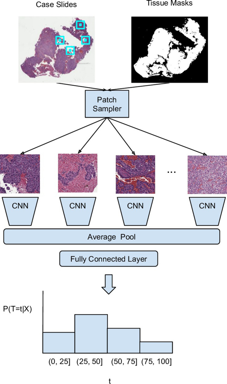
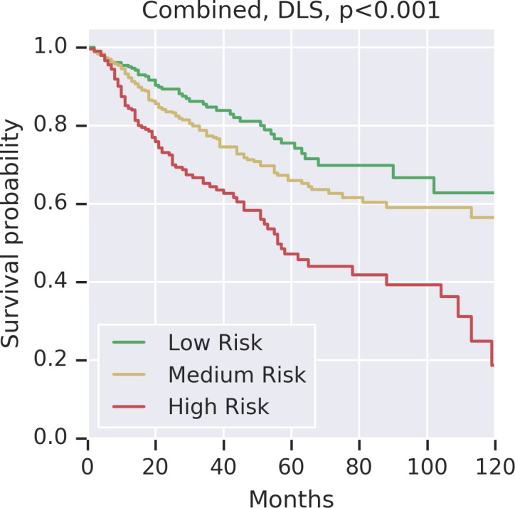
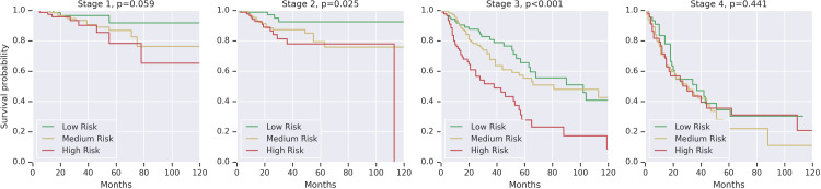
Providing prognostic information at the time of cancer diagnosis has important implications for treatment and monitoring. Although cancer staging, histopathological assessment, molecular features, and clinical variables can provide useful prognostic insights, improving risk stratification remains an active research area. We developed a deep learning system (DLS) to predict disease specific survival across 10 cancer types from The Cancer Genome Atlas (TCGA). We used a weakly-supervised approach without pixel-level annotations, and tested three different survival loss functions. The DLS was developed using 9,086 slides from 3,664 cases and evaluated using 3,009 slides from 1,216 cases. In multivariable Cox regression analysis of the combined cohort including all 10 cancers, the DLS was significantly associated with disease specific survival (hazard ratio of 1.58, 95% CI 1.28-1.70, p<0.0001) after adjusting for cancer type, stage, age, and sex. In a per-cancer adjusted subanalysis, the DLS remained a significant predictor of survival in 5 of 10 cancer types. Compared to a baseline model including stage, age, and sex, the c-index of the model demonstrated an absolute 3.7% improvement (95% CI 1.0-6.5) in the combined cohort. Additionally, our models stratified patients within individual cancer stages, particularly stage II (p = 0.025) and stage III (p<0.001). By developing and evaluating prognostic models across multiple cancer types, this work represents one of the most comprehensive studies exploring the direct prediction of clinical outcomes using deep learning and histopathology images. Our analysis demonstrates the potential for this approach to provide significant prognostic information in multiple cancer types, and even within specific pathologic stages. However, given the relatively small number of cases and observed clinical events for a deep learning task of this type, we observed wide confidence intervals for model performance, thus highlighting that future work will benefit from larger datasets assembled for the purposes for survival modeling.
Overview
The claims and statistics of this paper haven’t been checked yet.
Free: it reads the open-access text and checks the first 5 claims that cite a source we can check. Create a free account to check every claim.
Integrity signals
Registry
Cites
Statistics
Figures
Verification
Claims the paper makes
The claims and statistics of this paper haven’t been checked yet.
Free: it reads the open-access text and checks the first 5 claims that cite a source we can check. Create a free account to check every claim.
Claims other papers make about this one
Each is checked against this paper’s own text: does it say what the citing paper says it does?
The source backs part of the claim, not all of it — or backs it for a different population, condition or comparison.
In the citing paper
“Automated image analysis, feature recognition, data extraction and deep learning models are everyday reality for the tech giants but have only partially reached precision pathology (51–53).”
From Artificial Intelligence: Present and Future Potential for Solid Organ Transplantation (2022) · cites this paper as [52]
Experimental — accuracy not yet measuredThe cited source says what the claim attributes to it.
In the citing paper
“In fact, DL has the ability to enhance human lives by providing additional accuracy in diagnosis, including estimating natural disasters [40], the discovery of new drugs [41], and cancer diagnosis [42–44].”
From Review of deep learning: concepts, CNN architectures, challenges, applications, future directions (2021) · cites this paper as [43]
In the citing paper
“In addition, using multi-task learning 147 to train models to perform a variety of tasks rather than one narrowly defined task, such as multi-cancer detection from histopathology images 148 , makes them more generally useful and often more robust.”
From Deep learning-enabled medical computer vision (2021) · cites this paper as [148]
Self-check
Percentages, sums and reported differences re-computed from the paper’s own numbers.
The self-check needs the paper’s full text.
Citations
Cites
LeCun, Y., Bengio, Y., & Hinton, G. (2015) · Nature · doi:10.1038/nature14539 · Cited by 84,126
Regression Models and Life-Tables
Cox, D. R. (1972) · Journal of the Royal Statistical Society Series B: Statistical Methodology · doi:10.1111/j.2517-6161.1972.tb00899.x · Cited by 39,793
Regression Models and Life-Tables
Cox, D. R. (1992) · Springer Series in Statistics · doi:10.1007/978-1-4612-4380-9_37 · Cited by 26,297
MobileNets: Efficient Convolutional Neural Networks for Mobile Vision Applications
Howard, A. G., Zhu, M., Chen, B., Kalenichenko, D., Wang, W., Weyand, T., Andreetto, M., & Adam, H. (2017) · arXiv · doi:10.48550/arxiv.1704.04861 · Cited by 19,015 · cited 2×
“Our CNN consisted of layers of depth-wise separable convolution layers, similar to the MobileNet [23] CNN architecture.”
MethodInfluentialAdvances in Neural Information Processing Systems 19
(2007) · The MIT Press eBooks · doi:10.52202/085713 · Cited by 15,539
Nishimura, R., Osako, T., Nishiyama, Y., Tashima, R., Nakano, M., Fujisue, M., Toyozumi, Y., & Arima, N. (2013) · Oncology · doi:10.1159/000353099 · Cited by 13,166
The cancer genome atlas pan-cancer analysis project
The Cancer Genome Atlas Research Network, Weinstein, J. N., Collisson, E. A., Mills, G. B., Shaw, K. R. M., Ozenberger, B. A., Ellrott, K., Shmulevich, I., Sander, C., & Stuart, J. M. (2013) · Nature Genetics · doi:10.1038/ng.2764 · Cited by 9,092
Gulshan, V., Peng, L., Coram, M., Stumpe, M. C., Wu, D., Narayanaswamy, A., Venugopalan, S., Widner, K., Madams, T., Cuadros, J., et al. (2016) · JAMA · doi:10.1001/jama.2016.17216 · Cited by 7,697 · cited 1×
“In recent years, deep learning has been shown to recognize objects [6] and diagnose diseases from medical images [7,8] with impressive accuracy.”
BackgroundSelf-citationLiu, J., Lichtenberg, T., Hoadley, K. A., Poisson, L. M., Lazar, A. J., Cherniack, A. D., Kovatich, A. J., Benz, C. C., Levine, D. A., Lee, A. V., et al. (2018) · Cell · doi:10.1016/j.cell.2018.02.052 · Cited by 3,725 · cited 3×
“Clinical data (including approximated disease specific survival) were obtained from the TCGA Pan-Cancer Clinical Data Resource[21] and the Genomic Data Commons.”
MethodInfluentialSelf-citationEvaluating the Yield of Medical Tests
Harrell, F. E. (1982) · JAMA: The Journal of the American Medical Association · doi:10.1001/jama.1982.03320430047030 · Cited by 3,490 · cited 1×
“In this analysis, prognosis prediction performance was measured using the c-index, an extension of the AUC for binary outcomes without censorship [30].”
Method
Research works printed but not linked: 2
- Y Liu, 2018, Artificial Intelligence-Based Breast Cancer Nodal Metastasis Detection, Arch Pathol Lab Med
- T Tieleman, 2012, Lecture 6.5-rmsprop: Divide the gradient by a running average of its recent magnitude, COURSERA: Neural networks for machine learning, 4, 26
Cited by
Review of deep learning: concepts, CNN architectures, challenges, applications, future directions
Alzubaidi, L., Zhang, J., Humaidi, A. J., Al-Dujaili, A., Duan, Y., Al-Shamma, O., Santamaría, J., Fadhel, M. A., Al-Amidie, M., & Farhan, L. (2021) · Journal of Big Data · doi:10.1186/s40537-021-00444-8 · Cited by 7,906
Liu, J., Lichtenberg, T., Hoadley, K. A., Poisson, L. M., Lazar, A. J., Cherniack, A. D., Kovatich, A. J., Benz, C. C., Levine, D. A., Lee, A. V., et al. (2018) · Cell · doi:10.1016/j.cell.2018.02.052 · Cited by 3,725
Self-citationTowards a general-purpose foundation model for computational pathology
Chen, R. J., Ding, T., Lu, M. Y., Williamson, D. F. K., Jaume, G., Song, A. H., Chen, B., Zhang, A., Shao, D., Shaban, M., et al. (2024) · Nature Medicine · doi:10.1038/s41591-024-02857-3 · Cited by 1,560
Deep learning-enabled medical computer vision
Esteva, A., Chou, K., Yeung, S., Naik, N., Madani, A., Mottaghi, A., Liu, Y., Topol, E., Dean, J., & Socher, R. (2021) · npj Digital Medicine · doi:10.1038/s41746-020-00376-2 · Cited by 1,406
Self-citationDeep learning in histopathology: the path to the clinic
van der Laak, J., Litjens, G., & Ciompi, F. (2021) · Nature Medicine · doi:10.1038/s41591-021-01343-4 · Cited by 902
Metadata
- Authors
- Wulczyn, E., Steiner, D. F., Xu, Z., Sadhwani, A., Wang, H., Flament-Auvigne, I., Mermel, C. H., Chen, P.-H. C., Liu, Y., & Stumpe, M. C.
- Affiliations
- Google (United States) (US)
- Corresponding author
- Liu, Yun
- ORCID
- Venue
- PLOS ONE
- Publisher
- Public Library of Science (PLoS)
- Published
- 2020-06-17
- Volume
- 15
- Issue
- 6
- Pages
- e0233678
- ISSN
- 1932-6203 (ISSN-L)
- PMC
- PMC7299324
- OpenAlex
- W3036901136
- Cited by
- 266
- References
- 36
- Citations by year
- 2026: 39 · 2025: 33 · 2024: 53 · 2023: 36 · 2022: 51 · 2021: 46 · 2020: 7
- FWCI
- 13.75
- Type
- journal article
- Language
- en
- Open access
- gold
- doi.org
- Licence
- Locations
- PLoS ONE (publishedVersion, cc-by)
- PubMed Central (publishedVersion, cc-by)
- PubMed (publishedVersion)
- arXiv (Cornell University) (submittedVersion)
- RePEc: Research Papers in Economics (submittedVersion)
- DOAJ (DOAJ: Directory of Open Access Journals) (submittedVersion, cc-by-sa)
- arXiv (Cornell University)
- Funders
- Topics
- Physical Sciences › Computer Science › Artificial Intelligence › AI in cancer detection
- Health Sciences › Medicine › Radiology, Nuclear Medicine and Imaging › Radiomics and Machine Learning in Medical Imaging
- Physical Sciences › Computer Science › Artificial Intelligence › Machine Learning in Healthcare
- Keywords
- cancer survival prediction, histopathological images, disease-specific survival, prognostic model, weakly supervised learning, risk stratification, tumor staging, multiple cancer types, TCGA, deep learning
- MeSH terms
- Deep Learning, Adult, Age Factors, Feasibility Studies, Female, Humans, Image Processing, Computer-Assisted, Male, Middle Aged, Neoplasm Staging, Neoplasms, Prognosis, Risk Factors, Sex Factors, Survival Analysis, Risk Assessment, Datasets as Topic
About these checks
Registry, retraction, Cites and statistics results are computed, not judged, with one exception: a printed reference that no index lists is read once by a model before it counts as a citation error. Claims are extracted by a model and kept only when their quote appears verbatim in the text; each cited source is then checked, with a model’s help, for whether it says what the claim relies on. Those verdicts can be wrong — read the claim’s quote and the cited source before relying on one.
Statistics and arithmetic are recomputed, not judged.
Confidence bands are shown only for fields calibrated on our annotated test set. None is calibrated yet, so no band is shown.
What counts as a claim ›How we measure confidence ›How we check this ›
Are you an author, or do you see a problem with this page? Request a review
How to cite
Cite CoChat for the checks on this page, and the paper for its own findings.
- This page
- CoChat. (2026). Citation check: Deep learning-based survival prediction for multiple cancer types using histopathology images. https://app.cochat.ai/papers/10.1371/journal.pone.0233678
- The paper
- Wulczyn, E., Steiner, D. F., Xu, Z., Sadhwani, A., Wang, H., Flament-Auvigne, I., Mermel, C. H., Chen, P.-H. C., Liu, Y., & Stumpe, M. C. (2020). Deep learning-based survival prediction for multiple cancer types using histopathology images. PLOS ONE. https://doi.org/10.1371/journal.pone.0233678
Data sources
Bibliographic data: Semantic Scholar, OpenAlex, Crossref.
Contains information from the Semantic Scholar Academic Graph Datasets, made available under the ODC Attribution License (ODC-BY 1.0).
Semantic Scholar’s data is described in: Kinney, R., et al. (2023). The Semantic Scholar Open Data Platform. arXiv:2301.10140.
