

The DOI matches its Crossref record; 5 of the 5 references it cites were found in a registry or an index.
AlphaFold2 and RoseTTAFold predict posttranslational modifications. Chromophore formation in GFP-like proteins
Open accessPublished version of a preprint
Abstract
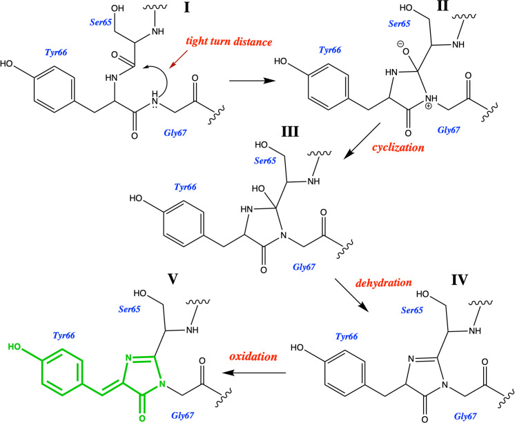
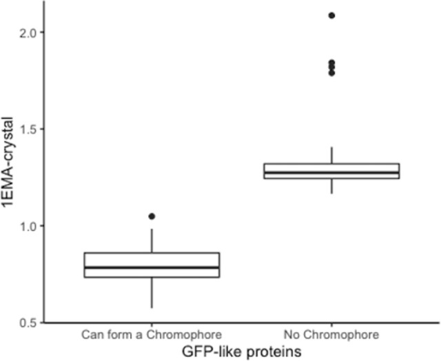
AlphaFold2 and RoseTTAfold are able to predict, based solely on their sequence whether GFP-like proteins will post-translationally form a chromophore (the part of the protein responsible for fluorescence) or not. Their training has not only taught them protein structure and folding, but also chemistry. The structures of 21 sequences of GFP-like fluorescent proteins that will post-translationally form a chromophore and of 23 GFP-like non-fluorescent proteins that do not have the residues required to form a chromophore were determined by AlphaFold2 and RoseTTAfold. The resultant structures were mined for a series of geometric measurements that are crucial to chromophore formation. Statistical analysis of these measurements showed that both programs conclusively distinguished between chromophore forming and non-chromophore forming proteins. A clear distinction between sequences capable of forming a chromophore and those that do not have the residues required for chromophore formation can be obtained by examining a single measurement—the RMSD of the overlap of the central alpha helices of the crystal structure of S65T GFP and the AlphaFold2 determined structure. Only 10 of the 578 GFP-like proteins in the pdb have no chromophore, yet when AlphaFold2 and RoseTTAFold are presented with the sequences of 44 GFP-like proteins that are not in the pdb they fold the proteins in such a way that one can unequivocally distinguish between those that can and cannot form a chromophore.
Overview
The claims and statistics of this paper haven’t been checked yet.
Run the check — free, no account needed. It reads the open-access text and checks the sources it cites.
Integrity signals
Registry
Cites
Statistics
Figures
Verification
Claims the paper makes
The claims and statistics of this paper haven’t been checked yet.
Run the check — free, no account needed. It reads the open-access text and checks the sources it cites.
Self-check
Percentages, sums and reported differences re-computed from the paper’s own numbers.
The self-check needs the paper’s full text.
Citations
Cites
Highly accurate protein structure prediction with AlphaFold
Jumper, J., Evans, R., Pritzel, A., Green, T., Figurnov, M., Ronneberger, O., Tunyasuvunakool, K., Bates, R., Žídek, A., Potapenko, A., et al. (2021) · Nature · doi:10.1038/s41586-021-03819-2 · Cited by 43,764
Accurate prediction of protein structures and interactions using a 3-track network
Baek, M., DiMaio, F., Anishchenko, I., Dauparas, J., Ovchinnikov, S., Lee, G. R., Wang, J., Cong, Q., Kinch, L. N., Schaeffer, R. D., et al. (2021) · doi:10.1101/2021.06.14.448402 · Cited by 5,549
Highly accurate protein structure prediction for the human proteome
Tunyasuvunakool, K., Adler, J., Wu, Z., Green, T., Zielinski, M., Žídek, A., Bridgland, A., Cowie, A., Meyer, C., Laydon, A., et al. (2021) · Nature · doi:10.1038/s41586-021-03828-1 · Cited by 3,025
Burley, S. K., Bhikadiya, C., Bi, C., Bittrich, S., Chen, L., Crichlow, G. V., Christie, C. H., Dalenberg, K., Di Costanzo, L., Duarte, J. M., et al. (2021) · Nucleic Acids Research · doi:10.1093/nar/gkaa1038 · Cited by 1,581
Harnessing protein folding neural networks for peptide–protein docking
Tsaban, T., Varga, J. K., Avraham, O., Ben-Aharon, Z., Khramushin, A., & Schueler-Furman, O. (2022) · Nature Communications · doi:10.1038/s41467-021-27838-9 · Cited by 1,205
Research works printed but not linked: 54
- SK Burley, 2018, RCSB Protein Data Bank: biological macromolecular structures enabling research and education in fundamental biology, biomedicine, biotechnology and energy, Nucleic Acids Research, 47, D464, 10.1093/nar/gky1004 · DOI
- H Bagdonas, 2021, The case for post-predictional modifications in the AlphaFold Protein Structure Database, Nature Structural & Molecular Biology, 28, 869, 10.1038/s41594-021-00680-9 · DOI
- ML Hekkelman, 2021, AlphaFill: enriching the AlphaFold models with ligands and co-factors, bioRxiv
- ZJ Wehrspan, 2022, Identification of Iron-Sulfur (Fe-S) Cluster and Zinc (Zn) Binding Sites Within Proteomes Predicted by DeepMind’s AlphaFold2 Program Dramatically Expands the Metalloproteome, Journal of Molecular Biology, 434, 167377, 10.1016/j.jmb.2021.167377 · DOI · Paper page
- The Nobel Prize in Chemistry 2008. Press Release. 2008:https://www.nobelprize.org/prizes/chemistry/2008/press-release/.
- The Nobel Prize in Chemistry 2014. Press Release. 2014:https://www.nobelprize.org/prizes/chemistry/2014/press-release/.
- M. Zimmer, 2005, Glowing Genes: A Revolution in Biotechnology
- M. Zimmer, 2015, Illuminating disease: An introduction to green fluorescent proteins
- V Pieribone, 2005, Aglow in the dark: the revolutionary science of biofluorescence
- M Chalfie, 1998, Green Fluorescent Protein: Properties, Applications, and Protocols
- RY Tsien, 1998, Green Fluorescent Protein, Annu Rev Biochem, 67, 509, 10.1146/annurev.biochem.67.1.509 · DOI
- M. Zimmer, 2002, Green fluorescent protein (GFP): Applications, structure, and related photophysical behavior, Chemical Reviews, 102, 759, 10.1021/cr010142r · DOI
- RN Day, 2009, The fluorescent protein palette: tools for cellular imaging, Chemical Society Reviews, 38, 2887, 10.1039/b901966a · DOI
- EA Rodriguez, 2017, The Growing and Glowing Toolbox of Fluorescent and Photoactive Proteins, Trends Biochem Sci, 42, 111, 10.1016/j.tibs.2016.09.010 · DOI
- OV Stepanenko, 2013, International Review of Cell and Molecular Biology, 221
- Y Nasu, 2021, Structure- and mechanism-guided design of single fluorescent protein-based biosensors, Nature Chemical Biology
- BR Branchini, 1998, A Computational Analysis of the Unique Protein-Induced Tight Turn That Results in Posttranslational Chromophore Formation in Green Fluorescent Protein, J Am Chem Soc, 120, 1, 10.1021/ja973019j · DOI
- DP Barondeau, 2007, The case of the missing ring: Radical cleavage of a carbon-carbon bond and implications for GFP chromophore biosynthesis, Journal of the American Chemical Society, 129, 3118, 10.1021/ja063983u · DOI
- WJH Ong, 2011, Function and structure of GFP-like proteins in the protein data bank, Molecular BioSystems, 7, 984, 10.1039/c1mb05012e · DOI
- NP Lemay, 2008, The role of the tight-turn, broken hydrogen bonding, Glu222 and Arg96 in the post-translational green fluorescent protein chromophore formation, Chemical Physics, 348, 152, 10.1016/j.chemphys.2008.02.055 · DOI
- BL Grigorenko, 2017, Molecular Modeling Clarifies the Mechanism of Chromophore Maturation in the Green Fluorescent Protein, Journal of the American Chemical Society, 139, 10239, 10.1021/jacs.7b00676 · DOI
- F Yang, 1996, The molecular structure of green fluorescent protein, Nature Biotechnol, 14, 1246, 10.1038/nbt1096-1246 · DOI
- M Ormoe, 1996, Crystal structure of the Aequorea victoria green fluorescent Protein, Science, 273, 1392, 10.1126/science.273.5280.1392 · DOI
- TI Wood, 2005, Defining the role of arginine 96 in green fluorescent protein fluorophore biosynthesis, Biochemistry, 44, 16211, 10.1021/bi051388j · DOI
- M Hopf, 2001, Mapping of binding sites for nidogens, fibulin-2, fibronectin and heparin to different IG modules of perlecan, Journal of Molecular Biology, 311, 529, 10.1006/jmbi.2001.4878 · DOI
- TJ Lambert, 2019, FPbase: a community-editable fluorescent protein database, Nature Methods, 16, 277, 10.1038/s41592-019-0352-8 · DOI
- WR Francis, 2016, Non-excitable fluorescent protein orthologs found in ctenophores, Bmc Evol Biol, 16, 167, 10.1186/s12862-016-0738-5 · DOI
- K Katoh, 2019, MAFFT online service: multiple sequence alignment, interactive sequence choice and visualization, Briefings in Bioinformatics, 20, 1160, 10.1093/bib/bbx108 · DOI · Paper page
- F Sievers, 2018, Clustal Omega for making accurate alignments of many protein sequences, Protein Science, 27, 135, 10.1002/pro.3290 · DOI · Paper page
- W. Kabsch, 1978, Discussion of Solution for Best Rotation to Relate 2 Sets of Vectors, Acta Crystallographica Section A, 34, 827, 10.1107/S0567739478001680 · DOI · Paper page
- W. Kabsch, 1976, Solution for Best Rotation to Relate 2 Sets of Vectors, Acta Crystallographica Section A, 32, 922, 10.1107/S0567739476001873 · DOI
- DP Barondeau, 2005, Understanding GFP chromophore biosynthesis: Controlling backbone cyclization and modifying post-translational chemistry, Biochemistry, 44, 1960, 10.1021/bi0479205 · DOI
- JL Tubbs, 2005, Crystallographic structures of Discosoma red fluorescent protein with immature and mature chromophores: Linking peptide bond trans-cis isomerization and acylimine formation in chromophore maturation, Biochemistry, 44, 9833, 10.1021/bi0472907 · DOI
- J Myšková, 2020, Directionality of light absorption and emission in representative fluorescent proteins, Proceedings of the National Academy of Sciences, 117, 32395, 10.1073/pnas.2017379117 · DOI
- R Ando, 2007, Highlighted Generation of Fluorescence Signals Using Simultaneous Two-Color Irradiation on Dronpa Mutants, Biophysical Journal, 92, L97, 10.1529/biophysj.107.105882 · DOI
- DM Chudakov, 2004, Photoswitchable cyan fluorescent protein for protein tracking, Nature Biotechnology, 22, 1435, 10.1038/nbt1025 · DOI
- JN Henderson, 2005, Crystal structures and mutational analysis of amFP486, a cyan fluorescent protein from Anemonia majano, Proceedings of the National Academy of Sciences of the United States of America, 102, 12712, 10.1073/pnas.0502250102 · DOI
- DA Shagin, 2004, GFP-like proteins as ubiquitous metazoan superfamily: Evolution of functional features and structural complexity, Molecular Biology and Evolution, 21, 841, 10.1093/molbev/msh079 · DOI
- R Heim, 1994, Wavelength mutations and posttranslational autoxidation of green fluorescent protein, Proc Natl Acad Sci USA, 91, 12501, 10.1073/pnas.91.26.12501 · DOI
- GG Lambert, 2020, Aequorea’s secrets revealed: New fluorescent proteins with unique properties for bioimaging and biosensing, PLOS Biology, 18, e3000936, 10.1371/journal.pbio.3000936 · DOI
- MV Matz, 1999, Fluorescent proteins from nonbioluminescent Anthozoa species, Nature Biotech, 17, 969, 10.1038/13657 · DOI
- A Shimizu, 2017, Molecular evolution of versatile derivatives from a GFP-like protein in the marine copepod Chiridius poppei, Plos One, 12, e0181186, 10.1371/journal.pone.0181186 · DOI
- YA Labas, 2002, Diversity and evolution of the green fluorescent protein family, Proceedings of the National Academy of Sciences of the United States of America, 99, 4256, 10.1073/pnas.062552299 · DOI
- H Shinoda, 2018, Acid-Tolerant Monomeric GFP from Olindias formosa, Cell Chemical Biology, 25, 10.1016/j.chembiol.2017.12.005 · DOI · Paper page
- BJ Bevis, 2002, Rapidly maturing variants of the Discosoma red fluorescent protein (DsRed), Nature Biotechnology, 20, 1159, 10.1038/nbt1102-1159 · DOI
- J Wiedenmann, EosFP, a fluorescent marker protein with UV-inducible green-to-red fluorescence conversion, Proceedings of the National Academy of Sciences of the United States of America
- EM Merzlyak, 2007, Bright monomeric red fluorescent protein with an extended fluorescence lifetime, Nature Methods, 4, 555, 10.1038/nmeth1062 · DOI
- S Kredel, 2008, Optimized and Far-Red-Emitting Variants of Fluorescent Protein eqFP611, Chemistry & Biology, 15, 224, 10.1016/j.chembiol.2008.02.008 · DOI
- OM Subach, 2008, Conversion of Red Fluorescent Protein into a Bright Blue Probe, Chemistry & Biology, 59, 1116, 10.1016/j.chembiol.2008.08.006 · DOI
- S Habuchi, 2008, mKikGR, a Monomeric Photoswitchable Fluorescent Protein, Plos One, 3, e3944, 10.1371/journal.pone.0003944 · DOI
- S Karasawa, 2004, Cyan-emitting and orange-emitting fluorescent proteins as a donor/acceptor pair for fluorescence resonance energy transfer, Biochem J, 381, 307, 10.1042/BJ20040321 · DOI
- NC Shaner, 2013, A bright monomeric green fluorescent protein derived from Branchiostoma lanceolatum, Nature Methods, 10, 407, 10.1038/nmeth.2413 · DOI
- T Kogure, 2006, A fluorescent variant of a protein from the stony coral Montipora facilitates dual-color single-laser fluorescence cross-correlation spectroscopy, Nature Biotechnology, 24, 577, 10.1038/nbt1207 · DOI
- DS Bindels, 2017, mScarlet: a bright monomeric red fluorescent protein for cellular imaging, Nature Methods, 14, 53, 10.1038/nmeth.4074 · DOI
Metadata
- Authors
- Hartley, S. M., Tiernan, K. A., Ahmetaj, G., Cretu, A., Zhuang, Y., & Zimmer, M.
- Affiliations
- Connecticut College (US)
- Corresponding author
- Zimmer, Marc
- Venue
- PLOS ONE
- Publisher
- Public Library of Science (PLoS)
- Published
- 2022-06-16
- Volume
- 17
- Issue
- 6
- Pages
- e0267560
- ISSN
- 1932-6203 (ISSN-L)
- PMC
- PMC9202861
- OpenAlex
- W4282969955
- Cited by
- 15
- References
- 5
- Citations by year
- 2026: 4 · 2025: 2 · 2024: 2 · 2023: 5 · 2022: 2
- FWCI
- 0.90
- Type
- journal article
- Language
- en
- Open access
- gold
- doi.org
- Licence
- Locations
- PLoS ONE (publishedVersion, cc-by)
- bioRxiv (Cold Spring Harbor Laboratory) (acceptedVersion, cc-by-nc-nd)
- PubMed Central (publishedVersion, cc-by)
- PubMed (publishedVersion)
- DOAJ (DOAJ: Directory of Open Access Journals) (submittedVersion, cc-by-sa)
- Topics
- Life Sciences › Biochemistry, Genetics and Molecular Biology › Molecular Biology › Protein Structure and Dynamics
- Life Sciences › Biochemistry, Genetics and Molecular Biology › Biophysics › Advanced Fluorescence Microscopy Techniques
- Life Sciences › Biochemistry, Genetics and Molecular Biology › Structural Biology › Enzyme Structure and Function
- Keywords
- AlphaFold2, RoseTTAFold, chromophore formation, green fluorescent protein, protein structure prediction, RMSD, geometric measurement
- MeSH terms
- Fluorescence, Luminescent Proteins, Protein Processing, Post-Translational, Green Fluorescent Proteins
About these checks
Registry, retraction, Cites and statistics results are computed, not judged, with one exception: a printed reference that no index lists is read once by a model before it counts as a citation error. Claims are extracted by a model and kept only when their quote appears verbatim in the text; each cited source is then checked, with a model’s help, for whether it says what the claim relies on. Those verdicts can be wrong — read the claim’s quote and the cited source before relying on one.
Statistics and arithmetic are recomputed, not judged.
Confidence bands are shown only for fields calibrated on our annotated test set. None is calibrated yet, so no band is shown.
What counts as a claim ›How we measure confidence ›How we check this ›
Are you an author, or do you see a problem with this page? Request a review
How to cite
Cite CoChat for the checks on this page, and the paper for its own findings.
- This page
- CoChat. (2026). Citation check: AlphaFold2 and RoseTTAFold predict posttranslational modifications. Chromophore formation in GFP-like proteins. https://app.cochat.ai/papers/10.1371/journal.pone.0267560
- The paper
- Hartley, S. M., Tiernan, K. A., Ahmetaj, G., Cretu, A., Zhuang, Y., & Zimmer, M. (2022). AlphaFold2 and RoseTTAFold predict posttranslational modifications. Chromophore formation in GFP-like proteins. PLOS ONE. https://doi.org/10.1371/journal.pone.0267560
