Cited by 173

The DOI matches its Crossref record; 1 of the 1 references it cites were found in a registry or an index.

Mechanism of Genomic Instability in Cells Infected with the High-Risk Human Papillomaviruses

Kadaja, Meelis, Isok-Paas, Helen, Laos, Triin, Ustav, Ene, Ustav, Mart·2009·PLoS Pathogens

Overview

The claims and statistics of this paper haven’t been checked yet.

Run the check — free, no account needed. It reads the open-access text and checks the sources it cites.

A few free checks a day · papers already verified open instantly · results are public

Integrity signals

Registry

Crossref · 10.1371/journal.ppat.1000397

Cites

1 of 1 references verified0 citation errors · 0 not checkable

Statistics

Figures

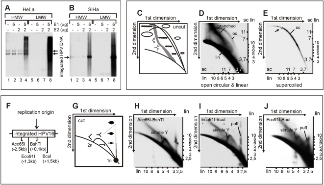
Figure 1 DNA replication initiated from the integrated HPV origin generates various low-molecular-weight DNA products. High-molecular-weight (HMW) DNA and low-molecular-weight (LMW) DNA were fractionated and purified by Hirt lysis 24 hrs post-transfection from HeLa cells (A) and from SiHa cells (B) that were both transfected as follows: mock-transfection (lanes 1 and 5); 5 µg of HPV18 E1 expression plasmid (lanes 2 and 6); 2 µg of E2 expression plasmid (lanes 3 and 7); 5 µg of HPV18 E1 and 2 µg of E2 expression plasmids (lanes 4 and 8). A 3 µg portion of HMW DNA and three times the respective amount of LMW DNA were digested with HindIII (A) or Acc65I/BshTI (B) and separated on a one-dimensional gel. The integrated HPV URR-specific signals were detected by Southern blot analysis. (C) Schematic presentation of the migration of dsDNA linear, supercoiled, and open circle molecules on 2D neutral-neutral gels. (D, E) 5 µg of HPV18 E1 and 2 µg of E2 expression plasmids were transfected into HeLa cells and LMW DNA was purified by Hirt lysis 48 hrs post-transfection. Extracted DNA was digested with DpnI and fractionated by the CsCl-ethidium bromide density gradient. The fraction of linear fragments (lin) and open circular molecules (oc) (D) and the fraction of supercoiled circular plasmids (sc) (E) were separated on a 2D gel, transferred to a nylon filter, and probed with the HPV18 genomic fragment (from nt 3917 to1575). Numbers shown on the axes represent the markers of linear (lin) and supercoiled circular (sc) DNA forms. Black arrowhead indicates the shift that was caused by mtDNA. (F) Schematic presentation of HPV16 integration locus within chromosome 13 in SiHa cells, where the cleavage sites of Acc65I, Eco91I, BshTI, and BcuI as well as their distances from HPV origin are presented. (G) Schematic presentation of the migration of replication forks and replication puffs on 2D neutral-neutral gels. (H,I) SiHa cells were co-transfected with 5 µg of HPV18 E1 and 2 µg of E2 expression plasmids. LMW DNA was extracted 24 hrs post-transfection and digested with Acc65I-BshTI (H) and Eco91I-BcuI (I). Respective HPV16 genome fragments were used as probes on 2D Southern blots. (J) SiHa cells were co-transfected with 1 µg of HPV18 E1 and 2 µg of E2 expression plasmids. Extracted LMW DNA was digested with Eco91I-BcuI and analyzed by 2D Southern blots.
Figure 1 DNA replication initiated from the integrated HPV origin generates various low-molecular-weight DNA products. High-molecular-weight (HMW) DNA and low-molecular-weight (LMW) DNA were fractionated and purified by Hirt lysis 24 hrs post-transfection from HeLa cells (A) and from SiHa cells (B) that were both transfected as follows: mock-transfection (lanes 1 and 5); 5 µg of HPV18 E1 expression plasmid (lanes 2 and 6); 2 µg of E2 expression plasmid (lanes 3 and 7); 5 µg of HPV18 E1 and 2 µg of E2 expression plasmids (lanes 4 and 8). A 3 µg portion of HMW DNA and three times the respective amount of LMW DNA were digested with HindIII (A) or Acc65I/BshTI (B) and separated on a one-dimensional gel. The integrated HPV URR-specific signals were detected by Southern blot analysis. (C) Schematic presentation of the migration of dsDNA linear, supercoiled, and open circle molecules on 2D neutral-neutral gels. (D, E) 5 µg of HPV18 E1 and 2 µg of E2 expression plasmids were transfected into HeLa cells and LMW DNA was purified by Hirt lysis 48 hrs post-transfection. Extracted DNA was digested with DpnI and fractionated by the CsCl-ethidium bromide density gradient. The fraction of linear fragments (lin) and open circular molecules (oc) (D) and the fraction of supercoiled circular plasmids (sc) (E) were separated on a 2D gel, transferred to a nylon filter, and probed with the HPV18 genomic fragment (from nt 3917 to1575). Numbers shown on the axes represent the markers of linear (lin) and supercoiled circular (sc) DNA forms. Black arrowhead indicates the shift that was caused by mtDNA. (F) Schematic presentation of HPV16 integration locus within chromosome 13 in SiHa cells, where the cleavage sites of Acc65I, Eco91I, BshTI, and BcuI as well as their distances from HPV origin are presented. (G) Schematic presentation of the migration of replication forks and replication puffs on 2D neutral-neutral gels. (H,I) SiHa cells were co-transfected with 5 µg of HPV18 E1 and 2 µg of E2 expression plasmids. LMW DNA was extracted 24 hrs post-transfection and digested with Acc65I-BshTI (H) and Eco91I-BcuI (I). Respective HPV16 genome fragments were used as probes on 2D Southern blots. (J) SiHa cells were co-transfected with 1 µg of HPV18 E1 and 2 µg of E2 expression plasmids. Extracted LMW DNA was digested with Eco91I-BcuI and analyzed by 2D Southern blots.CC BY · PubMed Central
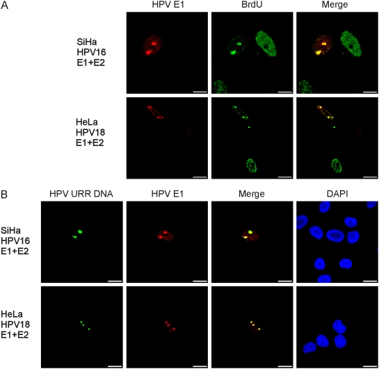
Figure 2 DNA replication of the integrated HPV takes place at specific nuclear foci. HeLa and SiHa cells were co-transfected with 5 µg of HPV18 E1 and 2 µg of E2 expression plasmids. Cells were analyzed 20 hrs post-transfection. (A) Co-immunostaining of E1 (Alexa Fluor 568, first column) and BrdU (FITC, second column). Cells were pulse-labeled with BrdU for 2 hrs prior to IF analysis. In the third column, the localizations of E1 and BrdU in the same cells are presented as a merged image. (B) Combined immunofluorescence and FISH analysis to detect the integrated HPV DNA (Alexa Fluor 488, first column) and the HPV E1 protein (Alexa Fluor 568, second column) in SiHa and HeLa cells. Localizations of the E1 and the integrated HPV DNA are also presented in the third column as a merged image. DNA was counterstained with DAPI (fourth column).
Figure 2 DNA replication of the integrated HPV takes place at specific nuclear foci. HeLa and SiHa cells were co-transfected with 5 µg of HPV18 E1 and 2 µg of E2 expression plasmids. Cells were analyzed 20 hrs post-transfection. (A) Co-immunostaining of E1 (Alexa Fluor 568, first column) and BrdU (FITC, second column). Cells were pulse-labeled with BrdU for 2 hrs prior to IF analysis. In the third column, the localizations of E1 and BrdU in the same cells are presented as a merged image. (B) Combined immunofluorescence and FISH analysis to detect the integrated HPV DNA (Alexa Fluor 488, first column) and the HPV E1 protein (Alexa Fluor 568, second column) in SiHa and HeLa cells. Localizations of the E1 and the integrated HPV DNA are also presented in the third column as a merged image. DNA was counterstained with DAPI (fourth column).CC BY · PubMed Central
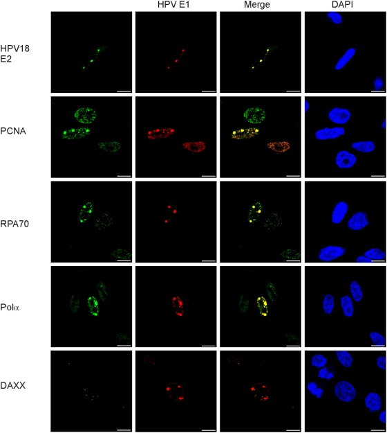
Figure 3 Cellular replication factors colocalize with the HPV 18 E1 in HeLa cells. Cells were transfected and analyzed as described previously. Co-immunostaining of HPV18 E1 (Alexa Fluor 568, second column) together with the following proteins are shown: HPV18 E2, PCNA, RPA, polymerase α/primase, and Daxx (Alexa Fluor 488, first column). Merged images are presented in the third column and DAPI stained nuclei in the fourth column.
Figure 3 Cellular replication factors colocalize with the HPV 18 E1 in HeLa cells. Cells were transfected and analyzed as described previously. Co-immunostaining of HPV18 E1 (Alexa Fluor 568, second column) together with the following proteins are shown: HPV18 E2, PCNA, RPA, polymerase α/primase, and Daxx (Alexa Fluor 488, first column). Merged images are presented in the third column and DAPI stained nuclei in the fourth column.CC BY · PubMed Central
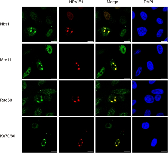
Figure 4 Replication centers of the integrated HPV recruit Mre11-Nbs1-Rad50 complex and Ku70/80 heterodimer. HeLa cells were transfected and analyzed as described previously. Co-immunostaining of HPV18 E1 (Alexa Fluor 568, second column) and the following proteins are presented: Mre11, Nbs1, Rad50, and Ku70/80 proteins (Alexa Fluor 488, first column). The localizations of the E1 and the respective DNA repair proteins are also shown in the third column as a merged image and DAPI stained nuclei are presented in the fourth column.
Figure 4 Replication centers of the integrated HPV recruit Mre11-Nbs1-Rad50 complex and Ku70/80 heterodimer. HeLa cells were transfected and analyzed as described previously. Co-immunostaining of HPV18 E1 (Alexa Fluor 568, second column) and the following proteins are presented: Mre11, Nbs1, Rad50, and Ku70/80 proteins (Alexa Fluor 488, first column). The localizations of the E1 and the respective DNA repair proteins are also shown in the third column as a merged image and DAPI stained nuclei are presented in the fourth column.CC BY · PubMed Central
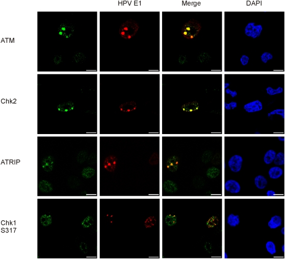
Figure 5 Repair of the amplified HPV18 integration locus is coordinated by the ATM and ATR signaling pathways. HeLa cells were transfected and analyzed as described previously. Co-immunostaining of HPV18 E1 (Alexa Fluor 568, second column) and the following proteins are shown at the figure: ATM, Chk2, ATRIP, and Chk1 (S317) (Alexa Fluor 488, first column). Merged images are shown in the third column and DAPI stained nuclei in the fourth column.
Figure 5 Repair of the amplified HPV18 integration locus is coordinated by the ATM and ATR signaling pathways. HeLa cells were transfected and analyzed as described previously. Co-immunostaining of HPV18 E1 (Alexa Fluor 568, second column) and the following proteins are shown at the figure: ATM, Chk2, ATRIP, and Chk1 (S317) (Alexa Fluor 488, first column). Merged images are shown in the third column and DAPI stained nuclei in the fourth column.CC BY · PubMed Central
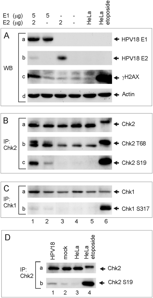
Figure 6 Activation of the ATM-Chk2 signaling pathway. (A–C) HeLa cells were transfected as follows: 5 µg of HPV18 E1 and 2 µg of HPV18 E2 expression plasmids (lane 1); 5 µg of HPV18 E1 expression plasmid (lane 2); 2 µg of E2 expression plasmid (lane 3); and a mock-transfection (lane 4). In every transfection, the amount of plasmid was adjusted to 10 µg with a carrier plasmid (pauxoMCF). Non-transfected HeLa cells are presented in lane 5 and HeLa cells that were treated 1 h with etoposide (50 µM) prior to the analysis in lane 6. Western blot analyses were performed at a 24 hrs time point to detect HPV18 E1 (A, panel a), HPV18 E2 (A, panel b), gamma histone H2AX (phosphorylated at S139) (A, panel c), and β-actin (A, panel d). Western blot analyses of Chk2 phosphorylated at Thr68 and Ser19 were performed after the immunoprecipitation with the anti-Chk2 antibody (B, panels a, b, c). Chk1 phosphorylated at Ser317 was detected from extracts that were immunoprecipitated with the anti-Chk1 antibody (C, panels a, b). (D) HeLa cells were transfected either with 2 µg of circular HPV18 genome, 2 µg of pBabePuro and 6 µg of carrier plasmid (lane 1), or with 2 µg of pBabePuro and 8 µg of carrier plasmid (lane 2). Untransfected cells were removed with puromycin treatment (2 µg/ml) 24–48 h posttransfection. Western blot analyses with anti-Chk2 (panel a) and anti-Chk2-Ser19 (panel b) antibodies were performed after the immunoprecipitation with the anti-Chk2 antibody at a 72 h time point. Untreated HeLa cells are shown in lane 3 and etoposide-treated HeLa cells in lane 4.
Figure 6 Activation of the ATM-Chk2 signaling pathway. (A–C) HeLa cells were transfected as follows: 5 µg of HPV18 E1 and 2 µg of HPV18 E2 expression plasmids (lane 1); 5 µg of HPV18 E1 expression plasmid (lane 2); 2 µg of E2 expression plasmid (lane 3); and a mock-transfection (lane 4). In every transfection, the amount of plasmid was adjusted to 10 µg with a carrier plasmid (pauxoMCF). Non-transfected HeLa cells are presented in lane 5 and HeLa cells that were treated 1 h with etoposide (50 µM) prior to the analysis in lane 6. Western blot analyses were performed at a 24 hrs time point to detect HPV18 E1 (A, panel a), HPV18 E2 (A, panel b), gamma histone H2AX (phosphorylated at S139) (A, panel c), and β-actin (A, panel d). Western blot analyses of Chk2 phosphorylated at Thr68 and Ser19 were performed after the immunoprecipitation with the anti-Chk2 antibody (B, panels a, b, c). Chk1 phosphorylated at Ser317 was detected from extracts that were immunoprecipitated with the anti-Chk1 antibody (C, panels a, b). (D) HeLa cells were transfected either with 2 µg of circular HPV18 genome, 2 µg of pBabePuro and 6 µg of carrier plasmid (lane 1), or with 2 µg of pBabePuro and 8 µg of carrier plasmid (lane 2). Untransfected cells were removed with puromycin treatment (2 µg/ml) 24–48 h posttransfection. Western blot analyses with anti-Chk2 (panel a) and anti-Chk2-Ser19 (panel b) antibodies were performed after the immunoprecipitation with the anti-Chk2 antibody at a 72 h time point. Untreated HeLa cells are shown in lane 3 and etoposide-treated HeLa cells in lane 4.CC BY · PubMed Central
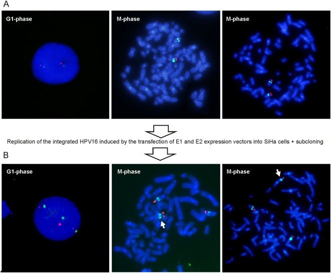
Figure 7 Re-replication of the integrated HPV induces the instability of chromosome structure. SiHa cells were transfected with 10 µg of HPV18 E1 and 5 µg of E2 expression plasmids. Single cell subcloning was performed 72 h after transfection [30]. SiHa cells and the subclones with novel restriction patterns were analyzed by FISH. (A) FISH analysis of the interphase nuclei (left panel) and metaphase chromosome spreads (the middle and the right panels) of SiHa cells. (B) Interphase FISH (left panel) and metaphase FISH (the middle and the right panels) analyses of the subclone with cross-chromosomal translocation. The integrated HPV16 is visible as green (Alexa Fluor 488), and the subtelomeric region of the chromosome 13 is visible as red (Texas Red). The translocation of 13q is indicated with the white arrowhead.
Figure 7 Re-replication of the integrated HPV induces the instability of chromosome structure. SiHa cells were transfected with 10 µg of HPV18 E1 and 5 µg of E2 expression plasmids. Single cell subcloning was performed 72 h after transfection [30]. SiHa cells and the subclones with novel restriction patterns were analyzed by FISH. (A) FISH analysis of the interphase nuclei (left panel) and metaphase chromosome spreads (the middle and the right panels) of SiHa cells. (B) Interphase FISH (left panel) and metaphase FISH (the middle and the right panels) analyses of the subclone with cross-chromosomal translocation. The integrated HPV16 is visible as green (Alexa Fluor 488), and the subtelomeric region of the chromosome 13 is visible as red (Texas Red). The translocation of 13q is indicated with the white arrowhead.CC BY · PubMed Central
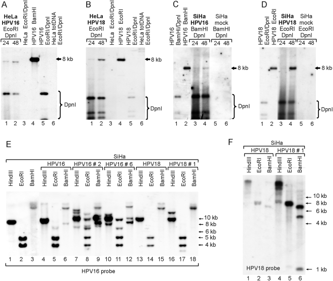
Figure 8 The HPV genome induces genomic instability in cells harboring the integrated HPV. Southern blot analyses of the HeLa (A, B) and SiHa cells (C, D) co-transfected with 1 µg of HPV16 genome and 1 µg of linearized pEGFPN-1 (A, C) or with 1 µg of HPV18 genome and 1 µg of linearized pEGFPN-1 (B, D). Low-molecular-weight DNA was extracted 24 h and 48 h after transfection and digested with restriction enzymes, as indicated in the figure. HPV plasmids were detected with a 32P-labeled HPV16 or HPV18 genome probe, respectively. (E, F) The restriction analyses of untreated SiHa cells (E, lanes 1–3), HPV16-transfected SiHa cells five weeks post-transfection (E, lanes 4–6), HPV18-transfected SiHa cells five weeks post-transfection (E, lanes 13–15; F, lanes 1–3), as well as the subclones of the HPV16-transfected SiHa cells (E, lanes 7–12) and HPV18-transfected SiHa cells (E, lanes 16–18; F, lanes 4–6). Digested total DNA was analyzed by Southern blotting, where the HPV16 (E) or HPV18 (F) genomes were used as probes. Restriction enzymes are indicated in the figure.
Figure 8 The HPV genome induces genomic instability in cells harboring the integrated HPV. Southern blot analyses of the HeLa (A, B) and SiHa cells (C, D) co-transfected with 1 µg of HPV16 genome and 1 µg of linearized pEGFPN-1 (A, C) or with 1 µg of HPV18 genome and 1 µg of linearized pEGFPN-1 (B, D). Low-molecular-weight DNA was extracted 24 h and 48 h after transfection and digested with restriction enzymes, as indicated in the figure. HPV plasmids were detected with a 32P-labeled HPV16 or HPV18 genome probe, respectively. (E, F) The restriction analyses of untreated SiHa cells (E, lanes 1–3), HPV16-transfected SiHa cells five weeks post-transfection (E, lanes 4–6), HPV18-transfected SiHa cells five weeks post-transfection (E, lanes 13–15; F, lanes 1–3), as well as the subclones of the HPV16-transfected SiHa cells (E, lanes 7–12) and HPV18-transfected SiHa cells (E, lanes 16–18; F, lanes 4–6). Digested total DNA was analyzed by Southern blotting, where the HPV16 (E) or HPV18 (F) genomes were used as probes. Restriction enzymes are indicated in the figure.CC BY · PubMed Central
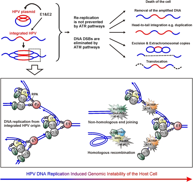
Figure 9 Mechanism of the genomic instability of the cells harboring the replication origin of integrated HPV. If HPV plasmid is present in the cells harboring integrated HPV, the DNA re-replication from the integrated HPV origin is initiated, and ATM and ATR signaling pathways respond, respectively, to the produced DSBs and ssDNA. In most cases, the cells will either become apoptotic or the damage sites will be properly repaired by homologous recombination and non-homologous end-joining. In addition, duplications within the HPV locus have previously been detected. In the current study, the excision and generation of extrachromosomal copies of the HPV locus and cross-chromosomal translocations were also detected. All these mutations require DSBs that could be generated, such as through head-to-tail fork collision or by encounter of the re-replication fork with the Okazaki fragments of the previous fork. Supported by our observations, we speculate that the cellular defense against the integrated HPV re-replication is primary coordinated by ATM. ATR pathways do not interfere with a properly working E1-driven replication fork but, rather, are responsible for the repair of the damage that is caused by fork collision and dissociation that reveals the RPA coated ssDNA without the generation of DSBs. DNA replication/repair factors that are shown to be within the HPV replication centers in the current work, are colored in the figure.
Figure 9 Mechanism of the genomic instability of the cells harboring the replication origin of integrated HPV. If HPV plasmid is present in the cells harboring integrated HPV, the DNA re-replication from the integrated HPV origin is initiated, and ATM and ATR signaling pathways respond, respectively, to the produced DSBs and ssDNA. In most cases, the cells will either become apoptotic or the damage sites will be properly repaired by homologous recombination and non-homologous end-joining. In addition, duplications within the HPV locus have previously been detected. In the current study, the excision and generation of extrachromosomal copies of the HPV locus and cross-chromosomal translocations were also detected. All these mutations require DSBs that could be generated, such as through head-to-tail fork collision or by encounter of the re-replication fork with the Okazaki fragments of the previous fork. Supported by our observations, we speculate that the cellular defense against the integrated HPV re-replication is primary coordinated by ATM. ATR pathways do not interfere with a properly working E1-driven replication fork but, rather, are responsible for the repair of the damage that is caused by fork collision and dissociation that reveals the RPA coated ssDNA without the generation of DSBs. DNA replication/repair factors that are shown to be within the HPV replication centers in the current work, are colored in the figure.CC BY · PubMed Central

Verification

Claims the paper makes

The claims and statistics of this paper haven’t been checked yet.

Run the check — free, no account needed. It reads the open-access text and checks the sources it cites.

A few free checks a day · papers already verified open instantly · results are public

Claims other papers make about this one

Each is checked against this paper’s own text: does it say what the citing paper says it does?

Self-check

Percentages, sums and reported differences re-computed from the paper’s own numbers.

The self-check needs the paper’s full text.

Citations

Cites

Show all 1 references

Research works printed but not linked: 76

  1. M Ustav, 1991, Transient replication of BPV-1 requires two viral polypeptides encoded by the E1 and E2 open reading frames., Embo J, 10, 449, 10.1002/j.1460-2075.1991.tb07967.x · DOI
  2. CM Chiang, 1992, Viral E1 and E2 proteins support replication of homologous and heterologous papillomaviral origins., Proc Natl Acad Sci U S A, 89, 5799, 10.1073/pnas.89.13.5799 · DOI
  3. F Sverdrup, 1994, Replication of human papillomavirus (HPV) DNAs supported by the HPV type 18 E1 and E2 proteins., J Virol, 68, 505, 10.1128/JVI.68.1.505-509.1994 · DOI
  4. M Kadaja, 2009, Papillomavirus DNA replication - from initiation to genomic instability., Virology, 384, 360, 10.1016/j.virol.2008.11.032 · DOI
  5. L Yang, 1993, The E1 protein of bovine papilloma virus 1 is an ATP-dependent DNA helicase., Proc Natl Acad Sci U S A, 90, 5086, 10.1073/pnas.90.11.5086 · DOI
  6. J Sedman, 1998, The papillomavirus E1 protein forms a DNA-dependent hexameric complex with ATPase and DNA helicase activities., J Virol, 72, 6893, 10.1128/JVI.72.8.6893-6897.1998 · DOI
  7. M Ustav, 1991, Identification of the origin of replication of bovine papillomavirus and characterization of the viral origin recognition factor E1., Embo J, 10, 4321, 10.1002/j.1460-2075.1991.tb05010.x · DOI
  8. E Ustav, 1993, The bovine papillomavirus origin of replication requires a binding site for the E2 transcriptional activator., Proc Natl Acad Sci U S A, 90, 898, 10.1073/pnas.90.3.898 · DOI
  9. M Remm, 1992, The E2 binding sites determine the efficiency of replication for the origin of human papillomavirus type 18., Nucleic Acids Res, 20, 6015, 10.1093/nar/20.22.6015 · DOI
  10. J Russell, 1995, cis-Acting components of human papillomavirus (HPV) DNA replication: linker substitution analysis of the HPV type 11 origin., J Virol, 69, 651, 10.1128/JVI.69.2.651-660.1995 · DOI
  11. A Stenlund, 2003, Initiation of DNA replication: lessons from viral initiator proteins., Nat Rev Mol Cell Biol, 4, 777, 10.1038/nrm1226 · DOI
  12. JB Ravnan, 1992, Random-choice replication of extrachromosomal bovine papillomavirus (BPV) molecules in heterogeneous, clonally derived BPV-infected cell lines., J Virol, 66, 6946, 10.1128/JVI.66.12.6946-6952.1992 · DOI
  13. M Piirsoo, 1996, Cis and trans requirements for stable episomal maintenance of the BPV-1 replicator., Embo J, 15, 1, 10.1002/j.1460-2075.1996.tb00328.x · DOI
  14. K Cooper, 1991, Episomal and integrated human papillomavirus in cervical neoplasia shown by non-isotopic in situ hybridisation., J Clin Pathol, 44, 990, 10.1136/jcp.44.12.990 · DOI
  15. E Kristiansen, 1994, Coexistence of episomal and integrated HPV16 DNA in squamous cell carcinoma of the cervix., J Clin Pathol, 47, 253, 10.1136/jcp.47.3.253 · DOI
  16. W Alazawi, 2002, Changes in cervical keratinocyte gene expression associated with integration of human papillomavirus 16., Cancer Res, 62, 6959
  17. P Peitsaro, 2002, Single copy heterozygote integration of HPV 33 in chromosomal band 5p14 is found in an epithelial cell clone with selective growth advantage., Carcinogenesis, 23, 1057, 10.1093/carcin/23.6.1057 · DOI
  18. P Peitsaro, 2002, Integrated human papillomavirus type 16 is frequently found in cervical cancer precursors as demonstrated by a novel quantitative real-time PCR technique., J Clin Microbiol, 40, 886, 10.1128/JCM.40.3.886-891.2002 · DOI
  19. S Andersson, 2005, Type distribution, viral load and integration status of high-risk human papillomaviruses in pre-stages of cervical cancer (CIN)., Br J Cancer, 92, 2195, 10.1038/sj.bjc.6602648 · DOI · Paper page
  20. H Arias-Pulido, 2006, Human papillomavirus type 16 integration in cervical carcinoma in situ and in invasive cervical cancer., J Clin Microbiol, 44, 1755, 10.1128/JCM.44.5.1755-1762.2006 · DOI
  21. SM Kulmala, 2006, Early integration of high copy HPV16 detectable in women with normal and low grade cervical cytology and histology., J Clin Pathol, 59, 513, 10.1136/jcp.2004.024570 · DOI
  22. MR Pett, 2006, Selection of cervical keratinocytes containing integrated HPV16 associates with episome loss and an endogenous antiviral response., Proc Natl Acad Sci U S A, 103, 3822, 10.1073/pnas.0600078103 · DOI
  23. M Pett, 2007, Integration of high-risk human papillomavirus: a key event in cervical carcinogenesis?, J Pathol, 212, 356, 10.1002/path.2192 · DOI
  24. KL Dall, 2008, Characterization of naturally occurring HPV16 integration sites isolated from cervical keratinocytes under noncompetitive conditions., Cancer Res, 68, 8249, 10.1158/0008-5472.CAN-08-1741 · DOI
  25. MR Pett, 2004, Acquisition of high-level chromosomal instability is associated with integration of human papillomavirus type 16 in cervical keratinocytes., Cancer Res, 64, 1359, 10.1158/0008-5472.CAN-03-3214 · DOI
  26. S Jeon, 1995, Integration of human papillomavirus type 16 into the human genome correlates with a selective growth advantage of cells., J Virol, 69, 2989, 10.1128/JVI.69.5.2989-2997.1995 · DOI
  27. H Romanczuk, 1992, Disruption of either the E1 or the E2 regulatory gene of human papillomavirus type 16 increases viral immortalization capacity., Proc Natl Acad Sci U S A, 89, 3159, 10.1073/pnas.89.7.3159 · DOI
  28. H Romanczuk, 1990, Mutational analysis of cis elements involved in E2 modulation of human papillomavirus type 16 P97 and type 18 P105 promoters., J Virol, 64, 2849, 10.1128/JVI.64.6.2849-2859.1990 · DOI
  29. M Kadaja, 2007, Genomic instability of the host cell induced by the human papillomavirus replication machinery., Embo J, 26, 2180, 10.1038/sj.emboj.7601665 · DOI
  30. SA Corden, 1999, The integration of HPV-18 DNA in cervical carcinoma., Mol Pathol, 52, 275, 10.1136/mp.52.5.275 · DOI
  31. M Kalantari, 2001, Physical state of HPV16 and chromosomal mapping of the integrated form in cervical carcinomas., Diagn Mol Pathol, 10, 46, 10.1097/00019606-200103000-00008 · DOI
  32. M Peter, 2006, MYC activation associated with the integration of HPV DNA at the MYC locus in genital tumors., Oncogene, 25, 5985, 10.1038/sj.onc.1209625 · DOI · Paper page
  33. S Duensing, 2004, Mechanisms of genomic instability in human cancer: insights from studies with human papillomavirus oncoproteins., Int J Cancer, 109, 157, 10.1002/ijc.11691 · DOI
  34. S Duensing, 2002, The human papillomavirus type 16 E6 and E7 oncoproteins independently induce numerical and structural chromosome instability., Cancer Res, 62, 7075
  35. S Duensing, 2003, Human papillomavirus type 16 E7 oncoprotein can induce abnormal centrosome duplication through a mechanism independent of inactivation of retinoblastoma protein family members., J Virol, 77, 12331, 10.1128/JVI.77.22.12331-12335.2003 · DOI · Paper page
  36. V Bechtold, 2003, Human papillomavirus type 16 E2 protein has no effect on transcription from episomal viral DNA., J Virol, 77, 2021, 10.1128/JVI.77.3.2021-2028.2003 · DOI
  37. B Hirt, 1967, Selective extraction of polyoma DNA from infected mouse cell cultures., J Mol Biol, 26, 365, 10.1016/0022-2836(67)90307-5 · DOI
  38. S Cohen, 2003, Extrachromosomal circular DNA of tandemly repeated genomic sequences in Drosophila., Genome Res, 13, 1133, 10.1101/gr.907603 · DOI
  39. PA Dijkwel, 1999, Physical and genetic mapping of mammalian replication origins., Methods, 18, 418, 10.1006/meth.1999.0800 · DOI
  40. A Mannik, 2002, Induction of the bovine papillomavirus origin “onion skin”-type DNA replication at high E1 protein concentrations in vivo., J Virol, 76, 5835, 10.1128/JVI.76.11.5835-5845.2002 · DOI
  41. P Meister, 2007, Replication foci dynamics: replication patterns are modulated by S-phase checkpoint kinases in fission yeast., Embo J, 26, 1315, 10.1038/sj.emboj.7601538 · DOI
  42. AA Philimonenko, 2006, The microarchitecture of DNA replication domains., Histochem Cell Biol, 125, 103, 10.1007/s00418-005-0090-0 · DOI
  43. H Leonhardt, 2000, Dynamics of DNA replication factories in living cells., J Cell Biol, 149, 271, 10.1083/jcb.149.2.271 · DOI
  44. CS Swindle, 1999, Human papillomavirus DNA replication compartments in a transient DNA replication system., J Virol, 73, 1001, 10.1128/JVI.73.2.1001-1009.1999 · DOI
  45. MK el Awady, 1987, Molecular analysis of integrated human papillomavirus 16 sequences in the cervical cancer cell line SiHa., Virology, 159, 389, 10.1016/0042-6822(87)90478-8 · DOI
  46. JD Meissner, 1999, Nucleotide sequences and further characterization of human papillomavirus DNA present in the CaSki, SiHa and HeLa cervical carcinoma cell lines., J Gen Virol, 80(Pt 7), 1725, 10.1099/0022-1317-80-7-1725 · DOI
  47. K Szuhai, 2000, Simultaneous molecular karyotyping and mapping of viral DNA integration sites by 25-color COBRA-FISH., Genes Chromosomes Cancer, 28, 92, 10.1002/(SICI)1098-2264(200005)28:1<92::AID-GCC11>3.0.CO;2-2 · DOI
  48. M Macville, 1999, Comprehensive and definitive molecular cytogenetic characterization of HeLa cells by spectral karyotyping., Cancer Res, 59, 141
  49. R Bernardi, 2007, Structure, dynamics and functions of promyelocytic leukaemia nuclear bodies., Nat Rev Mol Cell Biol, 8, 1006, 10.1038/nrm2277 · DOI
  50. HM Shih, 2007, Daxx mediates SUMO-dependent transcriptional control and subnuclear compartmentalization., Biochem Soc Trans, 35, 1397, 10.1042/BST0351397 · DOI
  51. M Shrivastav, 2008, Regulation of DNA double-strand break repair pathway choice., Cell Res, 18, 134, 10.1038/cr.2007.111 · DOI · Paper page
  52. E Sonoda, 2006, Differential usage of non-homologous end-joining and homologous recombination in double strand break repair., DNA Repair (Amst), 5, 1021, 10.1016/j.dnarep.2006.05.022 · DOI
  53. S Matsuoka, 2000, Ataxia telangiectasia-mutated phosphorylates Chk2 in vivo and in vitro., Proc Natl Acad Sci U S A, 97, 10389, 10.1073/pnas.190030497 · DOI
  54. X Xu, 2002, Chk2 activation and phosphorylation-dependent oligomerization., Mol Cell Biol, 22, 4419, 10.1128/MCB.22.12.4419-4432.2002 · DOI
  55. G Buscemi, 2006, DNA damage-induced cell cycle regulation and function of novel Chk2 phosphoresidues., Mol Cell Biol, 26, 7832, 10.1128/MCB.00534-06 · DOI
  56. J Couturier, 1991, Integration of papillomavirus DNA near myc genes in genital carcinomas and its consequences for proto-oncogene expression., J Virol, 65, 4534, 10.1128/JVI.65.8.4534-4538.1991 · DOI
  57. AA Brink, 2002, Simultaneous mapping of human papillomavirus integration sites and molecular karyotyping in short-term cultures of cervical carcinomas by using 49-color combined binary ratio labeling fluorescence in situ hybridization., Cancer Genet Cytogenet, 134, 145, 10.1016/S0165-4608(01)00620-3 · DOI
  58. MG Frattini, 1996, In vitro synthesis of oncogenic human papillomaviruses requires episomal genomes for differentiation-dependent late expression., Proc Natl Acad Sci U S A, 93, 3062, 10.1073/pnas.93.7.3062 · DOI
  59. SC Holmgren, 2005, The minor capsid protein L2 contributes to two steps in the human papillomavirus type 31 life cycle., J Virol, 79, 3938, 10.1128/JVI.79.7.3938-3948.2005 · DOI
  60. JJ Blow, 2005, Preventing re-replication of chromosomal DNA., Nat Rev Mol Cell Biol, 6, 476, 10.1038/nrm1663 · DOI
  61. YJ Machida, 2005, Right place, right time, and only once: replication initiation in metazoans., Cell, 123, 13, 10.1016/j.cell.2005.09.019 · DOI
  62. M Botchan, 1979, Studies on simian virus 40 excision from cellular chromosomes., Cold Spring Harb Symp Quant Biol, 43 Pt 2, 709, 10.1101/SQB.1979.043.01.079 · DOI
  63. D Hanahan, 1980, Characteristics of an SV40-plasmid recombinant and its movement into and out of the genome of a murine cell., Cell, 21, 127, 10.1016/0092-8674(80)90120-8 · DOI
  64. P Bullock, 1984, DNA sequence studies of simian virus 40 chromosomal excision and integration in rat cells., J Mol Biol, 174, 55, 10.1016/0022-2836(84)90365-6 · DOI
  65. DO Ferguson, 2000, The nonhomologous end-joining pathway of DNA repair is required for genomic stability and the suppression of translocations., Proc Natl Acad Sci U S A, 97, 6630, 10.1073/pnas.110152897 · DOI
  66. ZE Karanjawala, 1999, The nonhomologous DNA end joining pathway is important for chromosome stability in primary fibroblasts., Curr Biol, 9, 1501, 10.1016/S0960-9822(00)80123-2 · DOI
  67. NE Sharpless, 2001, Impaired nonhomologous end-joining provokes soft tissue sarcomas harboring chromosomal translocations, amplifications, and deletions., Mol Cell, 8, 1187, 10.1016/S1097-2765(01)00425-7 · DOI
  68. Y Zhang, 2006, Chromatin structural elements and chromosomal translocations in leukemia., DNA Repair (Amst), 5, 1282, 10.1016/j.dnarep.2006.05.020 · DOI
  69. C Vaziri, 2003, A p53-dependent checkpoint pathway prevents rereplication., Mol Cell, 11, 997, 10.1016/S1097-2765(03)00099-6 · DOI
  70. IS Mihaylov, 2002, Control of DNA replication and chromosome ploidy by geminin and cyclin A., Mol Cell Biol, 22, 1868, 10.1128/MCB.22.6.1868-1880.2002 · DOI
  71. W Zhu, 2004, Rereplication by depletion of geminin is seen regardless of p53 status and activates a G2/M checkpoint., Mol Cell Biol, 24, 7140, 10.1128/MCB.24.16.7140-7150.2004 · DOI
  72. E Liu, 2007, The ATR-mediated S phase checkpoint prevents rereplication in mammalian cells when licensing control is disrupted., J Cell Biol, 179, 643, 10.1083/jcb.200704138 · DOI
  73. MR Lentz, 2006, A phosphorylation map of the bovine papillomavirus E1 helicase., Virol J, 3, 13, 10.1186/1743-422X-3-13 · DOI
  74. I Ilves, 2003, Two separate replication modes of the bovine papillomavirus BPV1 origin of replication that have different sensitivity to p53., Virus Res, 96, 75, 10.1016/S0168-1702(03)00174-6 · DOI
  75. P Karakaidos, 2004, Overexpression of the replication licensing regulators hCdt1 and hCdc6 characterizes a subset of non-small-cell lung carcinomas: synergistic effect with mutant p53 on tumor growth and chromosomal instability–evidence of E2F-1 transcriptional control over hCdt1., Am J Pathol, 165, 1351, 10.1016/S0002-9440(10)63393-7 · DOI
  76. Y Tatsumi, 2006, Deregulation of Cdt1 induces chromosomal damage without rereplication and leads to chromosomal instability., J Cell Sci, 119, 3128, 10.1242/jcs.03031 · DOI

Cited by

Metadata

Authors
Kadaja, M., Isok-Paas, H., Laos, T., Ustav, E., & Ustav, M.
Venue
PLoS Pathogens
Published
2009-04-24
Volume
5
Issue
4
Pages
e1000397
ISSN
1553-7374
Cited by
173
References
1
Language
en

About these checks

Registry, retraction, Cites and statistics results are computed, not judged, with one exception: a printed reference that no index lists is read once by a model before it counts as a citation error. Claims are extracted by a model and kept only when their quote appears verbatim in the text; each cited source is then checked, with a model’s help, for whether it says what the claim relies on. Those verdicts can be wrong — read the claim’s quote and the cited source before relying on one.

Statistics and arithmetic are recomputed, not judged.

Confidence bands are shown only for fields calibrated on our annotated test set. None is calibrated yet, so no band is shown.

What counts as a claim ›How we measure confidence ›How we check this ›

Are you an author, or do you see a problem with this page? Request a review